3-Phosphate Dehydrogenase (GAPDH) and
b
-Actin Are
Targets of miR-644a
Kavleen Sikand1¤, Jagjit Singh1, Jey Sabith Ebron1, Girish C. Shukla1,2*
1Department of Biological, Geological and Environmental Sciences, Cleveland State University, Cleveland, Ohio, United States of America,2Center for Gene Regulation in Health and Disease, Cleveland State University, Cleveland, Ohio, United States of America
Abstract
Results of overexpression or downregulation of a microRNA (miRNA) on its target mRNA expression are often validated by reverse-transcription and quantitative PCR analysis using an appropriate housekeeping gene as an internal control. The possible direct or indirect effects of a miRNA on the expression of housekeeping genes are often overlooked. Among many housekeeping genes, expressions of glyceraldehyde-3-phosphate dehydrogenase (GAPDH) andb-actin have been used extensively for normalization of gene expression data. Here, we show that GAPDH andb-actin are direct targets of miR-644a. Our data demonstrate the unsuitability of GAPDH andb-actin as internal controls in miR-644a functional studies and emphasize the need to carefully consider the choice of a reference gene in miRNA experiments.
Citation:Sikand K, Singh J, Ebron JS, Shukla GC (2012) Housekeeping Gene Selection Advisory: Glyceraldehyde-3-Phosphate Dehydrogenase (GAPDH) andb -Actin Are Targets of miR-644a. PLoS ONE 7(10): e47510. doi:10.1371/journal.pone.0047510
Editor:Georg Stoecklin, German Cancer Research Center, Germany
ReceivedJune 16, 2012;AcceptedSeptember 12, 2012;PublishedOctober 16, 2012
Copyright:ß2012 Sikand et al. This is an open-access article distributed under the terms of the Creative Commons Attribution License, which permits unrestricted use, distribution, and reproduction in any medium, provided the original author and source are credited.
Funding:Research in GCS laboratory is supported by grants from National Science Foundation (NSF) and DoD Prostate Cancer Research Program. JS was supported by NSF grant 0842606 and JSE was supported by DoD-USAMRAA grant W81XWH-11-1-0204. The funders had no role in study design, data collection and analysis, decision to publish, or preparation of the manuscript.
Competing Interests:The authors have declared that no competing interests exist.
* E-mail: g.shukla@csuohio.edu
¤ Current address: Department of Translational and Regenerative Medicine, Postgraduate Institute of Medical Education and Research, Chandigarh, India
Introduction
The category of housekeeping genes consists of genes that are involved in the regulation of basic and ubiquitous cellular functions required for the survival of most cell types [1]. Due to their presumptive invariable expression, housekeeping genes have been used extensively as reference genes for normalization of gene expression data derived from a variety of cell types or experimen-tal treatments using microarray or quantitative reverse-transcrip-tase polymerase chain reaction (qRT-PCR). A reference gene is necessary to correct for basic sample differences, including differences in cellular input, RNA quality, efficiency of reverse transcription and batch to batch variation in reagents. Some of the housekeeping genes commonly used as expression controls include glyceraldehyde-3-phosphate dehydrogenase (GAPDH), b-actin, b2-microglobulin, cyclooxygenase 1, hypoxanthine phosphoribosyl transferase 1, glucose-6-phosphate dehydrogenase, cyclophilin A, tubulin, transferrin receptor and 18S ribosomal RNA [2,3].
Changes in mRNA expression profile in response to microRNA (miRNA) inhibition and/or overexpression provide important information about the function of a miRNA. The housekeeping genes, GAPDH andb-actin are routinely used for the normali-zation of data in qRT-PCR experiments estimating the effect of miRNAs on target mRNA expression. These studies assume unaltered expression of GAPDH and b-actin in the presence of ectopically overexpressed miRNAs without giving due consider-ation to the possibility of miRNA-mediated effects on the expression of these genes. A single miRNA has the potential to
target the expression of multiple genes. These target genes may include the housekeeping gene being considered for internal control, thus making it an inappropriate control gene in settings where the miRNA is the experimental treatment given to cells. Here, we show that miR-644a significantly represses GAPDH and b-actin expression. We also show that miR-644a functions by directly binding to its target site in the 39 untranslated region (UTR) of GAPDH andb-actin. Our results reinforce the earlier view of using caution and proper validation while selecting reference genes for a particular experimental setting.
Results and Discussion
to signal transducer and activator of transcription 2 (STAT2) expression. Our computational analysis confirmed that STAT2 open reading frame including 59and 39UTRs does not appear to contain any miR-644a target site that follows established miRNA-target mRNA base-pairing rules. Nevertheless, we investigated if STAT2 mRNA expression is affected by miR-644a transfection. As seen in figure 1A, miR-644a reduced GAPDH mRNA levels by 50% to 90% as compared to NC mimic in LNCaP, 293T and HeLa cells. Similar reduction was observed in b-actin mRNA expression in the three cell lines transfected with miR-644a mimic (Figure 1B). As expected, miR-644a failed to inhibit the expression of STAT2 mRNA (Figure 1C). These data confirmed that GAPDH and b-actin are targets of miR-644a and STAT2 is not. Furthermore, western blots showed the repression of GAPDH and b-actin protein levels by miR-644a transfection in LNCaP, 293T and HeLa cells (Figure 2). Taken together, these data provide evidence for miR-644a-mediated regulation of GAPDH and b-actin expression and hence, indicate the unsuitability of these housekeeping genes as internal controls in experiments involving miR-644a. A recent study [4] designed to evaluate the androgen receptor-targeting miRNAs has also noted the repressive effect of miR-644a on GAPDH mRNA expression; however, this study did not report any repressive effect of miR-644a onb-actin mRNA expression, which was used as an endogenous control instead of GAPDH. Also, no further attempts were made to evaluate if GAPDH is a direct target of miR-644a.
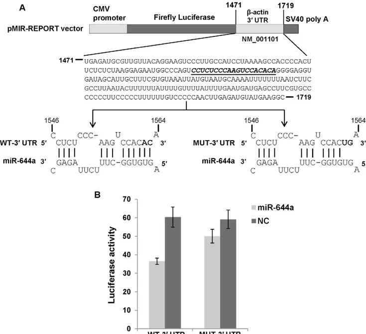
miR-644a Directly Targets GAPDH andb-actin 39UTRs Next, we asked if the observed reduction in GAPDH andb -actin mRNA levels is a consequence of miR-644a inter-acting with the 39UTRs of these mRNAs. A search for GAPDH- andb -actin-targeting miRNAs using TargetScan Human (release 6.1) revealed a potential binding site for miR-644a in GAPDH 39UTR andb -actin 39 UTR suggesting that miR-644a could be directly regulating the expression of these genes by binding to the predicted target sites. We checked the evolutionary conservation of miR-644a target site in GAPDH 39 UTR (Figure 3A) andb -actin 39UTR (Figure 3B) in seven mammalian genomes. The seed binding region of miR-644a target site (shown in bold, Figures 3A, B) was found to be highly conserved in both sets of 39UTRs. In order to validate the direct interaction of miR-644a with its cognate target site, we cloned GAPDH 39 UTR containing the wild type (WT) or mutated (MUT) miR-644a target site in a firefly luciferase reporter vector (Figure 4A). Similar luciferase reporter constructs were made using a segment of b-actin 39 UTR (Figure 5A). In the GAPDH MUT-39UTR construct, nucleotides 1183 to 1187 of the target site were mutated to their complementary nucleotides to disrupt any potential base-pairing interaction of miR-644a (Figure 4A). In theb-actin MUT-39UTR construct, nucleotides 1562 and 1563 of miR-644a binding site were mutated (Figure 5A). Each reporter construct (WT-39UTR or MUT-39UTR) was cotransfected with either miR-644a mimic or NC mimic in CHO-K1 cells and luciferase activity was measured 24 hours post-transfection. In experiments where miR-644a mimic was cotransfected with WT-39 UTR luciferase reporter construct, we observed a marked repression of luciferase activity (Figures 4B and 5B). As expected, in experiments where miR-644a mimic was cotransfected with MUT-39UTR construct, a reversal of luciferase expression was observed (Figures 4B and 5B). Taken together, these data show that miR-644a represses GAPDH and b-actin expression by directly interacting with its target sequence in the respective 39UTRs.
Our results show robust reduction in GAPDH and b-actin mRNA expression and luciferase reporter activity by
overex-pressed miR-644a. However, whether endogenous miR-644a plays a significant role in the modulation of GAPDH andb-actin expression needs further investigation. Interestingly, we could not detect the endogenous expression of miR-644a in several cancer cell lines, including LNCaP and HeLa using TaqMan MicroRNA Assays (Applied Biosystems, Foster City, CA) and qRT-PCR (data not shown). miR-644a is encoded in intron 14 of E3 ubiquitin-protein ligase itchy homolog (ITCH; NM_031483). Recent reports have shown that several miRNAs, including miR-644a repress androgen receptor expression [4,5]. Our data identify two target genes of miR-644a – GAPDH andb-actin. It would be interesting to investigate if miR-644a modulates the downstream functions of these two housekeeping genes and consequently, affects glycolysis, cytoskeleton dynamics and cell motility.
Housekeeping Genes and miRNAs
miRNAs orchestrate posttranscriptional regulation of gene expression mainly by binding to their target sites in the 39UTRs of mRNAs and triggering mRNA degradation or translational repression [6]. Most mammalian mRNAs have 39 UTRs containing target sites for multiple miRNAs. On the other hand, some genes appear to have evolved to avoid miRNA-mediated regulation. Housekeeping genes are considered the classic examples of miRNA non-target genes or ‘‘antitargets’’ [7,8]. Since housekeeping genes are required for basic cellular processes in all cells, miRNA-mediated repression of their expression would be unfavorable for cell survival. Studies indicate that housekeeping genes circumvent miRNA-mediated regulation by limiting their 39 UTR lengths [8,9]. Housekeeping genes appear to have relatively short 39UTRs and consequently, they can harbor fewer miRNA target sites. The miRNA target site prediction tools such as TargetScan and PicTar predict multiple miRNA binding sites in the 39 UTRs of housekeeping genes. However, whether these limited number of target sites and potentially interacting miRNAs play a role in the modulation of housekeeping genes’ expression remains to be determined. A significant biological question is whether the housekeeping gene-targeting miRNAs are expressed ubiquitously in order to maintain stable and constant expression of housekeeping genes in all biological contexts. Alternatively, are these miRNAs expressed at low levels or not expressed at all and consequently, have mild or no effect on the expression of housekeeping genes? Our data demonstrating the repression of GAPDH andb-actin expression by miR-644a suggest that some of the predicted miRNA target sites in the 39UTRs of housekeeping genes may indeed be functional. In addition, previous studies have shown the regulation of b-actin expression by miR-145 [10,11] and miR-206 [12]. Numerous studies have provided evidence for significant variation in the expression of housekeeping genes between different cell types, developmental stages and under different experimental conditions [2,3,13,14]. It is possible that miRNA-mediated regulation is responsible for a portion of this expression variability of housekeeping genes. Interestingly, some housekeeping genes have recently been classified as ‘‘disallowed genes’’ based on their profound repression in specific tissues. The observed disallowance of housekeeping genes has been attributed to epigenetic silencing and miRNA-mediated repression [15].
Several studies have demonstrated the roles of GAPDH in regulation of cytoskeleton [16], membrane fusion and transport [17–19], apoptosis [20,21], DNA repair, DNA replication [22,23] and regulation of transcription and translation [24–27]. In addition, GAPDH has been implicated in the pathophysiology
of neurodegenerative diseases [28]. Both GAPDH andb-actin are differentially expressed in several cancers [29–33]. b-actin expression has been shown to positively correlate with tumor invasiveness and metastatic potential [34,35]. Altered expression of b-actin has also been observed in Alzheimer’s disease and
Figure 1. miR-644a downregulates GAPDH andb-actin mRNA expression.(A and B) Quantitative real-time PCR analysis of GAPDH andb -actin mRNA expression in LNCaP, 293T and HeLa cells transfected with miR-644a mimic or negative control (NC) mimic. (C) In order to demonstrate that the repression of GAPDH andb-actin mRNA expression is a consequence of specific targeting by miR-644a, the effect of miR-644a was checked on a computationally predicted non-target gene, STAT2. STAT2 mRNA expression was determined by quantitative real-time PCR analysis in LNCaP, 293T and HeLa cells transfected with miR-644a mimic or NC mimic. GAPDH,b-actin and STAT2 mRNA expression was normalized to 18S rRNA expression. Data are plotted as mean6SE of three independent experiments.
Down’s syndrome patients [36,37]. A recent study reported the translocation of b-actin from cytoplasm to nucleus during macrophage differentiation of HL-60 cells [38]. The nuclear b -actin was found to regulate transcription during macrophage differentiation [38]. Several studies have reported considerable variation in the expression of GAPDH and b-actin between different tissue types and in response to several experimental treatments, demonstrating their differential regulation and hence, their inadequacy to function as reference genes for data normalization [13,29,33,39–43]. GAPDH expression has been
shown to be modulated by serum, epidermal growth factor, retinoic acid, insulin, norepinephrine, tri-iodothyronine, oestradi-ol, insulin growth factor 1, basic fibroblast growth factor, 1,25-dihydroxyvitamin D3 and some drugs such as bisphosphonates [39,43–46]. Likewise, some regulators ofb-actin include matrigel, hormones, serum, hyperglycemia, hypoxia and tumor necrosis factor-a [33]. Our study adds a new regulator, miR-644a to the growing list of GAPDH and b-actin regulators. In addition to miR-644a, several other miRNAs are predicted to bind GAPDH andb-actin 39UTRs and hence, possess the potential to regulate
Figure 2. miR-644a downregulates GAPDH and b-actin protein expression.(A, B and C) Representative western blots showing the expression of GAPDH,b-actin and STAT2 in LNCaP, 293T and HeLa cells treated with indicated amounts of miR-644a mimic or negative control (NC) mimic for 48 hours. STAT2 expression was used as a loading control. (D, E and F) Quantitation of GAPDH protein expression in the respective lanes as shown in A, B and C. (G, H and I) Quantitation ofb-actin protein expression in the respective lanes as shown in A, B and C. Three independent western blots were used for the quantification of protein expression. The signal intensities of bands were measured using ImageJ software. The GAPDH orb -actin expression in each lane was determined by normalizing GAPDH orb-actin band intensity to STAT2 band intensity. Data are plotted as mean6 SE of three independent experiments.
doi:10.1371/journal.pone.0047510.g002
their expression. It may be wise to consider the list of predicted miRNA binding sites in the 39UTR of a housekeeping gene before selecting it as an internal control in miRNA experiments. Also, it would be interesting to explore if a subset of miRNAs, which potentially target several housekeeping genes share common characteristics and can be grouped into a separate family.
In conclusion, we advise caution regarding the prevailing assumption of inconsequential effects of miRNAs on the expres-sion of housekeeping genes. Even though miRNAs may not play significant roles in the regulation of housekeeping genes under normal physiological conditions, they may exert measurable effect on housekeeping genes in ectopic overexpression experiments. Hence, in experiments where a miRNA is overexpressed in order to study its effect on target gene expression, careful consideration should be given to the selection of a reference gene.
Materials and Methods
Cell Culture
Human prostate cancer cell line, LNCaP was cultured in RPMI 1640 medium supplemented with 10% fetal bovine serum (FBS), 2 mM L-glutamine and antibiotics (100 units/ml of penicillin G sodium; 100mg/ml of streptomycin sulphate). Human cervical cancer cell line, HeLa and human embryonic kidney derived cell line, HEK 293T were cultured in DMEM supplemented with 10% FBS and antibiotics. Chinese hamster ovary derived cell line, CHO-K1 was cultured in DMEM supplemented with 5% FBS,
2 mM L-glutamine, 1 mM L-proline, 10 mM HEPES and antibiotics. All cell lines were obtained from American Type Culture Collection (Manassas, VA) and maintained in a humid-ified 5% CO2atmosphere at 37uC.
Determination of GAPDH,b-actin and STAT2 mRNA Expression by Quantitative Real-time PCR
LNCaP, 293T and HeLa cells were seeded in six-well plates one day prior to transfection. Cells were transfected with miR-644a mimic (50 nM in LNCaP and HeLa; 100 nM in 293T) or negative control (NC) mimic (50 nM) using Lipofectamine 2000 (Invitro-gen, Carlsbad, CA). Synthetic miRNA mimics were obtained from Dharmacon (Chicago, IL). Total RNA was isolated from these cells 48 hours post-transfection using Trizol reagent (Invitrogen). 5mg of total RNA was incubated with 5 units of DNase (Promega, Madison, WI) at 37uC for 40 minutes in order to remove DNA contamination. 1mg of DNase treated RNA was reverse transcribed into cDNA using the ImProm-II Reverse Transcrip-tion System (Promega). Primers for real-time PCR were as follows: (i) GAPDH Forward 59-ACCCACTCCTCCACCTTTGAC-39, Reverse 59-TGTTGCTGTAGCCAAATTCGTT-39; (ii) b-actin Forward 59-GCCGGGACCTGACTGACTAC-39, Reverse 59 -TTCTCCTTAATGTCACGCACGAT-39; (iii) STAT2 Forward 59-ACTGAGCCAATGGAAATCTTCAG-39, Reverse 59 -AAACCTCATCCACGGTGTTCTG-39; (iv) 18S rRNA For-ward 59-TCGGAACTGAGGCCATGATT-39, Reverse 59
-Figure 3. Conservation of miR-644a target site.Panels A and B show alignments of GAPDH andb-actin 39UTR sequences containing miR-644a binding site in 7 mammalian species. miR-644a target site sequence is shown in gray box and seed binding region is shown in bold. Stars indicate conserved nucleotides in the target sequence in at least 5 out of 7 species.
CTTTCGCTCTGGTCCGTCTT-39. The real-time PCR reac-tions were set up using the SYBR GreenER qPCR SuperMix Universal obtained from Invitrogen. In brief, a 20ml reaction was set up containing 1X SYBR Green Supermix (Invitrogen), 0.05mM of each of the forward and reverse primers, 500 nM ROX dye and 1ml of 1 in 5 diluted template cDNA. For amplification with 18S rRNA primers, 1ml of 1 in 100 diluted template cDNA was used per reaction. The reactions were dispensed into 96-well optical plates and amplification was carried out in StepOnePlus Real-time PCR System (Applied Biosystems, Foster City, CA) under the following conditions: 50uC for 2 minutes, 95uC for 10 minutes followed by 40 cycles of 95uC for 15 seconds and 60uC for 1 minute. Three replicates were performed per cDNA sample along with the ‘no reverse transcriptase’ and ‘no template’ controls. The specificity of amplification was confirmed by melting curve analysis and also by running PCR products on 3% agarose gels. Gene expression was quantified using the relative standard curve method. Different dilutions of cDNA synthesized
from RNA extracted from untreated LNCaP cells were used to plot the standard curves for each gene. GAPDH, b-actin and STAT2 mRNA expression was normalized to 18S rRNA expression. Mean normalized GAPDH, b-actin and STAT2 expression 6 SE was calculated from three independent exper-iments.
Western Blotting
Proteins were extracted from LNCaP, 293T and HeLa cells transfected with miR-644a mimic (50 nM, 100 nM) or NC mimic (50 nM) 48 hours post-transfection using the M-PER mammalian protein extraction reagent (Pierce, Rockford, IL) containing protease inhibitor and phosphatase inhibitor cocktail (Sigma-Aldrich, St. Louis, MO). The protein concentration in the total cell lysate was determined by Bradford protein assay. 2mg of protein was resolved on NuPAGE 4–12% Bis-Tris gels (Invitrogen) and electro-transferred to nitrocellulose membranes. Membranes were blocked with 5% nonfat dry milk for 1 hour at room temperature
Figure 4. GAPDH is a direct target of miR-644a.(A) Schematic representation of firefly luciferase reporter construct containing GAPDH 39UTR with either wild type (WT) or mutant (MUT) miR-644a target site. The miR-644a target site in GAPDH 39UTR is italicized and underlined. In the MUT-39 UTR construct, 5 nucleotides (1183–1187) in the seed binding region of the target site were mutated to their complementary nucleotides (shown in bold) in order to disrupt miR-644a binding. (B) Luciferase reporter assay in CHO-K1 cells cotransfected with WT-39UTR or MUT-39UTR construct and miR-644a mimic (2 nM) or negative control (NC) mimic (2 nM) as indicated. Renilla luciferase reporter plasmid was cotransfected in all cases as a control for transfection efficiency. Luciferase activity is plotted as a ratio of firefly to renilla luciferase activity. Each bar represents mean6SE of three independent experiments.
doi:10.1371/journal.pone.0047510.g004
and then incubated overnight with rabbit monoclonal anti-GAPDH antibody (1:20000, Cell Signaling Technology, Inc., Danvers, MA), mouse monoclonal anti-b-actin antibody (1:15000, Sigma-Aldrich, St. Louis, MO) and rabbit polyclonal anti-STAT2 antibody (1:1000, Santa Cruz Biotechnology, Santa Cruz, CA) at 4uC. Blots were washed and incubated with horseradish perox-idase conjugated anti-rabbit (1:5000, Santa Cruz Biotechnology) and anti-mouse (1:10000, GE Healthcare, Piscataway, NJ) secondary antibodies for 1 hour at room temperature. At the end of this incubation, blots were washed and treated with ECL Plus Western blotting detection reagent (GE Healthcare). Bands were visualized by exposing to X-ray films. The signal intensities of
bands were measured using ImageJ software. The level of GAPDH and b-actin protein expression in each lane was determined by normalizing GAPDH andb-actin band intensity to STAT2 band intensity.
Luciferase Assays
GAPDH 39 UTR (200 base pairs; accession number NM_002046.3) and a segment ofb-actin 39UTR (249 base pairs; accession number NM_001101.3) containing the predicted target site for miR-644a were cloned downstream of firefly luciferase coding region in pMIR-REPORT vector (Ambion, Austin, TX).
Figure 5.b-actin is a direct target of miR-644a.(A) Schematic representation of firefly luciferase reporter construct containingb-actin 39UTR with either wild type (WT) or mutant (MUT) miR-644a target site. The italicized and underlined sequence inb-actin 39UTR represents the miR-644a target site. In the MUT-39 UTR construct, 2 nucleotides (1562–1563) in the seed binding region of the target site were mutated to their complementary nucleotides (shown in bold) in order to disrupt miR-644a binding. (B) Luciferase reporter assay in CHO-K1 cells cotransfected with WT-39UTR or MUT-39UTR construct and miR-644a mimic (2 nM) or negative control (NC) mimic (2 nM) as indicated. Renilla luciferase reporter plasmid was cotransfected in all cases as a control for transfection efficiency. Luciferase activity is plotted as a ratio of firefly to renilla luciferase activity. Each bar represents mean6SE of three independent experiments.
These constructs were named WT-39UTR (WT: wild type). Site-directed mutagenesis of the putative target site for miR-644a in WT-39UTR constructs was carried out in order to generate the MUT-39UTR constructs using the Change-IT Multiple Mutation Site Directed Mutagenesis kit (USB Corporation, Cleveland, OH). In the GAPDH MUT-39UTR construct, 5 nucleotides in the seed matching region of the target site were mutated to their complementary sequence so as to abolish the putative miRNA:-target mRNA base-pairing. In the b-actin MUT-39 UTR construct, 2 nucleotides (AC) in the seed matching region of miR-644a target site were mutated to their complementary sequence (UG). Nucleotide sequences of the constructs were confirmed by DNA sequencing. For luciferase assays, CHO-K1 cells (30,000 cells/well) were plated in 24-well plates one day prior to transfection. Cells were co-transfected using Lipofectamine 2000 reagent (Invitrogen), with 100 ng of WT-39UTR or MUT-39 UTR firefly luciferase reporter construct, 0.5 ng of renilla luciferase reporter plasmid (Promega) and either miR-644a mimic
(2 nM) or NC mimic (2 nM). Cell lysates were assayed for firefly and renilla luciferase activities 24 hours after transfection using the Dual-Luciferase Reporter Assay System (Promega) and Victor 3 Multilabel Counter 1420 (PerkinElmer). Renilla luciferase activity served as a control for transfection efficiency. Data are represented as ratio of firefly luciferase activity to renilla luciferase activity. Luciferase assays were repeated at least three times with two replicates each time and substantially similar results were obtained.
Acknowledgments
Authors wish to thank Dr. Sailen Barik for critical reading of the manuscript.
Author Contributions
Conceived and designed the experiments: KS JS JSE GCS. Performed the experiments: KS JS JSE. Analyzed the data: KS JS JSE GCS. Contributed reagents/materials/analysis tools: GCS. Wrote the paper: KS JS JSE GCS.
References
1. Eisenberg E, Levanon EY (2003) Human housekeeping genes are compact. Trends Genet 19: 362–365.
2. De Kok JB, Roelofs RW, Giesendorf BA, Pennings JL, Waas ET, et al. (2005) Normalization of gene expression measurements in tumor tissues: comparison of 13 endogenous control genes. Lab Invest 85: 154–159.
3. Lee PD, Sladek R, Greenwood CM, Hudson TJ (2002) Control genes and variability: absence of ubiquitous reference transcripts in diverse mammalian expression studies. Genome Res 12: 292–297.
4. Ostling P, Leivonen SK, Aakula A, Kohonen P, Makela R, et al. (2011) Systematic analysis of microRNAs targeting the androgen receptor in prostate cancer cells. Cancer Res 71: 1956–1967.
5. Sikand K, Slaibi JE, Singh R, Slane SD, Shukla GC (2011) miR 488* inhibits androgen receptor expression in prostate carcinoma cells. Int J Cancer 129: 810–819.
6. Bartel DP (2004) MicroRNAs: genomics, biogenesis, mechanism, and function. Cell 116: 281–297.
7. Bartel DP, Chen CZ (2004) Micromanagers of gene expression: the potentially widespread influence of metazoan microRNAs. Nat Rev Genet 5: 396–400. 8. Stark A, Brennecke J, Bushati N, Russell RB, Cohen SM (2005) Animal
MicroRNAs confer robustness to gene expression and have a significant impact on 39UTR evolution. Cell 123: 1133–1146.
9. Cheng C, Bhardwaj N, Gerstein M (2009) The relationship between the evolution of microRNA targets and the length of their UTRs. BMC Genomics 10: 431.
10. Takagi T, Iio A, Nakagawa Y, Naoe T, Tanigawa N, et al. (2009) Decreased expression of microRNA-143 and -145 in human gastric cancers. Oncology 77: 12–21.
11. Szczyrba J, Loprich E, Wach S, Jung V, Unteregger G, et al. (2010) The microRNA profile of prostate carcinoma obtained by deep sequencing. Mol Cancer Res 8: 529–538.
12. Adams BD, Furneaux H, White BA (2007) The micro-ribonucleic acid (miRNA) miR-206 targets the human estrogen receptor-alpha (ERalpha) and represses ERalpha messenger RNA and protein expression in breast cancer cell lines. Mol Endocrinol 21: 1132–1147.
13. Schmittgen TD, Zakrajsek BA (2000) Effect of experimental treatment on housekeeping gene expression: validation by real-time, quantitative RT-PCR. J Biochem Biophys Methods 46: 69–81.
14. Ferreira E, Cronje MJ (2012) Selection of suitable reference genes for quantitative real-time PCR in apoptosis-induced MCF-7 breast cancer cells. Mol Biotechnol 50: 121–128.
15. Thorrez L, Laudadio I, Van DK, Quintens R, Hendrickx N, et al. (2011) Tissue-specific disallowance of housekeeping genes: the other face of cell differentiation. Genome Res 21: 95–105.
16. Huitorel P, Pantaloni D (1985) Bundling of microtubules by glyceraldehyde-3-phosphate dehydrogenase and its modulation by ATP. Eur J Biochem 150: 265– 269.
17. Glaser PE, Gross RW (1995) Rapid plasmenylethanolamine-selective fusion of membrane bilayers catalyzed by an isoform of glyceraldehyde-3-phosphate dehydrogenase: discrimination between glycolytic and fusogenic roles of individual isoforms. Biochemistry 34: 12193–12203.
18. Robbins AR, Ward RD, Oliver C (1995) A mutation in glyceraldehyde 3-phosphate dehydrogenase alters endocytosis in CHO cells. J Cell Biol 130: 1093–1104.
19. Tisdale EJ, Kelly C, Artalejo CR (2004) Glyceraldehyde-3-phosphate dehydro-genase interacts with Rab2 and plays an essential role in endoplasmic reticulum to Golgi transport exclusive of its glycolytic activity. J Biol Chem 279: 54046– 54052.
20. Ishitani R, Chuang DM (1996) Glyceraldehyde-3-phosphate dehydrogenase antisense oligodeoxynucleotides protect against cytosine arabinonucleoside-induced apoptosis in cultured cerebellar neurons. Proc Natl Acad Sci U S A 93: 9937–9941.
21. Hara MR, Snyder SH (2006) Nitric oxide-GAPDH-Siah: a novel cell death cascade. Cell Mol Neurobiol 26: 527–538.
22. Meyer-Siegler K, Mauro DJ, Seal G, Wurzer J, deRiel JK, et al. (1991) A human nuclear uracil DNA glycosylase is the 37-kDa subunit of glyceraldehyde-3-phosphate dehydrogenase. Proc Natl Acad Sci U S A 88: 8460–8464. 23. Baxi MD, Vishwanatha JK (1995) Uracil
DNA-glycosylase/glyceraldehyde-3-phosphate dehydrogenase is an Ap4A binding protein. Biochemistry 34: 9700– 9707.
24. Morgenegg G, Winkler GC, Hubscher U, Heizmann CW, Mous J, et al. (1986) Glyceraldehyde-3-phosphate dehydrogenase is a nonhistone protein and a possible activator of transcription in neurons. J Neurochem 47: 54–62. 25. Singh R, Green MR (1993) Sequence-specific binding of transfer RNA by
glyceraldehyde-3-phosphate dehydrogenase. Science 259: 365–368.
26. Nagy E, Rigby WF (1995) Glyceraldehyde-3-phosphate dehydrogenase selectively binds AU-rich RNA in the NAD(+)-binding region (Rossmann fold). J Biol Chem 270: 2755–2763.
27. Zheng L, Roeder RG, Luo Y (2003) S phase activation of the histone H2B promoter by OCA-S, a coactivator complex that contains GAPDH as a key component. Cell 114: 255–266.
28. Chuang DM, Hough C, Senatorov VV (2005) Glyceraldehyde-3-phosphate dehydrogenase, apoptosis, and neurodegenerative diseases. Annu Rev Pharma-col ToxiPharma-col 45: 269–290.
29. Schek N, Hall BL, Finn OJ (1988) Increased glyceraldehyde-3-phosphate dehydrogenase gene expression in human pancreatic adenocarcinoma. Cancer Res 48: 6354–6359.
30. Corbin IR, Gong Y, Zhang M, Minuk GY (2002) Proliferative and nutritional dependent regulation of glyceraldehyde-3-phosphate dehydrogenase expression in the rat liver. Cell Prolif 35: 173–182.
31. Blomberg J, Andersson M, Faldt R (1987) Differential pattern of oncogene and beta-actin expression in leukaemic cells from AML patients. Br J Haematol 65: 83–86.
32. Lupberger J, Kreuzer KA, Baskaynak G, Peters UR, le CP, et al. (2002) Quantitative analysis of beta-actin, beta-2-microglobulin and porphobilinogen deaminase mRNA and their comparison as control transcripts for RT-PCR. Mol Cell Probes 16: 25–30.
33. Ruan W, Lai M (2007) Actin, a reliable marker of internal control? Clin Chim Acta 385: 1–5.
34. Le PU, Nguyen TN, Drolet-Savoie P, Leclerc N, Nabi IR (1998) Increasedb -actin expression in an invasive moloney sarcoma virus-transformed MDCK cell variant concentrates to the tips of multiple pseudopodia. Cancer Res 58: 1631– 1635.
35. Nowak D, Skwarek-Maruszewska A, Zemanek-Zboch M, Malicka-Blaszkiewicz M (2005) Beta-actin in human colon adenocarcinoma cell lines with different metastatic potential. Acta Biochim Pol 52: 461–468.
36. Jabbour W, Pouplard-Barthelaix A, Houlgatte R, Emile J (1992) Abnormal expression of actin in lymphocytes of Alzheimer’s disease and Down’s syndrome patients. J Neuroimmunol 38: 199–208.
37. Gutala RV, Reddy PH (2004) The use of real-time PCR analysis in a gene expression study of Alzheimer’s disease post-mortem brains. J Neurosci Methods 132: 101–107.
38. Xu YZ, Thuraisingam T, Morais DA, Rola-Pleszczynski M, Radzioch D (2010) Nuclear translocation ofb-actin is involved in transcriptional regulation during macrophage differentiation of HL-60 cells. Mol Biol Cell 21: 811–820.
39. Revillion F, Pawlowski V, Hornez L, Peyrat JP (2000) Glyceraldehyde-3-phosphate dehydrogenase gene expression in human breast cancer. Eur J Cancer 36: 1038–1042.
40. Glare EM, Divjak M, Bailey MJ, Walters EH (2002) b-Actin and GAPDH housekeeping gene expression in asthmatic airways is variable and not suitable for normalising mRNA levels. Thorax 57: 765–770.
41. Bas A, Forsberg G, Hammarstrom S, Hammarstrom ML (2004) Utility of the housekeeping genes 18S rRNA, b-actin and glyceraldehyde-3-phosphate-dehydrogenase for normalization in real-time quantitative reverse transcrip-tase-polymerase chain reaction analysis of gene expression in human T lymphocytes. Scand J Immunol 59: 566–573.
42. Barber RD, Harmer DW, Coleman RA, Clark BJ (2005) GAPDH as a housekeeping gene: analysis of GAPDH mRNA expression in a panel of 72 human tissues. Physiol Genomics 21: 389–395.
43. Valenti MT, Bertoldo F, Dalle CL, Azzarello G, Zenari S, et al. (2006) The effect of bisphosphonates on gene expression: GAPDH as a housekeeping or a new target gene? BMC Cancer 6: 49.
44. Barroso I, Benito B, Garci-Jimenez C, Hernandez A, Obregon MJ, et al. (1999) Norepinephrine, tri-iodothyronine and insulin upregulate glyceraldehyde-3-phosphate dehydrogenase mRNA during Brown adipocyte differentiation. Eur J Endocrinol 141: 169–179.
45. Desprez PY, Poujol D, Saez S (1992) Glyceraldehyde-3-phosphate dehydroge-nase (GAPDH, E.C. 1.2.1.12.) gene expression in two malignant human mammary epithelial cell lines: BT-20 and MCF-7. Regulation of gene expression by 1,25-dihydroxyvitamin D3 (1,25-(OH)2D3). Cancer Lett 64: 219–224. 46. Matrisian LM, Rautmann G, Magun BE, Breathnach R (1985) Epidermal