Submitted23 May 2016
Accepted 30 September 2016
Published20 December 2016
Corresponding author
Chee Sian Kuan,
Academic editor
Dee Carter
Additional Information and Declarations can be found on page 16
DOI10.7717/peerj.2637
Copyright
2016 Ng et al.
Distributed under
Creative Commons CC-BY 4.0
OPEN ACCESS
Identification and characterization of
Daldinia eschscholtzii
isolated from skin
scrapings, nails, and blood
Kee Peng Ng1, Chai Ling Chan1, Su Mei Yew1, Siok Koon Yeo2, Yue Fen Toh1, Hong Keat Looi1, Shiang Ling Na1, Kok Wei Lee3, Wai-Yan Yee3and
Chee Sian Kuan1
1Department of Medical Microbiology, University of Malaya, Kuala Lumpur, Malaysia
2School of Biosciences, Taylor’s University, Lakeside Campus, Subang Jaya, Malaysia
3Codon Genomics, Selangor, Malaysia
ABSTRACT
Background.Daldinia eschscholtziiis a filamentous wood-inhabiting endophyte com-monly found in woody plants. Here, we report the identification and characterization of nineD. eschscholtziiisolates from skin scrapings, nail clippings, and blood.
Methods. The nine isolates were identified based on colony morphology, light microscopy, and internal transcribed spacer (ITS)-based phylogeny.In vitroantifungal susceptibility of the fungal isolates was evaluated by the Etest to determine the minimum inhibitory concentration (MIC).
Results. The nine isolates examined were confirmed asD. eschscholtzii. They exhibited typical features ofDaldiniasp. on Sabouraud Dextrose Agar, with white felty colonies and black-gray coloration on the reverse side. Septate hyphae, branching conidiophore with conidiogenous cells budding from its terminus, and nodulisporium-like conid-iophores were observed under the microscope. Phylogenetic analysis revealed that the nine isolates were clustered within theD. eschscholtziispecies complex. All the isolates exhibited low MICs against azole agents (voriconazole, posaconazole, itraconazole, and ketoconazole), as well as amphotericin B, with MIC of less than 1µg/ml.
Discussion. Early and definitive identification ofD. eschscholtziiis vital to reducing misuse of antimicrobial agents. Detailed morphological and molecular characterization as well as antifungal profiling ofD. eschscholtziiprovide the basis for future studies on its biology, pathogenicity, and medicinal potential.
SubjectsMycology, Infectious Diseases, Pathology
Keywords Daldinia eschscholtzii, Internal transcribed spacer, Plylogenetic analysis
INTRODUCTION
Members of the genusDaldiniaare pyrenomycetes, which are characterized by internal horizontally zonated stromata that develop conspicuously on woody plants (Stadler et al., 2014).Daldiniaspp. are initial colonizers as evident from the early appearance of stromata following stress or damage to the woody host plant. Initial colonization is a trait ofDaldinia
climatic stress, fire, or lightning (Johannesson, Læssøe & Stenlid, 2000;Srutka, Pazoutova & Kolarik, 2007). At this stage,Daldiniaspp. becomes wood-decaying in its habit, and produces anamorphic structures under favorable conditions of humidity and temperature to colonize the substrate further (Stadler et al., 2014).
D. eschscholtzii is a wood-inhabiting endophyte or wood-decaying fungus that is
widespread in warm tropical climate (Stadler et al., 2014). It is characterized by colonies that are white to smoky gray with a slight olivaceous-tone, and by conidiogenous structures with a nodulisporium-like branching pattern (Ju, Rogers & Martin, 1997;Stadler et al.,
2014). D. eschscholtziigrows preferentially on dead or decaying wood substrates, and
is commonly isolated from dead woody plants such as dicotyledonous crop plants, trees, and occasionally, marine algae (Karnchanatat et al., 2007; Tarman et al., 2012;
Zhang et al., 2008).
Compelling data in the last decade has demonstrated the presence of a wide array of secondary metabolites in this fungus, such as 1,1′
-binaphthalene-4,4′
-5,5′
-tetrol (BNT) (a polyketide derived from 1,8-dihydroxynaphthalene biosynthesis), cytochalasins (metabolites of mixed polyketide/NRPS origin), concentricols (terpenoids derived from the acetate-mevanolate pathway), dalesconol A and B (polyketides), and helicascolide C (polyketides) (Fang et al., 2012;Stadler et al., 2001a;Stadler et al., 2001b; Tarman et al., 2012;Zhang et al., 2008;Zhang et al., 2011). Some of these secondary metabolites are precursors of biologically active medicinal compounds. Dalesconol A and B have immunosuppressive activity (Zhang et al., 2008;Zhang et al., 2011) while helicascolide C exhibits antifungal activity against the phytopathogenic fungusCladosporium cucumerinum
(Tarman et al., 2012). In a previous study, genome analysis ofD. eschscholtziiclinical isolates showed that our isolates UM 1400 and UM 1020 are potentially rich in secondary metabolites (Chan et al., 2015). The presence of the gene encoding lovastatin nonaketide synthase suggests that these isolates can synthesize the drug lovastatin that is used to induce a hypocholesterolemic effect (Chan et al., 2015).
D. eschscholtzii had not been reported as a human pathogen until we isolated this
species from skin scrapings and the blood of patients with suspected fungal infections (Chan et al., 2015; Ng et al., 2012; Yew et al., 2014). To the best of our knowledge, all previous isolations of D. eschscholtzii from humans were by our group (Chan et al., 2015; Ng et al., 2012;Yew et al., 2014). Nevertheless, the clinical evidence of infection caused by this fungus remains unclear. In this study, we identified a total of nine D. eschscholtziiclinical isolates, including the aforementioned isolates in the past five years. Here, we present a detailed morphological, molecular, phenotypic characterization, and antifungal susceptibility profile ofD. eschscholtzii. These data may serve as a reference for the mycological research community for rapid detection ofD. eschscholtzii.
MATERIALS & METHODS
Ethical statementTable 1 Clinical isolates ofD. eschscholtziiisolated in this study.
Isolate Source Year GenBank
accession number
Reference
UM 1020a Blood 2010 JX966563.1 Chan et al. (2015);Ng
et al. (2012);Yew et al. (2014)
UM 230a Nail clipping 2011 JX966562.1 Yew et al. (2014)
UM 1400 Skin scraping 2012 JX966561.1 Chan et al. (2015);Ng et al. (2012)
UM 1094 Skin scraping 2014 KT936494 Present study
UM 1104 Skin scraping 2015 KT936495 Present study
UM 1134 Skin scraping 2015 KT936496 Present study
UM 1216 Nail clipping 2015 KT936497 Present study
UM 1217 Nail clipping 2015 KT936498 Present study
UM 1218 Nail clipping 2015 KT936499 Present study
Notes.
aInviable isolates, the morphological study and unique DNA signature evaluation for these two isolates were excluded in this study while the antifungal susceptibility Etest MIC readings were adopted from previous study (Yew et al., 2014).
Fungal isolates
UM 230, UM 1020, UM 1094, UM 1104, UM 1134, UM 1216, UM 1217, UM 1218, and UM 1400 were isolated from skin scrapings, nail clippings, and blood of patients with suspected fungal infection in the Mycology diagnostic laboratory, UMMC, Kuala Lumpur, Malaysia (Table 1). The isolates were processed according to the laboratory’s standard operating procedures (SOP) (Yew et al., 2014) with direct wet mount microscopy followed by culture on Sabouraud Dextrose Agar (SDA; Oxoid Ltd., Basingstoke, UK) for incubation at 30◦
C for seven days. The isolates were archived at 4◦
C in SDA slants and maintained by periodic subculturing on SDA slants at 30◦
C. UM 1020 and UM 230 isolates were not included in the morphological study as both isolates were no longer viable at the point of analysis.
Morphological study
Morphological and colony features such as color, texture, and topography of the isolates were examined on SDA, potato dextrose agar (PDA; Difco Laboratories, Detroit, MI), and V8 juice agar (V8; HiMedia Laboratories, Mumbai, India). The isolates were incubated at 30◦
C with alternate-day examination for fungal growth. Slide cultures of the fungi on SDA, PDA, and V8 agar were performed as previously described (Kuan et al., 2015). After a 7-day incubation at 30◦
C, the fungal slide cultures were stained with lactophenol cotton blue stain and examined under the light microscope (Leica DM3000 Led, Germany).
DNA extraction
using ZR Fungal/Bacterial DNA MiniPrepTM (Zymo Research, USA) according to the manufacturer’s protocol.
PCR amplification and DNA sequencing
The ITS1-5.8S-ITS2 region was PCR amplified from the isolates’ genomic DNA in a 25µl
reaction consisting of 10×PCR buffer, 10µM each of ITS1 (5′-TCCGTAGGTGAACCT
GCGG-3′) and ITS4 (5′-TCCTCCGCTTATTGATATGC-3′) primers (
White et al., 1990), 25 mM MgC12, 2 mM deoxynucleoside triphosphate, 2.5 unit of HotStarTaq DNA
polymerase, and 10 µg of each genomic DNA. The PCR was performed for 30 cycles at
94◦
C for 30 s, 58◦
C for 30 s, and 72◦
C for 60 s. The PCR products were then purified using ExpinTMPCR SV (GeneAll, Korea), and confirmed by Sanger sequencing (First Base Laboratories Kuala Lumpur, Malaysia). TraceTuner version 3.0.6 (Denisov, Arehart & Curtin, 2004) was used for base and quality calling of the sequenced ITS. The low-quality called bases (Phred value < 20) of both ends of the sequences were then trimmed by running Lucy version 1.20 (Chou & Holmes, 2001) and the included zapping.awk script. The processed ITS sequences were searched against the NCBI non-redundant (nr) nucleotide database using the nucleotide BLAST program.
Phylogenetic analysis
Unique ITS nucleotide sequences from the isolates, together with an additional 72 reference sequences for the ITS region ofDaldiniaspp., were compiled for ITS-based phylogenetic analysis (Table 2). TwoHypoxylon fragiformesequences were used as outgroup strains in the analysis (Table 2). Multiple sequence alignments of all ITS sequences were performed using M-Coffee (Moretti et al., 2007). The alignments were then trimmed using trimAl version 1.4.rev10 to remove the alignment regions with≥50% gaps (Capella-Gutierrez,
Silla-Martinez & Gabaldon, 2009). The trimmed alignments were subsequently used for phylogenetic analysis conducted using MrBayes version 3.2.1 (Huelsenbeck & Ronquist, 2001). Bayesian Markov chain Monte Carlo (MCMC) analysis was initiated by sampling across the entire general time reversible (GTR) model space. A total of 1,500,000 generations were run with a sampling frequency of 100, and diagnostics were calculated for every 1,000 generations. The first 2,500 trees were discarded with a burn-in setting of 25%. Convergence was assessed with a standard deviation of split frequencies below 0.01, no noticeable trend in the generation versus log probability of the data plot, and a potential scale reduction factor (PSRF) close to 1.0 for all parameters (Ronquist, Huelsenbeck & Teslenko, 2011).
In vitroantifungal susceptibility test
The Etest (bioMérieux, France) was performed according to the manufacturer’s instructions to determine the MICs of anidulafungin (ANID), amphotericin B (AMB), caspofungin (CAS), fluconazole (FLC), itraconazole (ITC), ketoconazole (KTC), posaconazole (PSC), and voriconazole (VRC). The concentration gradient of ANID, AMB, CAS, ITC, KTC, PSC, and VRC ranged from 0.002 to 32µg/ml, while that of FLC ranged from 0.016 to
256µg/ml. The test was performed on RPMI 1640 medium containing 2% glucose and
Table 2 Details of isolates subjected to ITS-based phylogenetic analysis.
Fungal species eIsolate GenBank accession no. References
Daldinia albofibrosa CBS117737 JX658518.1 Stadler et al. (2014)
Daldinia albofibrosa MUCL:43509 (T) JX658451.1 Stadler et al. (2014)
aDaldinia andina CBS114736 AM749918.1 Bitzer et al. (2008)
Daldinia asphalatum MUCL:47964 JX658544.1 Stadler et al. (2014)
Daldinia asphalatum MUCL:47966 JX658548.1 Stadler et al. (2014)
Daldinia australis ICMP 18263 (PT) JX658541.1 Stadler et al. (2014)
Daldinia australis CBS119013 (T) JX658450.1 Stadler et al. (2014)
Daldinia bambusicola CBS 122872 (T) JX658436.1 Stadler et al. (2014)
Daldinia barkalovii CBS116999 (T) JX658537.1 Stadler et al. (2014)
Daldinia caldariorum ATCC 36660 AM749933.1 Bitzer et al. (2008)
Daldinia caldariorum CBS122874 JX658452.1 Stadler et al. (2014)
Daldinia carpinicola CBS122880 (T) JX658442.1 Stadler et al. (2014)
Daldiniacf.australis MUCL:53761 JX658547.1 Stadler et al. (2014)
Daldiniacf.caldariorum CBS113045 JX658453.1 Stadler et al. (2014)
Daldiniacf.concentrica MUCL:45434 JX658473.1 Stadler et al. (2014)
Daldiniacf.dennisiivar.microspora ICMP18265 JX658539.1 Stadler et al. (2014)
Daldiniacf.eschscholtzii KC1690 JX658456.1 Stadler et al. (2014)
Daldiniacf.grandis IMCP18266 JX658543.1 Stadler et al. (2014)
Daldiniacf.mexicana Ww3844/MUCL JX658460.1 Stadler et al. (2014)
Daldiniacf.pyrenaica MUCL:47221 JX658515.1 Stadler et al. (2014)
Daldiniacf.pyrenaica MUCL:51700 JX658516.1 Stadler et al. (2014)
Daldinia childiae CBS116725 AM749932.1 Bitzer et al. (2008)
Daldinia childiae MUCL:48616 JX658464.1 Stadler et al. (2014)
Daldinia clavata MUCL:47436 JX658546.1 Stadler et al. (2014)
Daldinia concentrica CBS113277 AY616683.1 Triebel et al. (2005)
Daldinia concentrica MUCL:54179 JX658471.1 Stadler et al. (2014)
Daldinia decipiens MUCL:44610, CBS113046 JX658476.1 Stadler et al. (2014)
Daldinia decipiens CBS122879 (PT) JX658441.1 Stadler et al. (2014)
Daldinia dennisii CBS114741 (T) JX658477.1 Stadler et al. (2014)
Daldinia dennisii CBS114742 (PT) JX658479.1 Stadler et al. (2014)
Daldinia dennisiivar.microspora MUCL:45010 JX658478.1 Stadler et al. (2014)
Daldinia dennisiivar.microspora ICMP18264 JX658538.1 Stadler et al. (2014)
Daldinia eschscholtzii Not available AB284189.1 Karnchanatat et al. (2007)
Daldinia eschscholtzii CALP11206 (ET) HE590883.1 Stadler et al. (2014)
Daldinia eschscholtzii CBS113047 AY616684.1 Triebel et al. (2005)
Daldinia eschscholtzii MUCL:45434 JX658484.1 Stadler et al. (2014)
Daldinia eschscholtzii CBS113042 JX658497.1 Stadler et al. (2014)
Daldinia eschscholtzii CBS116032 JX658500.1 Stadler et al. (2014)
Daldinia eschscholtzii MUCL:38740 JX658493.1 Stadler et al. (2014)
Daldinia eschscholtzii MUCL:47965 JX658482.1 Stadler et al. (2014)
Daldinia gelatinoides MUCL 46173 GQ355621.1 Stadler et al. (2014)
Table 2(continued)
Fungal species eIsolate GenBank accession no. References
Daldinia gelatinosa UAMH 7406 JX658458.1 Stadler et al. (2014)
Daldinia gelatinosa CBS116730 JX658503.1 Stadler et al. (2014)
Daldinia govorovae CBS122883 (T) JX658443.1 Stadler et al. (2014)
Daldinia hausknechtii CBS119995 (T) JX658521.1 Stadler et al. (2014)
Daldinia lloydii CBS113483 JX658457.1 Stadler et al. (2014)
Daldinia loculata BJ Coppins 10274 (C), CBS 114738 (ET) AF176965.1 Johannesson, Læssøe & Stenlid (2000)
Daldinia loculata TL 4613 (C) AF176964.1 Johannesson, Læssøe & Stenlid (2000)
bDaldinia loculatoides BJ Coppins 8630 (E), CBS113279 (T) AF176982.1 Johannesson, Læssøe & Stenlid (2000)
Daldinia loculatoides PRM885050, CBS116729 AM407726.1 S Pazoutova, 2006, unpublished data Daldinia macaronesica Ww4196(M) (T) JX658506 Stadler et al. (2014)
Daldinia macaronesica CBS 113040 (PT) JX658504.1 Stadler et al. (2014)
Daldinia martinii CBS113041 (T) JX658507.1 Stadler et al. (2014)
Daldinia mexicana Ww3843/MUCL (T) JX658508.1 Stadler et al. (2014)
cDaldinia nemorosa UAMH 11227 HM114296.1 ML Davey, 2010, unpublished data
Daldinia novae-zelandiae CBS 114739 (PT) JX658509.1 Stadler et al. (2014)
Daldinia novae-zelandiae CBS 122873 JX658437.1 Stadler et al. (2014)
Daldinia palmensis CBS113039 (T) JX658510.1 Stadler et al. (2014)
Daldinia petriniae MUCL:49214, CBS119988 JX658512.1 Stadler et al. (2014)
Daldinia petriniae MUCL:51850 JX658513.1 Stadler et al. (2014)
Daldinia pyrenaica MUCL:43749 (T) AM749927.1 Bitzer et al. (2008)
Daldinia raimundi CBS 113038 (T) JX658517.1 Stadler et al. (2014)
Daldinia raimundi MUCL:51689 JX658446.1 Stadler et al. (2014)
Daldinia starbaeckii MUCL:45436 (T) JX658488.1 Stadler et al. (2014)
Daldinia starbaeckii CBS116727 JX658489.1 Stadler et al. (2014)
Daldinia steglichii MUCL:43512 (PT) JX658534.1 Stadler et al. (2014)
Daldinia steglichii MUCL:53886 JX658545.1 Stadler et al. (2014)
Daldinia theissenii BCRC34045, CBS122875 JX658468.1 Stadler et al. (2014)
dDaldinia theissenii CBS113044 AM749931.1 Bitzer et al. (2008)
Daldinia vanderguchtiae CBS113036 (T) JX658520.1 Stadler et al. (2014)
Daldinia vernicosa CBS 161.31 (T) JX658519.1 Stadler et al. (2014)
Daldinia vernicosa CBS119316 AM749925.1 Bitzer et al. (2008)
Hypoxylon fragiforme CBS114745 AY616690.1 Stadler et al. (2014)
Hypoxylon fragiforme YMJ 383 JN979420.1 Hsieh & Rogers (2005)
Notes.
aPreviously idetifies asD. grandisbyBitzer et al. (2008)and reclassified byStadler et al. (2014).
bPreviously identified asD. grandisbyJohannesson, Læssøe & Stenlid (2000)and reclassified byStadler et al. (2014). cPreviously identified asAnnelosporium nemorosumand reclassified byStadler et al. (2014).
spread 500µl fungal suspension evenly on a RPMI plate. Etest strips were placed on plates
that had been dried for at least 10 min at room temperature. The MICs were determined after 72 h of incubation at 30◦
C. Both on-scale and off-scale MICs were included in the data. For the calculation of geometric mean (GM), the high off-scale MICs (>32 and >256µg/ml) were rounded up to the next intermediate Etest dilution (48 and 384µg/ml),
while the low off-scale MICs (<0.002 and <0.016µg/ml) remained unchanged.
Nucleotide sequence accession numbers
The ITS nucleotide sequences of UM 1094, UM 1104, UM 1134, UM 1216, UM 1217, and UM 1218 were deposited in the GenBank database with the accession numbersKT936494, KT936495,KT936496,KT936497,KT936498, andKT936499, respectively (Table 1).
RESULTS
Morphological study
All seven isolates grew rapidly on SDA. Initially, white hyphae grew from the inoculated site and formed a felty azonate mycelium. With aging, it turned to smoky gray color with a slight olivaceous tone as the mycelium became fully differentiated (Fig. 1). The smoky gray coloration was indicative of sporulation. All isolates had a black-gray coloration on the reverse side. The cultural characteristics of all fungal isolates that grew on SDA were similar to those that growing on PDA plates. However, their growth rates on PDA were different. The strains UM 1134, UM 1216, and UM 1218 grew faster (five days to reach the periphery of the 9 cm plate) than UM 1400, UM 1094, UM 1104, and UM 1217 (seven days) (Fig. 2). On V8 agar, all the colonies reached the edge of plate after a 5-day incubation; they had a felty to fluffy texture appearance (Fig. 3). The surfaces of the colony changed from white to gray or black after 5 days of incubation. The reverse side of V8 agar plate was initially colorless, and became slightly black after culture for five days. UM 1134 showed denser mycelial growth on SDA, PDA, and V8 agar as compared to the other isolates (Figs. 1D,2Dand3D).
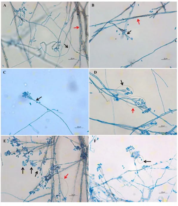
Light microscope analysis revealed that all isolates that grew on SDA, PDA, and V8 agar had similar morphology. The septate hyphae could be hyaline thin-walled or melanized thick-walled (Fig. 4A). The thick-walled hyphae showed black exudates on their surfaces (Fig. 4B). Septate conidiophores were irregularly branched into mononematous, dichotomous or trichotomous structures with one to three conidiogenous cells originating from the terminus (Figs. 4C–4E). The conidiophores were hyaline and black with occasional pigmented exudates. The conidiogenous cells were hyaline and cylindrical. On the apex of conidiogenous cells, conidia were produced holoblastically in a sympodial sequence (Fig. 4E). The conidia were hyaline and ellipsoid with an attenuated base as shown in Fig. 4F.Table 3summarizes the morphological features of this fungal species.
Molecular study
Figure 1 Colonial morphology ofD. eschscholtziiisolates on SDA. (A) UM 1400, (B) UM 1094, (C) UM 1104, (D) UM 1134, (E) UM 1216, (F) UM 1217, and (G) UM 1218 were incubated at 30◦C for 5
Figure 2 Colonial morphology ofD. eschscholtziiisolates on PDA.(A) UM 1400, (B) UM 1094, (C) UM 1104, and (G) UM 1218 were incubated at 30◦C for 7 days. (D) UM 1134, (E) UM 1216, and (F) UM
Figure 3 Colonial morphology ofD. eschscholtziiisolates on V8 agar.(A) UM 1400, (B) UM 1094, (C) UM 1104, (D) UM 1134, (E) UM 1216, (F) UM 1217, and (G) UM 1218 were incubated at 30◦C for 5
Table 3 Key morphological features of clinically isolatedD. eschscholtzii.
Culture medium Macroscopic features Microscopic features
Sabouraud dextrose agar Colonies attain a diameter of 9 cm agar plate in 5 days of incubation at 30◦C.
Colonies are felty and azonate.
At first colonies are whitish, turning smoke gray with slight olivaceous tone in age. Reverse appears black in color. Potato dextrose agar Colonies attain a diameter of 9 cm agar
plate in 5–7 days of incubation at 30◦C.
Colonies are felty, azonate or zonate. At first colonies are whitish, turning smoke gray with slight olivaceous tone with age. Reverse appears black in color.
V8 juice agar Colonies attain a diameter of 9 cm agar plate in 5 days of incubation at 30◦C.
Colonies are felty to fluffy, azonate.
At first colonies are whitish, later turning gray or black, some with slight olivaceous tone. Reverse is initially uncolored, later becoming slight blackish.
Hyphae are septate, thin-walled and hyaline to thick-walled and melanized, with thick-walled hyphae often have blackish exudates. Conidiophores are septate, hyaline to melanized, some with blackish exudates, mononematously, dichotomously or trichotomously irregular branched, occasionally branched from conidiogenous region, bearing one to three conidiogenous cells on its terminus, up to 167µm length×2.2–3.3µm diameter. Conidiogenous cells are cylindrical and hyaline, bearing conidia on its apical region, 8.9–27.8µm length×1.1–2.2µm diameter. Conidia are ellipsoid, aseptate, solitary, hyaline, with attenuated base, produced holoblastically in sympodial sequence, 4.4–6.7µm length×1.7–2.2µm diameter.
group (Group I),D. concentricagroup (Group II),D. vernicosa/loculatagroup (Group III), D. childiaegroup (Group IV), andD. petriniae group (Group V). In this study, all the isolates were clustered within the D. eschscholtziigroup, forming a cluster with the reference isolates ofD. eschscholtzii.
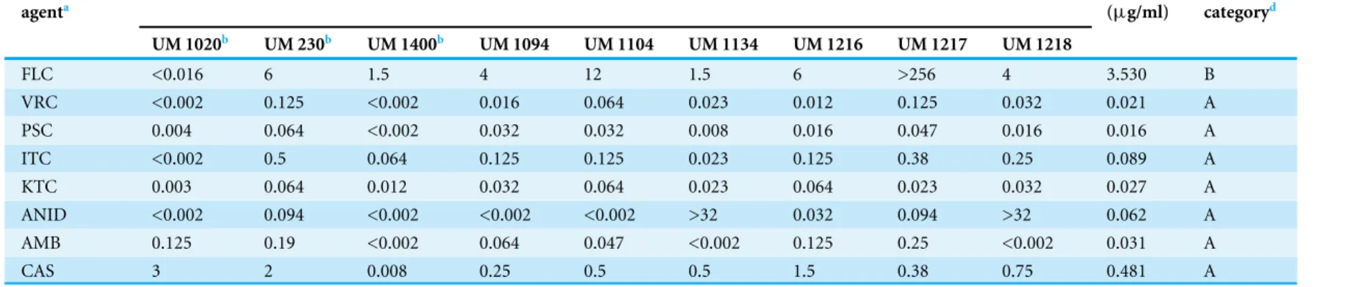
In vitroantifungal susceptibility test
The MICs of the nine isolates tested are shown inTable 4. The antifungal susceptibility profiles obtained were isolate-dependent. In general, VRC, PSC, ITC, KTC, and AMB displayed very low MICs to D. eschscholtzii, with all the isolates exhibiting MICs of ≤1µg/ml. ANID also showed low MICs to (≤1µg/ml) the different isolates, with the
exception of UM 1134 and UM 1218 (>32µg/ml). Similarly, low MIC values (≤1µg/ml)
were obtained for CAS, with two thirds of the isolates eliciting MICs of≤1µg/ml, and
the remaining eliciting MICs of >1µg/ml. Overall, PSC exhibited the highestin vitro
anti-fungal activity (GM MIC 0.016µg/ml) against all isolates, followed by VRC (GM
MIC 0.021µg/ml), KTC (GM MIC 0.027µg/ml), AMB (GM MIC 0.031µg/ml), ANID
(GM MIC 0.062µg/ml), ITC (GM MIC 0.089µg/ml), and CAS (GM MIC 0.481µg/ml).
Relatively high MICs againstD. eschscholtzii(88.89% with MIC >1µg/ml; GM MIC value
of 3.530µg/ml) were found for FLC, indicating potential resistance ofD. eschscholtziito
this drug.
DISCUSSION
Daldinia eschscholtzii is a filamentous fungus commonly found as an endophyte or a
Table 4 Minimum inhibitory concentration (MICs) for the isolates determined by Etest.
Antifungal agenta
Etest MIC (µg/ml) GMc
(µg/ml)
MIC categoryd
UM 1020b UM 230b UM 1400b UM 1094 UM 1104 UM 1134 UM 1216 UM 1217 UM 1218
FLC <0.016 6 1.5 4 12 1.5 6 >256 4 3.530 B
VRC <0.002 0.125 <0.002 0.016 0.064 0.023 0.012 0.125 0.032 0.021 A
PSC 0.004 0.064 <0.002 0.032 0.032 0.008 0.016 0.047 0.016 0.016 A
ITC <0.002 0.5 0.064 0.125 0.125 0.023 0.125 0.38 0.25 0.089 A
KTC 0.003 0.064 0.012 0.032 0.064 0.023 0.064 0.023 0.032 0.027 A
ANID <0.002 0.094 <0.002 <0.002 <0.002 >32 0.032 0.094 >32 0.062 A
AMB 0.125 0.19 <0.002 0.064 0.047 <0.002 0.125 0.25 <0.002 0.031 A
CAS 3 2 0.008 0.25 0.5 0.5 1.5 0.38 0.75 0.481 A
Notes.
aFLC, fluconazole; VRC, voriconazole; PSC, posaconazole; ITC, itraconazole; KTC, ketoconazole; ANID, anidulafungin; AMB, amphotericin B; CAS, caspofungin. bEtest MIC values were retrieved from previous study (Yew et al., 2014).
cGM, geometric mean.
dMIC categories: Category A:≤1µg/ml, Category B: >1–32µg/ml or >1–256µg/ml, Category C: >32µg/ml or >256µg/ml.
Ng
e
t
al.
(2016),
P
eerJ
,
DOI
from humans, it is unclear whether it is the cause of an actual infection, or if it merely exists as a harmless colonizer living in the nail plate or skin surface damaged by trauma or other diseases. In this study, we obtained nineD. eschscholtziiisolates from blood specimens, skin scrapings, and nail clippings. While the clinical significance ofD. eschscholtziiremains in question, repeated isolation of this fungal species from humans recently suggests that it is not a mere environmental contaminant in patients.Chan et al. (2015)report that the genomes ofD. eschscholtziiharbor several stress adaptation mechanisms for their survival in human hosts. Hence, it would not be surprising for the species to have undergone rapid evolution to select for fitness attributes as well as virulence factors related to pathogenicity in humans.
Filamentous fungi are routinely identified by colony morphology and microscopy. The former would not precisely identifyD. eschscholtziiowing to their natural variation among the isolates and tendency towards media-dependency, as evident from their macroscopic appearances. Identification to species level based on morphological examination alone would be difficult as many species ofDaldiniaare morphologically very similar (Ju, Rogers & Martin, 1997;Stadler et al., 2014), and hence considerable expertise and experience are required of the examiner in this regard.
The ITS region of the nuclear rDNA can be used to examine species level relationship in fungi due to its higher degree of variation. Thus, PCR-based ITS sequence analysis has been widely used to identify D. eschscholtzii(Chan et al., 2015;Hu et al., 2014;Tarman et al., 2012;Yuyama et al., 2013). Despite recent studies reporting limitations of the ITS region in distinguishing between the species complexes ofDaldiniaspp., this region has the broadest taxa covered inDaldinia(Stadler et al., 2014). The phylogenetic analysis showed that our clinical isolates and reference environmental isolates ofD. eschscholtziiformed a cluster. However, further studies based on protein coding genes are needed to segregate members of theD. eschscholtziispecies complex reliably.
The Etest is a simple, reliable, and reproducible assay that has been shown to correlate with the Clinical and Laboratory Standards Institute (CLSI) method in antifungal susceptibility testing of filamentous fungi (Espinel-Ingroff, 2001; Szekely, Johnson & Warnock, 1999). In line with this, we applied the Etest to study the antifungal profiles of our isolates. The results showed thatD. eschscholtziielicited low MICs in all the antifungal agents tested, except for FLC. Among these antifungals, VRC, PSC, ITC, KTC, and AMB were the more active in vitro, with all isolates inhibited by concentrations of less than 1 µg/ml. Since there is no available information on antifungal susceptibility profiles
forD. eschscholtzii, this work will contribute towards establishing an optimal antifungal
precautionary treatment for this fungus.
CONCLUSIONS
In this paper, we report the isolation ofD. eschscholtziifrom superficial sites in humans, predominantly skin and nails. If these fungi are confirmed to be of clinical importance,
thein vitroantifungal activities determined here might be useful in clinical practice. The
D. eschscholtzii, and to provide clues on how they are evolutionarily adapted to the human host. The data in this study will serve as a foundation for future research on pathogenicity ofD. eschscholtziiin humans.
ADDITIONAL INFORMATION AND DECLARATIONS
Funding
This study was supported by High Impact Research MoE Grant UM.C/625/1/HIR/ MOHE/MED/31 (Account no. H-20001-00-E000070) from the Ministry of Education Malaysia. Codon Genomics SB provided support in the form of salaries for Kok Wei Lee and Wai-Yan Yee. The funders had no role in study design, data collection and analysis, decision to publish, or preparation of the manuscript.
Grant Disclosures
The following grant information was disclosed by the authors: High Impact Research MoE: UM.C/625/1/HIR/MOHE/MED/31.
Competing Interests
Kok Wei Lee and Wai-Yan Yee are employed by Codon Genomics SB. All other authors have declared that no competing interest exists. These affiliations do not alter our adherence to all the policies on sharing data and materials.
Author Contributions
• Kee Peng Ng conceived and designed the experiments, contributed reagents/materials/-analysis tools, wrote the paper, reviewed drafts of the paper.
• Chai Ling Chan performed the experiments, analyzed the data, wrote the paper, prepared figures and/or tables.
• Su Mei Yew performed the experiments, wrote the paper, reviewed drafts of the paper.
• Siok Koon Yeo analyzed the data, wrote the paper, reviewed drafts of the paper.
• Yue Fen Toh and Shiang Ling Na performed the experiments.
• Hong Keat Looi analyzed the data, wrote the paper, prepared figures and/or tables.
• Kok Wei Lee analyzed the data, contributed reagents/materials/analysis tools.
• Wai-Yan Yee analyzed the data, contributed reagents/materials/analysis tools, reviewed drafts of the paper.
• Chee Sian Kuan conceived and designed the experiments, performed the experiments, analyzed the data, contributed reagents/materials/analysis tools, wrote the paper, prepared figures and/or tables, reviewed drafts of the paper.
DNA Deposition
REFERENCES
Bitzer J, Læssøe J, Fournier T, Kummer V, Decock C, Tichy H-V, Piepenbring M, Peršoh D, Stadler M. 2008.Affinities of Phylacia and the daldinoid Xylariaceae, inferred from chemotypes of cultures and ribosomal DNA sequences.Mycological
Research112:251–270 DOI 10.1016/j.mycres.2007.07.004.
Capella-Gutierrez S, Silla-Martinez JM, Gabaldon T. 2009.trimAl: a tool for auto-mated alignment trimming in large-scale phylogenetic analyses.Bioinformatics 25:1972–1973DOI 10.1093/bioinformatics/btp348.
Chan CL, Yew SM, Ngeow YF, Na SL, Lee KW, Hoh CC, Yee WY, Ng KP. 2015.
Genome analysis ofDaldinia eschscholtziistrains UM 1400 and UM 1020, wood-decaying fungi isolated from human hosts.BMC Genomics16:966 DOI 10.1186/s12864-015-2200-2.
Chou HH, Holmes MH. 2001.DNA sequence quality trimming and vector removal.
Bioinformatics17:1093–1104DOI 10.1093/bioinformatics/17.12.1093.
Denisov GA, Arehart AB, Curtin MD. 2004.System and method for improving the accuracy of DNA sequencing and error probability estimation through application of a mathematical model to the analysis of electropherograms. Paracel, Inc. US Patent 6,681,186.Available athttps:// www.google.com/ patents/ US6681186.
Espinel-Ingroff A. 2001.Comparison of the E-test with the NCCLS M38-P method for antifungal susceptibility testing of common and emerging pathogenic filamentous fungi.Journal of Clinical Microbiology39:1360–1367
DOI 10.1128/JCM.39.4.1360-1367.2001.
Fang W, Ji S, Jiang N, Wang W, Zhao GY, Zhang S, Ge HM, Xu Q, Zhang AH, Zhang YL, Song YC, Zhang J, Tan RX. 2012.Naphthol radical couplings determine structural features and enantiomeric excess of dalesconols inDaldinia eschscholzii.
Nature Communications3:Article 1039DOI 10.1038/ncomms2031.
Hsieh H-M, Rogers JD. 2005.Molecular phylogeny ofHypoxylonand closely related genera.Mycologia94:844–865.
Hu ZX, Xue YB, Bi XB, Zhang JW, Luo ZW, Li XN, Yao GM, Wang JP, Zhang YH. 2014.
Five new secondary metabolites produced by a marine-associated fungus,Daldinia
eschscholzii.Marine Drugs12:5563–5575DOI 10.3390/md12115563.
Huelsenbeck JP, Ronquist F. 2001.MRBAYES: bayesian inference of phylogenetic trees.
Bioinformatics17:754–755DOI 10.1093/bioinformatics/17.8.754.
Johannesson HS, Læssøe T, Stenlid J. 2000.Molecular and morphological inves-tigation ofDaldiniain northern Europe.Mycological Research104:275–280 DOI 10.1017/S0953756299001719.
Ju YM, Rogers JD, San Martin F. 1997.A revision of the genusDaldinia.Mycotaxon 61:243–293.
eschscholzii(Ehrenb.:Fr.) Rehm.FEMS Microbiology Letters270:162–170 DOI 10.1111/j.1574-6968.2007.00662.x.
Karnchanatat A, Petsom A, Sangvanich P, Piapukiew J, Whalley AJS, Reynolds CD, Gadd GM, Sihanonth P. 2008.A novel thermostable endoglucanase from the wood-decaying fungusDaldinia eschscholzii(Ehrenb.:Fr.) Rehm.Enzyme and Microbial
Technology42:404–413DOI 10.1016/j.enzmictec.2007.11.009.
Kuan CS, Yew SM, Toh YF, Chan CL, Lim SK, Lee KW, Na SL, Hoh CC, Yee WY, Ng KP. 2015.Identification and characterization of a rare fungus,Quambalaria cyanescens, isolated from the peritoneal fluid of a patient after nocturnal intermittent peritoneal dialysis.PLoS ONE10:e0145932DOI 10.1371/journal.pone.0145932.
Moretti S, Armougom F, Wallace IM, Higgins DG, Jongeneel CV, Notredame C. 2007.The M-Coffee web server: a meta-method for computing multiple sequence alignments by combining alternative alignment methods.Nucleic Acids Research 35:W645–W648DOI 10.1093/nar/gkm333.
Ng KP, Ngeow YF, Yew SM, Hassan H, Soo-Hoo TS, Na SL, Chan CL, Hoh CC, Lee KW, Yee WY. 2012.Draft genome sequence ofDaldinia eschscholziiisolated from blood culture.Eukaryotic Cell11:703–704DOI 10.1128/EC.00074-12.
Ronquist F, Huelsenbeck J, Teslenko M. 2011. Draft MrBayes version 3.2 manual: tutorials and model summaries.
Srutka P, Pazoutova S, Kolarik M. 2007.Daldinia decipiensandEntonaema cinnabarina
as fungal symbionts of Xiphydria wood wasps.Mycological Research111:224–231 DOI 10.1016/j.mycres.2006.10.006.
Stadler M, Baumgartner M, Grothe T, Mühlbauer A, Seip S, Wollweber H. 2001a.
Concentricol, a taxonomically significant triterpenoid fromDaldinia concentrica.
Phytochemistry56:787–793 DOI 10.1016/S0031-9422(01)00032-2.
Stadler M, Laessoe T, Fournier J, Decock C, Schmieschek B, Tichy HV, Persoh D. 2014.
A polyphasic taxonomy ofDaldinia(Xylariaceae).Studies in Mycology77:1–143 DOI 10.3114/sim0016.
Stadler M, Wollweber H, Mu A, Hashimoto T, Rogers JD, Ju Y, Wetzstein H, Tichy H. 2001b.Molecular chemotaxonomy ofDaldiniaand otherXylariaceae.Mycological
Research105:1191–1205DOI 10.1016/S0953-7562(08)61990-5.
Szekely A, Johnson EM, Warnock DW. 1999.Comparison of E-test and broth microdi-lution methods for antifungal drug susceptibility testing of molds.Journal of Clinical
Microbiology37:1480–1483.
Tarman K, Palm GJ, Porzel A, Merzweiler K, Arnold N, Wessjohann LA, Unterseher M, Lindequist U. 2012.Helicascolide C, a new lactone from an Indonesian marine algicolous strain ofDaldinia eschscholzii(Xylariaceae, Ascomycota).Phytochemistry
Letters5:83–86DOI 10.1016/j.phytol.2011.10.006.
Triebel D, Peršoh D, Wollweber H, Stadler M. 2005.Phylogenetic relationships among
Daldinia,Entonaema, andHypoxylonas inferred from ITS nrDNA analyses of
Xylariales.Nova Hedwigia80:25–43DOI 10.1127/0029-5035/2005/0080-0025.
White TJ, eds.PCR protocol a guidance to methods application. New York: Academic Press, 315–322.
Yew SM, Chan CL, Lee KW, Na SL, Tan R, Hoh CC, Yee WY, Ngeow YF, Ng KP. 2014.
A five-year survey of dematiaceous fungi in a tropical hospital reveals potential opportunistic species.PLoS ONE9:e104352DOI 10.1371/journal.pone.0104352.
Yuyama K, Pereira J, Maki C, Ishikawa N. 2013.Daldinia eschscholtzii(Ascomycota, Xylariaceae) isolated from the Brazilian Amazon: taxonomic features and mycelial growth conditions.Acta Amazonica43:1–8DOI 10.1590/S0044-59672013000100001.
Zhang YL, Ge HM, Zhao W, Dong H, Xu Q, Li SH, Li J, Zhang J, Song YC, Tan RX. 2008.Unprecedented immunosuppressive polyketides fromDaldinia eschscholzii, a mantis-associated fungus.Angewandte Chemie International Edition in English 47:5823–5826DOI 10.1002/anie.200801284.