Biometric study of human teeth
*
Carlos Alberto Gregório Cabrera**, Arnaldo Pinzan***, Marise de Castro Cabrera****,
José Fernando Castanha Henriques*****, Guilherme Janson******, Marcos Roberto de Freitas*******
Objectives: To determine the biometric dimensions of human teeth in the mesiodistal, buc-colingual and occlusal/incisal-cervical directions. Methods: It was used a sample of dental casts from 57 patients, i.e., 31 females with a mean age of 15 years and 5 months, and 26 males with a mean age of 16 years and 6 months. The sample was previously qualified by adopting the criteria established by Andrews’ six keys to normal occlusion, whose values were matched to the variations obtained by Bolton. Two examiners used a digital caliper with original (short) and modified (long) tips. Results and Conclusions: After statistical analysis of the data it was concluded that the teeth were shown to be symmetrical in the dental arches of both genders. Tooth dimensions are smaller in females than in males and should therefore be studied separately. Overall mean values were obtained and used to build tables distinguishing such dimensions according to gender. Mean values for the three tooth dimensions, occurrence rates of these dimensions and their standard deviations were also calculated. These values allowed the development of an equation called “C” equation as well as “C” percentile tables. With the aid of both, it became possible to measure only one dimension of a given tooth to find the other two “probable” dimensions of the other teeth in the dental arches.
Abstract
Keywords: Tooth dimensions. Tooth proportions. Tooth size.
** PhD in Orthodontics, Bauru School of Dentistry, São Paulo University (USP).
*** Associate Professor, Department of Pediatric Dentistry, Orthodontics and Public Health, Bauru School of Dentistry - USP.
**** PhD in Orthodontics, Bauru School of Dentistry, São Paulo University (USP). Head of the Orthodontics Specialization Course – Cabrera/Herrero. ***** Full Professor, Department of Pediatric Dentistry, Orthodontics and Public Health, Bauru School of Dentistry - USP.
****** Full Professor and Head of the Department of Pediatric Dentistry, Orthodontics and Public Health, Bauru School of Dentistry - USP. Head of the Masters Course, FOB-USP.
******* Full Professor, Department of Pediatric Dentistry, Orthodontics and Public Health, Bauru School of Dentistry - USP. Head of the Doctoral course, FOB-USP.
intROduCtiOn
In view of the difficulty to accommodate me-siodistal, buccolingual and occlusal/incisal-cer-vical volumes of the dental masses in restricted locations available in the jaws, orthodontists are ultimately hard pressed to resort to alternative therapies to change the perimeter of the dental arches, either reducing them through extractions
and stripping, or expanding them by proclining the teeth. Although these alternatives have been uncontroversially established, decision-making can sometimes prove challenging. If on the one hand, extractions, when needed, can assist in ad-justing the dental arches and promoting function, on the other hand, retoclined upper central inci-sors may cause cosmetic damage, with consequent
» The authors report no commercial, proprietary, or inancial interest in the products or companies described in this article.
How to cite this article: Cabrera CAG, Pinzan A, Cabrera MC, Henriques
JFC, Janson G, Freitas MR. Biometric study of human teeth. Dental Press J Orthod. 2011 July-Aug;16(4):111-22.
prominence of the nose, particularly if lip retrac-tion is excessive.
Given the uncertainty in deciding whether or not to perform tooth extractions or stripping, this study aimed to determine the biometric dimen-sions of orthodontic patients’ teeth properly fin-ished in the mesiodistal, buccolingual and occlu-sal/incisal-cervical direction in both genders, with a view to providing mathematical support for this decision.
LitERAtuRE REViEW
Literature review disclosed that many re-searchers have sought to address issues that have been accepted but not yet well understood. This constant search stems from the investigative spirit of human beings who, not satisfied with the in-formation currently available, seek out conceptual definitions that can be supported by existing sci-entific methods.
To this end one sees studies focused on several areas, such as dentistry,15 endodontics,9 orthodon-tics,1,5,12,17,18,22 prosthesis16 and forensics.6,21
As the first author to publish a table of mea-surements of human teeth, Black4 is credited as having conducted the first and most detailed study of dental morphology and anatomical no-menclature of all times.
One hundred and one years after Black’s pub-lication4 in 1902, Harris and Burris,11 in 2003, em-phasized that the most often cited tooth dimen-sions in literature were those published by Black.4 However, they also argue that these values differ from modern values and should therefore be reas-sessed. The authors of the present study were moti-vated by this contention to undertake this research.
MAtERiAL And MEthOds Material
sample
This study used a sample of orthodontic plas-ter models of 57 patients distributed between the two genders of the human species, 31 were female
with mean age of 15 years and 5 months, and 26 males with mean age of 16 years and 6 months. No racial, cultural or socio-economic criteria were established. All cases were treated with standard Straight-Wire orthodontic appliances (“A” Com-pany). Cases had no extractions, no pre- and post-treatment interproximal stripping and were all well finished.
Sample qualiication (Andrews and Bolton) The goal consisted in finishing all cases with the six keys to normal occlusion recommended by Andrews.2 Results received an “A” qualifica-tion, the highest quality grade for this method. Additionally, all cases showed proportionality between the 12 maxillary teeth and the 12 man-dibular ones, and between the 6 upper anterior teeth and the 6 lower ones when compared to the values described by Bolton5 (Fig 1).
digital caliper
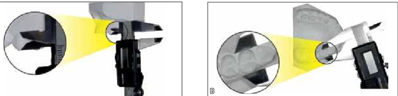
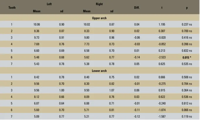
In seeking to emulate the method adopted by Yamaguto22 and Castro7 some modifications were made (Fig 2) to the tips of the original digital caliper. They were replaced by two lon-ger tips to enable measurements in areas of difficult physical access, i.e., to measure ex-clusively the mesiodistal dimensions (Fig 3) of dental crowns on plaster models and thereby determine whether or not errors occurred in the methods, i.e., original tips vs. modified tips. Original tips were referred to as “short” and the modified tips, “long”.
Methods
Methods used to perform measurements in the models
a
D
B
E
C
a
a
a
B
B
B
FIGURE 1 - Images of case used for sample qualification.
FIGURE 2 - Images showing digital caliper with a) its original tips (short) and B) the modified tips (long).
FIGURE 3 - Mesiodistal measurement of a first upper premolar using the digital caliper with a) its original tips (short) and B) the modified tips (long).
statistical study
In strict compliance with all scientific proto-cols, the results and values obtained from tooth size in the sample were subjected to statistical tests. In comparing gender variables Student’s t-test was applied whereas to assess the tions between measurements Pearson’s correla-tion coefficient was employed. Paired t-test was used to identify intra-examiner, inter-examiners and intmethods systematic errors. Random er-ror was calculated using the method proposed by Dahlberg8 as described by Houston.13 In all statis-tical tests a significance level of 5% was adopted.23
The tests were performed using the software Sta-tistics for Windows v. 5.1 (Statsoft, USA).
REsuLts
This study made use of a sample of 57 patients examining the differences between the genders. It required measuring all teeth in three dimen-sions, resulting in 6,620 measurements, which resulted in 59 tables, with 19 of these tables be-ing named primary, 22 secondary and 18 tertiary or consequent. It should be underlined that due to the format constraints of this publication only a few tables were made available.
TABLE 1 - Mean values in mm of mesiodistal, buccolingual and occlusal/incisal-cervical dimensions of the upper and lower arches in males and females.
Values in millimeters
Male Female
Tooth M-distal B-lingual I-cervical M-distal B-lingual I-cervical
Upper arch
1 9.18 7.79 10.33 8.93 7.33 9.79 2 7.26 6.98 8.52 7.04 6.55 8.20 3 8.29 8.4 10.05 7.92 7.96 9.53 4 7.28 9.60 7.90 7.06 9.46 7.55 5 7.10 9.82 6.72 6.82 9.58 6.48 6 10.87 11.39 5.65 10.31 11.05 5.45
∑ 49.98 48.08
Lower arch
1 5.63 6.42 8.72 5.44 6.15 8.15 2 6.18 6.60 8.60 6.01 6.32 8.04 3 7.34 7.50 9.99 6.91 7.04 9.14 4 7.50 8.20 8.41 7.13 7.76 7.85 5 7.53 8.76 6.97 7.20 8.49 6.79 6 11.31 10.70 5.77 10.95 10.43 5.56
disCussiOn
Variables under study
intra-examiner and inter-examiners errors In the statistical analysis, two examiners were used to prevent potential distortions in the measurement methods:
» Intra-examiner error: Examiner 1 measured the materials (models) twice with a 60-day interval to ensure that the results would not become inductive and eventually allow hu-mor factors to disqualify the outcomes. » Inter-examiners error: Examiner 2 used
the same materials for measuring in order to compare his values with those of exam-iner 1, since any natural inclination or af-finity with the work performed by exam-iner 1 did not interfere with the outcome.
» Inter-method error: With the purpose of ascertaining whether there was error in the methods, the same investigator used calipers with different tips to measure the mesiodistal dimensions of the teeth. One had the original tips, and was called “Short” and one had modified tips and was named “Long”. Ten cases were measured, 5 male and 5 female.
In checking intra-examiner and inter-ex-aminers systematic and random errors results revealed that only two of the 42 measures showed statistically significant differences. However, in checking the means it was found that these differences lay below the random er-ror, i.e., tenths of a millimeter, and therefore should be ignored as operational values.
TABLE 2 - Mean percentage values of mesiodistal, buccolingual and occlusal/incisal-cervical dimensions of the upper and lower arches in males and females.
Percentage values
Male Female
Tooth M-distal B-lingual I-cervical M-distal B-lingual I-cervical
Upper arch
1 18.37 14.40 21.02 18.57 14.09 20.84 2 14.52 12.92 17.32 14.63 12.57 17.42 3 16.58 15.64 20.44 16.48 15.33 20.29 4 14.57 17.78 16.06 14.68 18.23 16.07 5 14.21 18.18 13.69 14.18 18.47 13.79 6 21.74 21.08 11.47 21.46 21.31 11.60
∑ 100% 100%
Lower arch
1 12.37 13.33 18.01 12.47 13.31 17.90 2 13.59 13.69 17.75 13.77 13.67 17.67 3 16.13 15.56 20.61 15.83 15.22 20.06 4 16.48 17.02 17.38 16.33 16.81 17.24 5 16.56 18.19 14.36 16.49 18.40 14.93 6 24.86 22.21 11.89 25.10 22.60 12.19
inter-methods systematic and random inter-examiners errors
In examining systematic and random inter-method errors when using a digital caliper in two different manners, i.e., with original tips (short) and modified tips (long), no statisti-cally significant difference was found between these two techniques.
OutCOME AnALYsis
sexual dimorphism between genders
This investigation revealed that 27 of the 42 measurements taken between the genders displayed statistically significant differences, with women’s measurements showing lower values than men’s. Therefore, in absolute terms
it is suggested that men and women be stud-ied separately. These results agree with most authors,10,14,19,20,22 with the sole exception of Baum and Cohen,3 who, in attempting to assess the occurrence of dimorphism, found striking similarities between patients of both genders.
symmetry
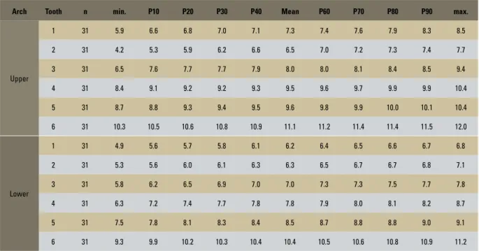
Ghose and Baghdady10 identified statistically non-significant variations between the mesio-distal diameters of teeth after comparing the right and left sides. In the present study, the re-sults showed that only 3 of 42 measures yielded statistically significant differences. It cannot therefore be implied that there is any difference between the sides (Tables 3, 4 and 5).
TABLE 3 - Comparison between mesiodistal measurements in the left and right sides.
Left Right
Diff. t p
Tooth Mean sd Mean sd
Upper arch
1 9.04 0.54 9.05 0.53 -0.01 -0.200 0.842 ns 2 7.09 0.41 7.18 0.44 -0.09 -3.022 0.004 *
3 8.06 0.51 8.12 0.48 -0.06 -1.742 0.087 ns 4 7.18 0.41 7.14 0.37 0.04 1.526 0.133 ns 5 6.93 0.46 6.96 0.48 -0.03 -0.990 0.327 ns 6 10.57 0.64 10.56 0.70 0.01 0.212 0.833 ns 7 10.00 0.60 9.89 0.49 0.11 2.112 0.042 *
Lower arch
1 5.52 0.36 5.54 0.39 -0.02 -1.013 0.316 ns 2 6.08 0.39 6.10 0.38 -0.02 -0.710 0.481 ns 3 7.11 0.44 7.11 0.47 0.00 -0.010 0.992 ns 4 7.28 0.42 7.31 0.40 -0.03 -1.342 0.185 ns 5 7.35 0.41 7.35 0.44 0.00 0.014 0.989 ns 6 11.12 0.54 11.11 0.53 0.01 0.609 0.545 ns 7 10.52 0.57 10.47 0.56 0.05 0.950 0.349 ns ns = no statistically significant difference.
TABLE 4 - Comparison between buccolingual measurements in the left and right sides.
TABLE 5 - Comparison between occlusal/incisal-cervical measurements in the left and right sides. ns = no statistically significant difference.
* Statistically significant difference (p<0.05)
Tooth
Left Right
Diff. t p
Mean sd Mean sd
Upper arch
1 7.52 0.67 7.55 0.65 -0.03 -1.083 0.283 ns 2 6.72 0.80 6.78 0.83 -0.06 -0.896 0.374 ns 3 8.18 0.59 7.19 0.58 -0.02 -0.371 0.712 ns 4 9.52 0.44 9.53 0.45 -0.01 -0.479 0.634 ns 5 9.69 0.47 9.69 0.49 0.00 0.025 0.980 ns 6 11.20 0.54 11.21 0.56 -0.02 -0.545 0.588 ns 7 11.15 0.63 11.19 0.66 -0.04 -0.169 0.249 ns
Lower arch
1 6.29 0.48 6.26 0.51 0.04 1.411 0.164 ns 2 6.46 0.49 6.43 0.49 0.02 0.869 0.389 ns 3 7.26 0.54 7.28 0.56 -0.02 -0.684 0.497 ns 4 7.91 0.51 8.02 0.63 -0.11 -1.839 0.071 ns 5 8.60 0.51 8.63 0.48 -0.03 -0.749 0.457 ns 6 10.56 0.63 10.55 0.64 0.01 0.384 0.702 ns 7 10.26 0.63 10.15 0.69 0.11 1.480 0.146 ns
Tooth
Left Right
Diff. t p
Mean sd Mean sd
Upper arch
1 10.06 0.90 10.02 0.87 0.04 1.195 0.237 ns 2 8.36 0.87 8.33 0.90 0.02 0.387 0.700 ns 3 9.73 0.91 9.80 0.96 -0.06 -0.820 0.416 ns 4 7.69 0.76 7.73 0.73 -0.03 -0.852 0.398 ns 5 6.60 0.69 6.58 0.70 0.01 0.213 0.832 ns 6 5.48 0.68 5.62 0.77 -0.14 -2.523 0.015 *
7 5.43 0.78 5.38 0.78 0.05 0.625 0.535 ns
Lower arch
1 8.42 0.78 8.40 0.75 0.02 0.666 0.508 ns 2 9.56 0.70 8.30 0.82 -0.01 -0.275 0.784 ns 3 9.56 1.00 9.50 1.07 0.06 0.915 0.364 ns 4 8.12 0.66 8.09 0.76 0.03 0.622 0.536 ns 5 6.87 0.64 6.88 0.71 -0.01 -0.240 0.812 ns 6 5.60 0.70 5.71 0.81 -0.11 -1.874 0.066 ns 7 5.09 0.77 5.21 0.77 -0.12 -1.587 0.119 ns ns = non statistically significant difference.
dEVELOPMEnt OF “C” FORMuLA
In view of the fact that the dimensional reference tables (Tables 1 and 2) depict the mean values as well as the percentages of mesiodistal, buccolingual and occlusal/incisal-cervical dimensions in both genders, the following equation — named the “C” Formula — was developed based on the dimensions of only one tooth so that allows one to calculate the likely dimen-sions of the other teeth on the same quadrant:
“C” PERCENTILE TABLES
To facilitate searching and reading the ref-erence values that correspond to the three tooth dimensions in each gender, 6 (six) Tables (6, 7, 8, 9, 10 and 11) were developed from the “C” formula. These show respectively the mini-mum values, percentiles 10, 20, 30, 40, means, 60, 70, 80 and 90, and maximum values (in mm) and ∑ mesiodistal values (1-6) of each quadrant in both genders.
Tables 6, 7 and 8 depict respectively female percentile values for mesiodistal, buccolingual and occlusal/incisal-cervical measurements.
Tables 9, 10 and 11 depict respectively the male percentile values for mesiodistal, buccolin-gual and occlusal/incisal-cervical measurements.
TABLE 6 - Percentile values of mesiodistal measurements / Female.
arch Tooth n min. P10 P20 P30 P40 Mean P60 P70 P80 P90 max.
Upper
1 31 8.0 8.2 8.4 8.6 8.8 8.9 9.3 9.3 9.4 9.5 10.0 2 31 6.1 6.4 6.7 6.8 7.0 7.0 7.2 7.3 7.4 7.5 7.8 3 31 7.3 7.5 7.7 7.7 7.8 7.9 8.1 8.1 8.1 8.4 8.5 4 31 6.2 6.6 6.8 6.9 7.0 7.1 7.2 7.2 7.4 7.5 7.8 5 31 5.6 6.2 6.5 6.6 6.7 6.8 7.0 7.1 7.1 7.3 7.8 6 31 9.3 9.8 9.9 10.1 10.2 10.3 10.4 10.5 10.7 10.9 11.3 1 - 6 31 44.6 45.5 45.9 47.0 47.9 48.1 48.9 49.2 49.4 50.5 51.4
Lower
1 31 4.7 5.0 5.2 5.3 5.4 5.4 5.5 5.5 5.7 5.8 6.1 2 31 5.4 5.5 5.8 5.9 5.9 6.0 6.1 6.2 6.3 6.6 6.8 3 31 6.2 6.4 6.5 6.8 6.8 6.9 7.1 7.1 7.3 7.4 7.5 4 31 6.4 6.7 6.8 6.9 7.0 7.1 7.2 7.2 7.5 7.6 7.9 5 31 6.3 6.7 6.9 7.0 7.1 7.2 7.3 7.4 7.6 7.7 8.1 6 31 9.7 10.4 10.7 10.7 10.8 11.0 11.1 11.2 11.4 11.6 11.8 1 - 6 31 39.9 41.6 42.1 42.7 43.2 43.6 44.1 44.2 45.4 46.0 47.7
Wx = Wk x Px = R Pk
Wx = Width of unknown tooth Wk = Width of known tooth
TABLE 7 - Percentile values of buccolingual measurements / Female.
arch Tooth n min. P10 P20 P30 P40 Mean P60 P70 P80 P90 max.
Upper
1 31 5.9 6.6 6.8 7.0 7.1 7.3 7.4 7.6 7.9 8.3 8.5 2 31 4.2 5.3 5.9 6.2 6.6 6.5 7.0 7.2 7.3 7.4 7.7 3 31 6.5 7.6 7.7 7.7 7.9 8.0 8.0 8.1 8.4 8.5 9.4 4 31 8.4 9.1 9.2 9.2 9.3 9.5 9.6 9.7 9.9 9.9 10.4 5 31 8.7 8.8 9.3 9.4 9.5 9.6 9.8 9.9 10.0 10.1 10.4 6 31 10.3 10.5 10.6 10.8 10.9 11.1 11.2 11.4 11.4 11.5 12.0
Lower
1 31 4.9 5.6 5.7 5.8 6.1 6.2 6.4 6.5 6.6 6.7 6.8 2 31 5.3 5.6 6.0 6.1 6.3 6.3 6.5 6.7 6.7 6.8 7.1 3 31 5.8 6.2 6.5 6.9 7.0 7.0 7.3 7.3 7.5 7.7 7.8 4 31 6.3 7.2 7.4 7.7 7.8 7.8 7.9 8.0 8.1 8.2 8.7 5 31 7.5 7.8 8.1 8.3 8.4 8.5 8.7 8.8 8.8 9.0 9.1 6 31 9.3 9.9 10.2 10.3 10.4 10.4 10.5 10.6 10.8 10.9 11.2
TABLE 8 - Percentile values of occlusal/incisal-cervical measurements / Female.
arch Tooth n min. P10 P20 P30 P40 Mean P60 P70 P80 P90 max.
Upper
1 31 8.4 8.9 9.1 9.2 9.4 9.8 10.0 10.2 10.6 10.8 11.4 2 31 6.8 7.2 7.5 7.6 8.0 8.2 8.5 8.6 8.9 9.2 10.2 3 31 7.7 8.6 8.8 8.9 9.3 9.5 9.8 10.0 10.1 10.6 11.3 4 31 6.5 6.7 7.0 7.2 7.3 7.5 7.7 7.8 7.9 8.4 9.2 5 31 5.4 5.9 6.1 6.2 6.3 6.5 6.5 6.6 6.8 7.2 8.1 6 31 4.4 4.7 5.1 5.1 5.3 5.5 5.5 5.8 5.9 6.2 6.9
Lower
TABLE 10 - Percentile values of buccolingual measurements / Male.
arch Tooth n min. P10 P20 P30 P40 Mean P60 P70 P80 P90 max.
Upper
1 26 6.8 7.3 7.4 7.5 7.6 7.8 7.9 8.1 8.3 8.3 8.9 2 26 5.9 6.3 6.5 6.6 7.0 7.0 7.1 7.3 7.4 7.6 8.1 3 26 7.6 7.9 8.1 8.1 8.4 8.4 8.6 8.7 8.9 9.0 9.3 4 26 8.7 9.1 9.3 9.5 9.5 9.6 9.7 9.8 9.9 10.2 10.5 5 26 9.1 9.4 9.4 9.5 9.6 9.8 9.9 10.1 10.2 10.4 10.8 6 26 9.6 10.9 11.0 11.1 11.2 11.4 11.5 11.7 11.8 12.2 12.4
Lower
1 26 5.6 5.9 6.1 6.2 6.4 6.4 6.5 6.6 6.7 7.0 7.2 2 26 5.6 6.1 6.3 6.5 6.6 6.6 6.7 6.7 6.8 7.2 7.3 3 26 6.6 6.9 7.1 7.3 7.3 7.5 7.5 7.9 8.0 8.1 8.3 4 26 7.3 7.6 7.7 7.9 8.1 8.2 8.2 8.5 8.7 8.9 9.1 5 26 7.6 8.0 8.4 8.7 8.7 8.8 8.9 9.0 9.2 9.4 9.5 6 26 7.8 10.1 10.4 10.5 10.8 10.7 11.1 11.1 11.1 11.3 11.7 TABLE 11 - Percentile values of occlusal/incisal-cervical measurements / Male.
arch Tooth n min. P10 P20 P30 P40 Mean P60 P70 P80 P90 max.
Upper
1 26 8.2 9.4 9.5 9.9 10.1 10.3 10.7 10.8 11.0 11.5 11.7 2 26 7.3 7.6 7.7 8.0 8.2 8.5 8.6 8.7 9.0 9.9 10.3 3 26 7.8 9.2 9.4 9.8 9.9 10.0 10.2 10.6 10.7 11.1 11.8 4 26 6.0 7.1 7.4 7.6 7.7 7.9 8.1 8.2 8.4 8.5 10.3 5 26 5.5 6.0 6.3 6.3 6.4 6.7 6.9 7.0 7.2 7.3 8.7 6 26 4.2 5.0 5.2 5.2 5.4 5.7 5.7 5.8 6.3 6.4 7.9
Lower
1 26 7.5 7.7 8.0 8.2 8.6 8.7 8.9 9.3 9.4 9.6 10.0 2 26 7.2 7.7 8.0 8.2 8.2 8.6 8.8 8.9 9.2 9.6 10.3 3 26 8.1 8.9 9.2 9.5 9.7 10.0 10.1 10.4 10.8 11.3 12.1 4 26 7.0 7.6 8.1 8.2 8.3 8.4 8.5 8.7 8.8 9.1 10.2 5 26 4.7 6.1 6.6 6.9 6.9 7.0 7.2 7.3 7.5 7.6 8.7 6 26 3.8 5.0 5.3 5.4 5.5 5.8 6.0 6.1 6.2 6.7 7.6 TABLE 9 - Percentile values of mesiodistal measurements / Male.
arch Tooth n min. P10 P20 P30 P40 Mean P60 P70 P80 P90 max.
Upper
1 26 8.0 8.5 8.7 8.8 9.2 9.2 9.3 9.5 9.6 9.7 10.0 2 26 6.5 6.7 6.9 7.1 7.2 7.3 7.4 7.5 7.6 7.7 7.9 3 26 7.1 7.6 7.8 8.0 8.3 8.3 8.5 8.6 8.7 9.0 9.3 4 26 6.4 6.8 7.1 7.1 7.2 7.3 7.5 7.6 7.6 7.6 7.8 5 26 6.1 6.6 6.9 7.0 7.0 7.1 7.2 7.4 7.5 7.6 7.7 6 26 9.5 9.9 10.3 10.4 10.7 10.9 11.1 11.3 11.5 11.7 12.4 1 - 6 26 46.2 46.4 46.9 49.1 49.5 50.0 50.8 51.5 52.0 52.6 54.3
Lower
Example of application and use of “C” percentile tables
Assuming a patient with the following char-acteristics: Female, with missing first premolars. Planning involved prosthetic reconstruction of the first upper premolars with implant support. What should the mesiodistal dimension of the first maxillary premolars be?
First step: Measure the dimensions of any one of the teeth either in the casts or clinically in the patient. Assuming that the mesiodistal size of one of the upper central incisors was measured and found to be 9.5 mm, the value that corre-sponds to this dimension is then checked in Table 6 in the “C” percentile table for females. Since the value of 9.5 mm is in column P90, the prob-able value of the first upper premolars is 7.5 mm. Additionally, the following conclusions can be drawn. The mesiodistal, buccolingual and oc-clusal/incisal-cervical dimensions shown in the P90 columns in the Tables 6, 7 and 8 are the probable dental dimensions of the remaining teeth whenever the female upper incisors show a mesiodistal distance of 9.5 mm.
Note also that the sum of the upper and low-er quadrants is shown in the respective columns. One could also measure the distances from in-cisors to canines, and from inin-cisors to second premolars, adding to the respective values.
CLiniCAL COnsidERAtiOns
“C” percentile tables may be applied along with other preexisting methods to determine tooth size and thus assist in various areas of dentistry, such as in morphological, esthetic and functional reconstructions. In orthodon-tics, they could be used to determine individu-al, collective and inter-arch discrepancies. They can also contribute as an auxiliary method in forensic investigations.
The feasibility of the clinical applications and hypotheses suggested in this study can only be confirmed, denied or amended by
means of longitudinal applications and assess-ments of the outcomes. However, one should note that often human dental arches exhibit morphological variations and disproportionate tooth dimensions. As a result of these events it is suggested that given the size variations a more conservative alternative should be tried first rather than hastily propose a reduction in dental materials.
COnCLusiOns
Based on the materials and methods used and the results obtained in this study, the following could be established:
» The biometric mesiodistal, buccolingual and occlusal/incisal-cervical dimensions of human teeth are distinguishable be-tween genders in terms of the mean, mini-mum and maximini-mum coefficients, standard deviations, variation coefficients and per-centages of each tooth in their respective dimension.
» Tooth dimensions are smaller in females than in males and should therefore be studied separately.
» The teeth in their mesiodistal, buccolingual and occlusal/incisal-cervical dimensions proved to be symmetrical in both genders. Through the overall values obtained, it was possible to build tables to distinguish these di-mensions according to gender.
The mean values of mesiodistal, buccolin-gual and occlusal/incisal-cervical tooth dimen-sion were provide along with the percentage of occurrence between these dimensions and their standard deviations.
Contact address
Carlos Alberto Gregório Cabrera Rua Lamenha Lins , 62, 4º Andar CEP: 80.250-020 – Curitiba/PR, Brazil E-mail: [email protected]
REFEREnCEs
Submitted: August 7, 2008
Revised and accepted: January 26, 2009
1. Andrews LF. The six keys to normal occlusion. Am J Orthod. 1972;62(3):296-309.
2. Andrews LF. The Straight-Wire appliance: explained and compared. J Clin Orthod. 1976;10(3):174-95.
3. Baum BJ, Cohen MM. Decreased odontometric sex difference in individuals with dental agenesis. Am J Phys Anthropol. 1973;38(3):739-42.
4. Black GV. Descriptive anatomy of the human teeth. 4th ed.
Philadelphia S.S. White Dental; 1902.
5. Bolton WA. Disharmony in tooth size and its relation to the analysis and treatment of malocclusion [thesis]. Seattle (WA): University of Washington; 1952.
6. Burnes KR. Forensic anthropology. Training manual. University of Carolina at Charlotte. New Jersey: Prentice Hall; 1999. p. 109-27.
7. Castro RCFR. Utilização do índice morfológico das coroas dos incisivos inferiores para predição da recidiva em casos tratados com extrações. [dissertação]. Bauru (SP): Faculdade de Odontologia, Universidade de São Paulo; 2005. 8. Dahlberg GG. Statistical methods for medical and biological
students. New York: Interscience; 1940.
9. De Deus Q. Endodontia. Rio de Janeiro: Medsi; 1992. 10. Ghose L, Baghdady VS. Analyses of the Iraqi dentition;
mesiodistal crown diameters of permanent teeth. J Dent Res. 1979;58(3):1047-50.
11. Harris FE, Burris GB. Contemporary permanent tooth dimensions, with comparisons to G.V. Blacks data. J Tenn Dent Assoc. 2003;83(4):25-9.
12. Henrique JFC. Determinação de um índice morfológico das coroas dos incisivos inferiores, em adolescentes brasileiros, com oclusão normal para predição da recidiva do
apinhamento pós-contenção [tese]. Bauru (SP): Universidade de São Paulo; 1992.
13. Houston WJ. The analysis of errors in orthodontic measurements. Am J Orthod. 1983;83(5):382-90.
14. Ling JYK, Hagg U. Tooth size discrepancies among different occlusion groups of southern Chinese children. Am J Orthod Dentofacial Orthop. 2001;120(5):556-8.
15. Mondelli J. Estética e cosmética em clínica integrada restauradora. São Paulo: Quintessence; 2003. 16. Okada MS. Estudo do tamanho dos dentes naturais
superiores e dentes artiiciais de diferentes marcas
comerciais [dissertação]. São Paulo (SP): Universidade de São Paulo; 2005.
17. Pinzan A, Martins DR, Freitas MR. Análise da discrepância de tamanho dentário de Bolton. Ortodontia. 1991;24(1):61-4. 18. Ramos AL, Suguino R, Furquim LZ, Silva Filho OG.
Considerações sobre análise da discrepância dentária de
Bolton e a inalização ortodôntica. Rev Dent Press de Ortod
Ortop Facial. 1996;1(2):86-106.
19. Richardson ER, Malhotra SK. Mesiodistal crown dimension of the permanent dentition of American Negroes. Am J Orthod. 1975;68(2):157-64.
20. Santoro M, Ayoub ME, Pardi VA, Canialosi TJ. Mesiodistal crown dimension and tooth size discrepancy of the permanent dentition of Dominican Americans. Angle Orthod. 2000;70(4):303-7.
21. Taylor KT. Forensic art and illustration. Boca Raton: CRC Press LLC; 2001.
22. Yamaguto OT. Determinação das medidas dentárias mésio-distais em indivíduos brasileiros leucodermas com oclusão normal. [dissertação]. São Bernardo do Campo (SP): Universidade Metodista de São Paulo; 2003.