Multiple or severe root resorptions
are not associated with systemic factors,
individual susceptibility, family tendency
or individual predisposition
Alberto Consolaro*, Telma Regina Gobbi Franscischone**, Laurindo Zanco Furquim***
Multiple or severe root resorptions, still of-ten assigned to systemic changes, particularly endocrinopathies, are seen when the severity of alveolar bone loss is great, especially during
orthodontic movements.18,19,20
In bone turnover, matrix deposition alter-nates continuously with bone resorption at different sites and times. This dynamic process enables the bone to adapt to the functional demands of each skeletal area and to actively participate in the maintenance of the mineral homeostasis to control serum calcium and phosphorus concentrations.2,3,6,15 The skeleton
renews completely at variable time intervals ac-cording to the patient’s age.22
Teeth, particularly root structures, do not have bone turnover,1,8,12,17 a consequence of the
activity of osteoblasts, osteocytes, macrophages and osteoclasts. These cells organize in basic multicellular units (BMUs), or bone remodel-ing cells, and receive stimuli from systemic and local mediators found in membrane surface re-ceptors of several cell types, particularly osteo-blasts and macrophages.2,7,22
Cementoblasts, the cells that populate the root surface, do not have receptors in sufficient number and significance to become bone turn-over mediators.6,21 Cementoblasts are “deaf" to
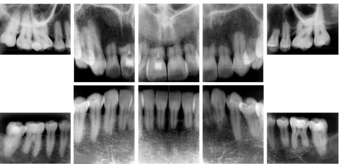
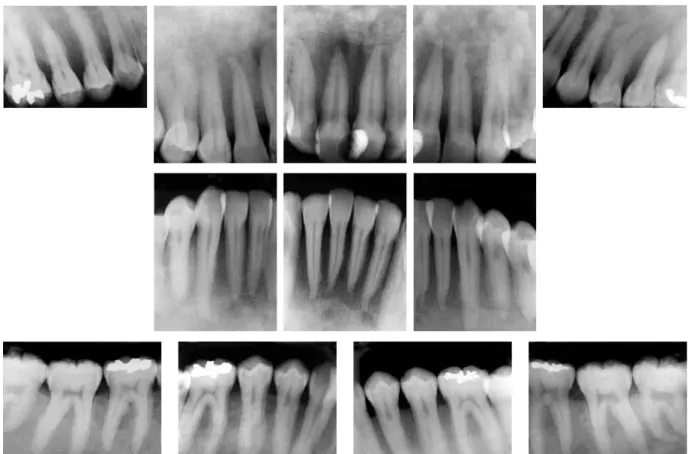
the messages sent by the bone turnover media-tors, even when found in large concentrations in periodontal tissues, as is the case in hyper-parathyroidism. In periodontal inflammation, the local levels of mediators that induce bone resorption are also high, but cementoblasts do not respond to them and, therefore, teeth are protected from bone turnover (Figs 1 and 2).
The "absence” of cementoblast surface re-ceptors for bone turnover mediators compli-cates any attempt to assign a systemic origin,
such as endocrinopathies, to root resorptions.21
In other words, cementoblasts are not affected by systemic mediators or hormones. Cemento-blasts remain on the root surfaces no matter whether the concentration of systemic media-tors is high or low.
The causes of root resorption should be as-sociated with the loss of cementoblasts from the root surface, which may have its origin in trauma
* Head Professor of Pathology, FOB-USP and FORP-USP Postgraduate Program. ** Endocrinologist, PhD in Pathology at FOB-USP, Brazil.
and chemical or biological processes, but which is a local event7 (Figs 1 and 2). In human
pathol-ogy, the absence or decrease of cementoblasts on the root surface does not characterize any systemic disease. For root resorption to occur, the first necessary step is the removal or local loss of cementoblasts.
The definition of a cause of root resorption requires a careful investigation of the patient’s history to retrieve information about previous events, addictions, accidents, types of sports and leisure activities, previous treatments and associated local diseases. In the investigation of history, priority should be assigned to relevant details, such as mild trauma (concussion, sub-luxation), which the patient does not always re-member and which cannot be detected by the clinical examiner. Mild trauma may occur due to several reasons: biting very hard objects dur-ing mastication; impact durdur-ing leisure activities; surgical trauma due to leverage for extraction of neighboring teeth; and laryngoscope impact
during procedures for general anesthesia. When the local cause of root resorption can-not be determined after all resources have been used, its etiology may be classified as idiopathic, a term which defines the impossibility of detect-ing the cause without assigndetect-ing any systemic or iatrogenic origin to it.
In root resorptions assigned to a probable systemic cause, it is necessary to refer the pa-tient to an endocrinologist,9,10,16 because
en-docrinopathies at an advanced stage may lead to death. The dentist is not expected to make an accurate diagnosis of endocrinopathies, but should refer patients to an endocrinologist for evaluation and final identification of the prob-lem when it is suspected (Figs 1 and 2).
of hereditary involvement or specific genetic changes, the patient should be referred to a ge-neticist for a careful evaluation and identifica-tion of the problem.
In terms of individual susceptibility, family tendencies and individual predisposition, local factors should not be ignored, such as root mor-phology, root apex shape, shape of the alveolar bone crest and crown-to-root ratio, which sub-stantially affect the prognosis of root resorptions during future orthodontic treatment.11 These
morphological factors, which may predict great-er or lowgreat-er root resorption during orthodontic treatment, may be inherited or acquired during odontogenesis due to environmental factors. However, when individual susceptibility, fam-ily tendency and individual predisposition are
mentioned in root resorption in orthodontics in general, they are often classified as systemic, and not local, factors.
The publication of the characteristics of large orthodontic samples may be an important contribution to the literature:
1. How many patients are carefully exam-ined during evaluation and diagnosis to check systemic conditions associated with diabetes and the functions of the thyroid, parathyroid, pituitary, ovaries and the other endocrine glands?
2. How many patients are seen by the endo-crinologist before orthodontic treatment is started?
odontic treatment, were discharged, and did not have any problems in the amount of bone and rate of root resorp-tions. How many of these patients un-derwent systemic evaluations later on, when endocrinopathies were detected? 4. How many had multiple or severe root
resorptions, were evaluated by an endocri-nologist and had any endocrine disorder? If there is no evidence that systemic chang-es, individual susceptibility, family tendency and individual predisposition are causes of root resorptions, to what should the multiple or severe root resorptions seen in orthodontic clinics be assigned?
First explanation
Cementoblasts should be removed, eliminat-ed or simply vanish from the region for root re-sorption to begin on the root surface. Cemento-blasts are closely associated with the cement sur-face and, simultaneously, are laterally protected by collagen fibers that insert into or fusion with root cement. Trauma, surgical procedures and bacterial and chemical products applied to the root surface may remove cementoblasts.
In injured teeth, the large lesions of the ce-mentoblast layer may be reconstructed, partly with cells from the osteoblastic line, which have receptors for the mediators of bone turn-over. They will be cementoblast-like, but start an early local resorption process when there is a local inflammatory stimulus, and promote a greater loss of root structure every time tooth movement is induced than surfaces without ce-mentoblast-like cells.7 Therefore, the following
assumption may be made: injured teeth, when moved during orthodontic treatment, have a greater chance of more frequent and more se-vere root resorptions. This does not translate into a contra-indication, but greater care should be taken when applying orthodontic forces to teeth in such conditions, particularly, whenever
possible, their homogeneous distribution along the root and special attention to their intensity.
Secondexplanation
Cementoblast removal may also occur during induced tooth movement. An excessive force to prevent the periodontal ligament, compressed between the bone and the teeth, to receive blood supply. The cells of this compressed liga-ment segliga-ment, such as the celiga-mentoblasts, move away from the region and leave the surface or die before migrating, and consequently the root is exposed. Cell scattering or death leaves only a hyalinized extracellular matrix in the region. The root surface, without the protection of ce-mentoblasts, becomes the site of resorptions in the process of periodontal reorganization in the region after the dissipation of the force applied. After a few days, reorganization is over and a new layer of cementoblasts is produced and again protects the root from resorption. The surface may acquire an irregular outline, but the periodontal ligament is back to its normal function in the region. This root will not be sus-ceptible to future root resorptions.
After about 21 days, the process described above may resume, and a new loss of mineral-ized dentinal tissue adds up to the previous loss. However, if the force is adequate and compat-ible with the life of cementoblasts, new root resorptions will not occur. Root resorption diag-nosed at the end of the orthodontic treatment is the sum of the resorptions at each activation time, and does not represent a single process during orthodontic movement (Fig 2).
between the multiple or severe root resorption and their systemic status or family history.
Although there are no associations of root re-sorptions with systemic factors, individual suscep-tibility, family tendencies and individual predis-position, cases of patients with controlled or un-controlled systemic diseases and who underwent orthodontic treatment should be carefully se-lected and published to enrich the literature with evidence that root resorptions are not part of the clinical signs and symptoms of endocrinopathies.
Final considerations
Cases of multiple or severe root resorption have been less often assigned to systemic fac-tors or diseases, individual susceptibility, family tendency and individual predisposition. When any of these factors is suspected and affects the appearance and progression of root resorptions, patients should be referred to endocrinologists or geneticists for adequate medical examination. When this approach is adopted, patients usually return with the result that there is no association
1. Albright F, Aub JC, Bauer W. Hyperparathyroidism: common and polymorphic conditions as illustrated by seventeen proved case from one clinic. J Am Med Assoc. 1934 Apr;102:1276-87.
2. Aurbach GD, Marx SJ, Speegl AM. Parathyroid hormone, calcitonin and the calciferol. In: Wilson JD, Foster DW, editors. Williams textbook of Endocrinology. 8th ed.
Philadelphia: Saunders; 1992. cap. 27, p. 1397-476. 3. Baumrind S, Korn EL, Boyd RL. Apical root resorption in
orthodontically treated adults. Am J Orthod Dentofacial Orthop. 1996 Sep;110(3):311-20.
4. Brezniak N, Wasserstein A. Root resorption after orthodontic treatment: part 1: literature review. Am J Orthod Dentofacial Orthop. 1993 Jan;103(1):62-6.
5. Brezniak N, Wasserstein A. Root resorption after orthodontic treatment: part 2: literature review. Am J Orthod Dentofacial Orthop. 1993 Feb;103(2):138-46.
6. Cho MI, Lin WL, Garant PR. Occurrence of epidermal growth-binding sites during differentiation of cementoblasts and
periodontal ligament ibroblasts of young rat: a light and
electron microscopic radioautographic study. Anat Rec. 1991 Sep;231(1):14-24.
7. Consolaro A. Reabsorções dentárias nas especialidades clínicas. 2ª ed. Maringá: Dental Press; 2005.
8. Fish EW. An experimental investigation of enamel, dentine and the dental pulp. London: John Bale; 1932.
9. Francischone PC. Avaliação da perda óssea maxilar pela
análise da radiograia panorâmica digitalizada, comparando
com a densitometria óssea lombar e femural [dissertação]. Bauru (SP): Universidade de São Paulo;1999.
10. Francischone TRCG. Reabsorção dentária: determinação de sua freqüência em pacientes com endocrinopatias [tese]. Bauru (SP): Universidade de São Paulo; 2002.
11. Furquim LZ. Peril endocrinológico de pacientes ortodônticos
com e sem reabsorções dentárias [tese]. Bauru (SP): Universidade de São Paulo; 2002.
12. Gies WJ. Studies of internal secretions in their relation to the development and condition of the teeth. J Nat Dent Assoc. 1918;5:527-31.
13. Harris EF, Kineret SE, Tolley EA. A heritable component for external apical root resorption in patients treated orthodontically. Am J Orthod Dentofacial Orthop. 1997 Mar;111(3):301-9.
REFERENCES
Contact address
Alberto Consolaro
E-mail: [email protected]
14. Harris EF, Robinson QC, Woods MA. An analysis of causes of apical root resorption in patients not treated orthodontically. Quintessence Int. 1993 Jun;24(6):417-28.
15. Hill PA, Orth M. Bone remodelling. Br J Orthod. 1998 May; 25(2):101-7.
16. Krall EA, Dawson-Hughes B, Hannan MT, Wilson PW, Kiel DP. Postmenopausal estrogen replacement and tooth retention. Am J Med. 1997 Jun;102(6):536-42.
17. Kronfeld R, Boyle PE. Histopatologia dos dentes. 3ª ed. Rio
de Janeiro: Cientíica; 1955. p. 37-46.
18. Levander E, Malmgren O. Evaluation of the risk of root resorption during orthodontic treatment: a study of upper incisors. Eur J Orthod. 1988 Feb;10(1):30-8.
19. Levander E, Malmgren O, Eliasson S. Evaluation of root resorption in relation to two orthodontic treatment regimes. A clinical experimental study. Eur J Orthod. 1994 Jun;16(3):223-8. 20. Levander E, Malmgren O, Stenback K. Apical root resorption
during orthodontic treatment of patients with multiple aplasia: a study of maxillary incisors. Eur J Orthod. 1998 Aug;20(4):427-34.
21. Lindskog S, Blomlöf L, Hammarström L. Comparative effects of parathyroid hormone on osteoblasts and cementoblasts. J Clin Periodontol. 1987 Aug;14(7):386-9.
22. Manolagas SC. Birth and death of bone cells: basic regulatory mechanisms and implications for the pathogenesis and treatment of osteoporosis. Endocr Rev. 2000 Apr;21(2):115-37. 23. Miller SC. Hormonal regulation of osteogenesis. In: