Ki Beom Kim
An interview with
• Associate Professor and Clinical Director of the Department of Orthodontics, New Southeastern University, Fort Lauderdale (Florida), United States.
• Adjunct Professor, Department of Orthodontics, Saint Louis University, United States, from 2005 to 2011. • Graduated in the American Board of Orthodontics, 2011.
• Residency in Orthodontics at Vanderbilt University (Nashville), United States, 2005. • Doctor of Orofacial Pain by Dankook University, South Korea, 2002.
• Graduated in the American Board of Orofacial Pain, 2000. • Master in Dentistry by Dankook University, South Korea, 1995.
• Residency in Oral Medicine and Temporomandibular Disorders by Dankook University, South Korea, 1995.
It is a great honor and I am glad to bring to the readers a little bit of the scientiic experience and knowl -edge from one of the most important current researchers and clinician in the United States: Prof. Dr. Ki
Beom Kim. We have conirmed that Prof. Kim dedicated a profound attention when answering the ques
-tions from the interviewers, proving respect for us, the readers of the Dental Press Journal of Orthodontics. During this interview, he discussed with a lot of property important current matters and of great relevance, such as the use of 3D image technologies, self-ligating brackets, mini-implants and orthodontic treatment on patients with temporomandibular disorder. A deep lover and fond of sports and photography, Prof. Kim, despite working with state of the art means available in Orthodontics, he believes in diagnostic and in strict and individual planning as the main way to reach success in orthodontic treatments. Married and father of 2 sons, Prof. Kim was born in South Korea and there he graduated in Dentistry and post-graduated in
Orofacial pain. After this, he moved to the United States and never left. Nowadays, he is an Associated Pro
-fessor and Clinical Director on the Department of Orthodontics at the New Southeastern University (Fort Lauderdale, Florida/United States), where he is the Clinical Director of the Post-Graduation Program. I hope you all enjoy this opportunity of knowing some of his point of view and the way this great researcher and clinician works.
Marcelo Castellucci
How to cite this interview: Ki Beom Kim. Interview. Dental Press J Orthod. 2012 Mar-Apr;17(2):18-26.
Submitted: October 20, 2011 - Revised and accepted: March 1, 2012.
What has been your experience with the self-ligating brackets? What are their real beneits and disadvantages? Is there a particular proile of the patient or professional to whom this type of bracket its best or is it worthwhile for every-one? (Marcelo Castellucci)
I have been using self-ligating brackets for about 10 years. In spite of many claims from the bracket companies, I don’t see a big difference. Currently there is no scientiic evidence supporting the beneit of the self-ligating brackets.
There have been many studies presenting lower friction with the self-ligating brackets but all of them were done in the lab setting. If we consider the force decay of the elastomeric ties, perturbation from the mastication, 1st, 2nd, and 3rd order misalignments of the teeth, the lower friction of self-ligated brackets is meaningless. We oversimpliied and overestimated the results from the lab studies.
Some people claim that self-ligation brackets are
more hygienic than the conventional brackets.1 How
-ever, the other study reported that the self-ligating brackets do not have an advantage over conventional brackets with respect to the periodontal status of the
mandibular anterior teeth.2 There is not enough evi
-dence to support the claim that conventional brack
-ets are less hygienic than self-ligating brack-ets. Few studies indicated that the self-ligating brack
-et systems have quicker wire removal and place
-ment.3,4 In a recent systemic review, only 2 outcomes,
chair time and inal mandibular incisor inclination,
showed signiicant differences.5
The only beneits in my opinion are shorter chair time and possible longer appointment interval.
The self-ligated bracket system industry tries to disseminate the idea of reduced treatment time. Based on the principle that teeth biological re-sponse is the same, whatever is used to move them, how could treatment time be reduced? (Orlando Tanaka)
A lot of studies tried to compare the eficiency of the self-ligation brackets, but I haven’t come across any ar
-intelligence, they are just handles to move teeth. We are just brain washed from all those commercials from the companies. It is simply not true.
According to AJO-DO reader’s forum, in 2010 August, only two advantages of self-ligated bracket systems are scientiically proven, which are chair time reduction and lower incisor torque control. Do you agree with that? What about the expansion stability, also defended by these brackets sellers? Why are these bracket systems being so much used, despite of their higher price? (Orlando Tanaka)
You can decrease the chair time once you get
used to the bracket system. You can decrease 3 sec
-onds per bracket for every appointment.6 If we use
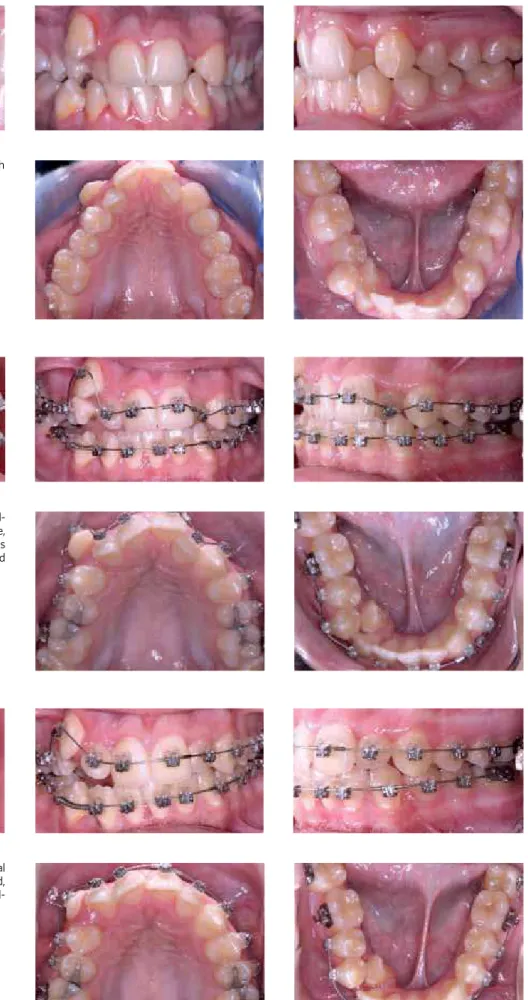
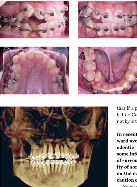
-Figure 1 - Patient shows severe crowding in both arches.
Figure 2 - Passive self-ligated brackets were bond-ed. If lower friction in self-ligated brackets is true, the NiTi wire should slide through the brackets with low pressure and upper right canine should extrude without intrusion of adjacent teeth.
The expansion is related to the arch form. One company is using a very broad arch form, so the arch expansion is a result from the arch wire, not
from the self-ligated brackets. You can do arch ex
-pansion with any bracket system. It doesn’t mat -ter what kind of brackets you use. One company stated that their system doesn’t need RME but we do know if we just expand through the arch wire, it is going to be unstable and also create potential periodontal consequences.
Figure 4 - Three months follow up.
that if a product costs more, the product should be better. Current orthodontics is driven by industries not by orthodontists ourselves.
In recent years, some clinicians have tended to-ward overuse of mini-implants in clinical orth-odontic treatment. Yet, literature still lacks some information about the long-term response of surrounding tissues (i.e. roots) and the stabil-ity of some clinical results. Do you think, based on the current literature, we should treat with caution or dive in headirst? (Luiz Gandini Jr.)
Some of the very dificult cases with traditional biomechanics can now be successfully treated with mini-implants. We as orthodontists are so excited about the possibility of the mini-implant, and we have tried many different applications. As a faculty of post graduate orthodontic program, I often noticed
that our residents believe mini-implants will magi
-cally solve all the biomechanical problems for them. Rather than trying to igure out the conventional treatment mechanics, they simply mention “I will use mini-implants.”
However, we must remember the same bio
-mechanical rule also applies to the patients with mini-implants. We have to consider the risk of root damage, infection, and soft tissue inflammation.
Mini-implants are one of the main topics in cur-rent orthodontics journals and meetings. How predictable, efective and stable do you believe are the vertical (posterior intrusion) and trans-verse dimensions (maxillary expansion) when managing an orthognathic adult patient with mini-implants? (Luiz Gandini Jr.)
It has been only a couple of years using mini-implants for molar intrusion and expansion. It may take a while to have a long-term studies for stability and relapse.
But based on the studies that are currently avail
-able and case reports, it seems very effective. Just the possibility to correct the difficult problems
without going through a major orthognathic sur
-gery itself is very exciting.
According to the study from South Korea, a re
-lapse rate of 23% at the 3-year follow-up in long-term stability of anterior open bite cases by in
-trusion of maxillary posterior teeth.7 But we can’t
make a conclusion based on just one study. Until we have more data, we cannot jump to conclusions.
With the TAD (Temporary Anchorage Device), 3D cone beam images and the technology pres-ent in new brackets and wires, is the technique becoming more important than orthodontic di-agnosis? With the TAD, procedures such as “sur-gery irst” will be the rule or the conventional treatment will still be necessary in surgery cas-es treatment? Did the extraction frequency re-duce? Has the stability results improved? (Orlando Tanaka)
It is obvious that this new technological advance
-ment gives us additional diagnostic information. We can see many unseen anatomical structures with cone beam computed tomography and evaluate the three-dimensional topographic structures with the soft tissue scanning technique.
Now we are applying these new image techniques
to re-evaluate the treatment effectiveness and effi
-ciencies. This will help us understand many differ
-ent aspects of the diagnosis, treatm-ent and stability.
However, the new technology can not make a di
-agnostic decision for us. We have to consider all the other diagnostic information including 3D images, make a diagnosis and develop a treatment plan by
ourselves. None of the technology can replace this very subjective diagnostic procedure.
I agree that there are potential beneits for some patients in surgery-irst approach. But we can’t as -sume all the patients will show the same treatment
response even with new imaging techniques, com
-puter simulation and very sophisticated articulators. If we are dealing with the mechanical objects, then I would also try surgery-irst approach because I can
expect the outcome very precisely, but we are treat
-ing a human be-ing, not a mechanical object.
Orthognathic surgery is not a reversible proce
-dure, it is an invasive and irreversible procedure.
If I can minimize the uncertainty out of the equa
-tion, I would chose the traditional approach rather than risky surgery-first approach.
With the advent of 3D cone beam technology, 2D cephalometric analysis in conventional ortho-dontics may be facing a paradigm shift. What is the current state of 3D technology in orthodon-tics and where do you see us going in the near fu-ture? (Luiz Gandini Jr.)
As I mentioned before, we can obtain very large amounts of information through various 3D imaging techniques. We can precisely locate the relationship between the anatomical structures and teeth. In two-dimensional cephalometrics, we can only look at the
changes of the midline structures or overlapped bilat
-eral structures, but now we can evaluate all the struc -tures separately without overlapping other struc-tures. This is a quite revolutionized development.
Although we are in a very exciting moment, we are not fully ready to use this newly available information. Not all cephalometric norms based on the two-di -mensional radiographs can be directly applied to the three-dimensional images. We need to establish a new method for understanding the craniofacial structures. In two-dimensional radiographs, it is not very difi -cult to locate the landmarks on the lines of the images. But locating reliable points on the three-dimensional surfaces is not easy. Another dificulty is that it’s very hard to describe the three-dimensional changes.
It is easy to explain the difference in two dimen
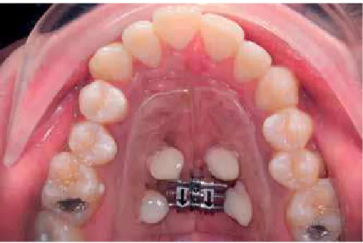
Figure 6 - Implant-supported RME: Pre-expansion. Figure 7 - Implant-supported RME: Post-expansion.
Figure 8 - Pre-expansion: Frontal view. Figure 9 - Post-expansion: Frontal view.
Figure 11 - Post-expansion transverse section at the palatal plane.
MRI can be the answer for the future imaging technique. Currently it is a lot more expensive than the CBCT machine and needs a larger space to in -stall and takes minutes to obtain the images. But MRI doesn’t use the ionized radiation to attain the images. If we have a smaller, cheaper and faster MRI machine, it will replace the CBCT technology soon.
Do you think that a 3D cephalometric analysis may arise and become so used and recognized such as the 2D analysis already established? (Marcelo Castellucci)
Currently, we don’t have an agreed method for a three-dimensional cephalometrics. Before we develop an analysis, we need to reconsider all the landmarks that we have been using for a two-di -mensional cephalometrics. Some of the landmarks are constructed landmarks which are imaginary
points. Therefore we have to re-establish the land
-marks for the three-dimensional cephalometric first. As I mentioned before locating a landmark in a three-dimensional image is not easy and it is a time consuming process. Every single landmark
should be evaluated in three different planes. Au
-tomatic landmark positioning would be very help
-ful for orthodontists because we can save time and
it will give us more reliable, reproducible and pre
-cise measurements.
There have been a couple of articles that sug
-gested methods for the three-dimensional super
-imposition. It needs to be evaluated for the accu
-racy and effectiveness.
We have to change our view from the two-dimen
-sional way of thinking which we look at the length
and angulation to a three-dimensional way of think
-ing which is space or volumetric analysis.
Do you use any protocol for the treatment of TMD patients seeking orthodontic treatment? Orthodontic treatment can be started immedi-ately or the patient must go through any other therapy before it? (Marcelo Castellucci)
Some of the patients with minor masticatory problems can start the orthodontic treatment right away, but general rule is that don’t start orthodontic treatment before the problems are identified and diagnosed correctly.
If a patient is having pain, any type of orthodontic
treatment shouldn’t be started. When TMD symp
-toms are present, the orthodontist should attempt to determine what problems are contributing to the
TMD. Usually an occlusal appliance is delivered to re
-duce the TMD symptoms along with pain medications, physical therapy and behavioral modiication. Patient is advised to use an appliance for 24 hours per day ini -tially, then decrease to part-time use, most commonly at night. Once the patient’s symptoms are gone, the or -thodontist should ask the patient to reduce the use of the appliance. If the symptoms don’t return, then the orthodontic treatment can be started.
Patients should be pain free before the orthodon
-tic treatment, but that doesn’t mean that all the joint noises need to disappear.
If we consider removing the joint noises as one of the treatment goals, then treatment success rate is going to be only 20–30%. Many studies have been suggested that we should focus on the pain not the joint noises.
Especially in osteoarthritic patients, the orth -odontic treatment should be postponed until all the symptoms are gone and also patients’ condyles are stable which means degenerative change is stopped and the condyle shape is stable and unchanged.
It is not clear when orthodontic treatment can be started from that point. If we wait longer then it would be safer but there is no consensus among the orthodontists how long we need to wait. I would wait at least 3 months after all the symptoms are gone and indirectly evaluate the condyle through the occlusal contacts on the occlusal appliance.
And for chronic muscle orofacial pain patients, what medicine protocol do you suggest?
(Paulo Rocha)
Chronic muscle pain conditions are very dificult
to manage. Because of the chronic nature, many pa
-tients suffer depression. Tricyclic antidepressants
(TCA) are usually prescribed along with some mus
-cle relaxants but the eficiency of mus-cle relaxants are now being questioned. Many side effects may be related to the antimuscarinic properties of the TCAs.
Such side effects are relatively common and may in
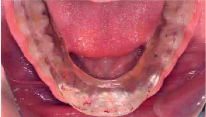
Figure 12 - Patient presented anterior open bite with osteoarthritic joints on both TMJs.
Figure 13 - Mandibular stabilization splint was delivered.
Figure 14 - Progress photograph: Anterior open bite got worse. Figure 15 - All the TMD symptoms disappeared. Anterior open bite continued to get worse (Compare to the initial photograph).
What do you think about the use of botulinum toxin in the control of patients with muscle hy-peractivity? (Paulo Rocha)
Injecting hyperactive muscles with minimum quantities of botulinum toxin would result in de -crease muscle activity by blocking the release of acetylcholine from the neuron. This will effectively weaken the muscle for a period of three to six months.
If a patient has a normal facial height and nor -mal incisor position but shows an excessive gingival
display because of the muscle hyperactivity, then
botulinum toxin injection can be very helpful. How
-ever, the treatment effect is only temporary. Patients
need repetitive injections every 3 to 6 months. Cur
-rently there is no guideline for the amount of toxin and location of the injection. There is no long-term studies of the effectiveness of this type of treatment. Further studies need to establish the guidelines for the injection locations, injection amount of botuli -num toxin and long-term treatment effects.
Luiz G. Gandini Jr.
» Full Professor, Faculty of Dentistry of Araraquara-UNESP.
» Adjunct Professor of Departmento of Orthodon
-tics, Baylor College of Dentistry and Saint Louis University.
Marcelo Castellucci
» MSc in Dentistry, UFBA.
» Specialist in Orthodontics, PUC-MG.
» Professor of Specialization course in Orthodontics, UFBA.
Orlando Tanaka
» Associate Professor of Graduation and Post Gradua
-tion in Dentistry, PUC-PR.
» Diplomate by Brazilian Board of Orthodontics and Facial Orthopedics.
» CADE Post-doctorate student, St. Louis University.
Paulo Vicente Rocha
» PhD and Adjunct Professor of Integrated Clinic,
FO-UFBA.
» MSc and PhD in Oral Rehabilitation, FOB- USP. » Specialist in TMD, CFO.
» Coordinator of Specialization course in Prosthodon
-tics, ABO-Bahia.
1. Pellegrini P, Sauerwein R, Finlayson T, McLeod J, Covell DA Jr, Maier T, et al. Plaque retention by self-ligating vs elastomeric orthodontic brackets: Quantitative comparison of oral bacteria and detection with adenosine triphosphate-driven bioluminescence. Am J Orthod Dentofacial Orthop. 2009;135(4):426.e1-9; discussion 426-7.
2. Pandis N, Vlachopoulos K, Polychronopoulou A, Madianos P, Eliades T. Periodontal condition of the mandibular anterior dentition in patients with conventional and self-ligating brackets. Orthod Craniofac Res. 2008;11(4):211-5.
3. Turnbull NR, Birnie DJ. Treatment efficiency of conventional vs self-ligating brackets: effects of archwire size and material. Am J Orthod Dentofacial Orthop. 2007;131(3):395-9.
4. Chen SS, Greenlee GM, Kim JE, Smith CL, Huang GJ. Systematic review on self ligating brackets. Am J Orthod Dentofacial Orthop. 2010;137(6):726.e1-18; discussion 726-7.
5. Chen SS, Greenlee GM, Kim JE, Smith CL, Huang GJ. Systematic review on self-ligating brackets. Am J Orthod Dentofacial Orthop. 2010;137(6):726.e1-8. 6. Turnbull NR, Birnie DJ. Treatment efficiency of conventional vs self-ligating
brackets: Effects of archwire size and material. Am J Orthod Dentofacial Orthop. 2007;131(3):395-9.
7. Baek MS, Choi YJ, Yu HS, Lee KJ, Kwak J, Park YC. Long-term stability of anterior open-bite treatment by intrusion of maxillary posterior teeth. Am J Orthod Dentofacial Orthop. 2010;138(4):396.e1-9; discussion 396-8.