Evidence-Based
Speech-Language
Pathology and Audiology
Fonoaudiologia Baseada
em Evidências
Luciana da Silva Barberena
1Brunah de Castro Brasil
1,2Roberta Michelon Melo
1Carolina Lisbôa Mezzomo
1,3Helena Bolli Mota
1,3Márcia Keske-Soares
1,3Descritores
Ultrassom Fala Fonética Fonoaudiologia Voz Deglutição Terapia Miofuncional
Keywords
Ultrasonics Speech Phonetics Speech, Language and Hearing Sciences Voice Swallow Myofunctional Therapy
Correspondence address: Roberta Michelon Melo
Rua Marechal Floriano Peixoto, 1.751, Centro, Santa Maria (RS), Brasil, CEP: 97015-372.
E-mail: [email protected]
Received: 12/23/2013
Accepted: 09/01/2014
Study carried out at the Graduate Program in Human Communication Disorders, Universidade Federal de Santa Maria – UFSM – Santa Maria (RS), Brazil.
(1) Graduate Program in Human Communication Disorders, Universidade Federal de Santa Maria – UFSM – Santa Maria (RS), Brazil.
(2) Universidade Federal do Rio Grande do Sul – UFRGS – Porto Alegre (RS), Brazil.
(3) Speech Language Pathology and Audiology Course, Universidade Federal de Santa Maria – UFSM – Santa Maria (RS), Brazil.
Financial support: Fundação de Amparo à Pesquisa do Estado do Rio Grande do Sul – FAPERGS – e Coordenação de Aperfeiçoamento de Pessoal de Nível Superior – CAPES.
Conlict of interests: nothing to declare.
Ultrasound applicability in Speech Language
Pathology and Audiology
Aplicabilidade da ultrassonograia na Fonoaudiologia
ABSTRACT
Purpose: To present recent studies that used the ultrasound in the ields of Speech Language Pathology and Audiology, which evidence possibilities of the applicability of this technique in different subareas. Research strategy: A bibliographic research was carried out in the PubMed database, using the keywords “ultrasonic,” “speech,” “phonetics,” “Speech, Language and Hearing Sciences,” “voice,” “deglutition,” and “myofunctional therapy,” comprising some areas of Speech Language Pathology and Audiology Sciences. The keywords “ultrasound,” “ultrasonography,” “swallow,” “orofacial myofunctional therapy,” and “orofacial myology” were also used in the search. Selection criteria: Studies in humans from the past 5 years were selected. In the preselection, duplicated studies, articles not fully available, and those that did not present direct relation between ultrasound and Speech Language Pathology and Audiology Sciences were discarded. Data analysis: The data were analyzed descriptively and classiied subareas of Speech Language Pathology and Audiology Sciences. The following items were considered: purposes, participants, procedures, and results. Results: We selected 12 articles for ultrasound versusspeech/phonetics subarea, 5 for ultrasound versusvoice, 1 for ultrasoundversusmuscles of mastication, and 10 for ultrasound versus swallow. Studies relating “ultrasound” and “Speech Language Pathology and Audiology Sciences” in the past 5 years were not found. Conclusion: Different studies on the use of ultrasound in Speech Language Pathology and Audiology Sciences were found. Each of them, according to its purpose, conirms new possibilities of the use of this instrument in the several subareas, aiming at a more accurate diagnosis and new evaluative and therapeutic possibilities.
RESUMO
INTRODUCTION
The Speech Language Pathology and Audiology clinic uses
various instruments to validate its actions. It is common to use
assessment protocols, often subjective, to deine the
differ-ent steps and procedures of Speech Language Pathology and
Audiology therapy. In recent years, there is a growing search
for instruments and more direct and quantifying analysis,
seeking greater accuracy of the data collected, as well as of
the diagnostics and prognostics.
The auditory–perceptual analysis is used widely in the areas
of speech and language and can be complemented by other
instrumental evaluations, so that the participant is evaluated and
treated in all respects of its communicative disorder. Acoustic
analysis of speech and voice has been used for a long time as
tool for analysis of altered cases, for promoting more accurate
diagnoses, and as the monitoring procedure of the
therapeu-tic process, both by speech language pathologist and by the
patient
(1-4). The articulatory analysis in the area of speech and
orofacial motricity emerges as a new possibility, especially at
the national level
(5-10), for integration of these analyses already
mentioned, using, for example, the ultrasonography for
evalu-ation of tongue movements
(11-22), and of the hyoid bone
(19,23-27),
without inserting devices within the oral cavity.
The investigation of tongue movements is one of the
possi-bilities of using this type of articulation assessment, which is not
considered invasive and is available with minimum interference in
the visualization of intraoral movements
(28,29), enabling research
in various subareas of Speech Language Pathology and Audiology.
The knowledge of speech language pathologists about the
pos-sibilities of use of ultrasound in a variety of Speech Language
Pathology and Audiology alterations (the focus of this article) can
affect important research in the area and consequent relevant
ind-ings. The use of ultrasound allows the speech language pathologist
to perform articulatory data analysis of the participant assessed and
of the patient undergoing therapeutic procedure
(11,12,30).
PURPOSE
To present studies of the past 5 years that used
ultrasonogra-phy in the ield of Speech Language Pathology and Audiology,
which can show possibilities of applicability of this technique in
different subareas of Speech Language Pathology and Audiology.
RESEARCH STRATEGY
A narrative review of the literature based on the question
that guided this study was conducted: “In what areas and in
what way the ultrasound data can contribute to the knowledge
of Speech Language Pathology and Audiology?”
To answer this question, a literature review was
con-ducted from May to July of 2013. It was performed initially
in the PubMed international database, which is maintained
by the National Center for Biotechnology Information (NCBI) at
the National Institute of Health (NIH). This search strategy was
chosen because the tool catalogs scientiic articles published in
journals indexed in major global scientiic collections.
The keywords provided by Health Sciences Descriptors
(DeCS) used in the research were “ultrasonic,” “speech,”
“phonet-ics,” “Speech, Language and Hearing Sciences,” “voice,”
“degluti-tion,” and “myofunctional therapy,” covering some of the areas of
the Speech Language Pathology and Audiology. The terms
“ultra-sound,” “ultrasonography,” “swallow,” “orofacial myofunctional
therapy,” and “orofacial myology” were also researched in the
examination, even though they are not descriptors provided by
the Regional Library of Medicine (BIREME), because they are
found several times in articles related to the area. The
combina-tions of the descriptors and terms were as follows: ultrasonic AND
speech, ultrasound AND speech, ultrasonography AND speech,
and thus successively with the other terms and descriptors.
After the search for articles with the descriptors and terms
quoted, the abstracts of the studies were searched and, if they
were of interest to this study, the full text of the respective
articles were searched.
In this way, to get some full articles not available in
PubMed, the
Coordenação de Aperfeiçoamento de Pessoal de
Nível Superior
(CAPES) journals portal was also used, through
the search for the topic with the job title.
The databases cited were prioritized due to the their easy
access, and it was done by speciic agreements made by the
Universidade Federal de Santa Maria, of which the authors of
this article are part of.
SELECTION CRITERIA
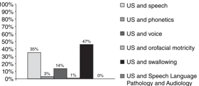
The following were selected as search criteria in the database:
studies of the past 5 years, and studies in humans. In this way,
320 abstracts were found in the survey conducted through the
PubMed database. Figure 1 shows the total number of articles
for each area of the Speech Language Pathology and Audiology
before the adoption of the criteria for inclusion and exclusion of
articles that comprised the present literature review.
As stated in the Research Strategy section, after the search in
the database, there was a preselection of the material collected
from the abstracts. The inclusion criteria of the work in this review
were: to use ultrasound as a tool for obtaining data; to relate the
ul-trasound information to some of the areas of interest of the Speech
Language Pathology and Audiology — speech, phonetics, voice,
orofacial motricity, and swallowing; to ind the availability of the
full text, either in PubMed or in the CAPES journals portal.
Figure 1. Total of selected abstracts in the PubMed database as the intersection of the descriptors
Caption: US = Ultrasound
100% 80% 90% 70% 60% 50% 40% 30% 20% 10% 0%
US and speech
US and phonetics
US and voice
US and orofacial motricity
US and swallowing
US and Speech Language Pathology and Audiology
35%
3% 14%
1% 47%
In the preselection, duplicate studies; articles not available
in its entirety; and the ones that presented no direct relation of
ultrasound with the Speech Language Pathology and Audiology,
for example, articles that used ultrasound to investigate
biop-sies, carcinomas, and esophageal swallowing, were discarded.
At the end of the survey, 12 articles for the subarea ultrasound
versus speech/phonetics, 5 for ultrasound versus voice; 1 for
ultrasound versus masticatory muscles, and 10 for ultrasound
versus swallowing were selected. Although they are
descrip-tors used in this review, no studies were found when crossing
the terms “ultrasound” and “Speech Language Pathology
and Audiology” (Speech, Language and Hearing Sciences).
Therefore, we selected 28 articles, being the oldest of 2008
and the latest of 2013, for this review.
DATA ANALYSIS
The data were analyzed in a descriptive manner and
sepa-rately for each subarea of Speech Language Pathology and
Audiology, as described earlier.
The items analyzed were the following:
• purposes,
• subjects considered in the study,
• important procedures, and
• the main results of the study.
RESULTS
Ultrasound and speech/phonetics
The studies related to the research “Ultrasound and Speech”
aimed, in general,
• to apply the ultrasound images for speech therapy
(11,12,30);
• to investigate the coarticulatory effects, for example, of the
syllable CV (consonant–vowel) versus CVC (consonant–
vowel–consonant), or vowel context
(13,18,31);
• to describe some descriptors of the tongue movements,
among them, the speed and the distance covered
(14,16);
• characterize the gestures involved in the production of
consonant segments, as for instance the phoneme /r/
(15);
• to describe compensatory articulatory strategies and hidden
movements
(32); and
• to propose articulatory measures via ultrasonography
(22).
The deinition of the sample was restricted to the purposes of
the searches. Therefore, the articles that applied ultrasound data
for the investigation of speech focused on participants with
typi-cal
(13,15,16,18,31)and atypical
(11,12,14,16,30,32)speech. Speech alterations
were due to speech disorder
(11,12), glossectomy
(14,16), hearing loss
(30),
and cleft palate
(32). The search for this subarea had on average 8.5
participants; of these, 64.9% were adults and 35.1% children.
As the characterization of the group of subjects, the
ob-served methodological aspects are quite heterogeneous, also
restricted to the research proposal (Chart 1).
The main results of the articles are also presented in Chart 1.
The various possibilities of application of tongue ultrasound
images in the speech subarea can be observed, because the
identiication of gestural patterns in an evaluative procedure to
the insertion of this technology as a therapeutic procedure is
avail-able to the Speech Language Pathology and Audiology Clinic.
With the crossing of the descriptors related to “ultrasound”
and “phonetic,” a study
(21)directed to the research of speech
seg-ments was found. The article aims to analyze the images of the
forms of the tongue of an individual, native speaker of a dialect
of Nepal, during the production of plosives and affricates, voiced
and voiceless, dental, retrolexes, and palatals. In this study,
the authors believe that the spatial and dynamic information
obtained via ultrasound image of the tongue can supplement
the data obtained with the static electropalatography, related to
the location and type of constriction of the tongue on the palate.
The results showed differences in the forms of the tongue,
even if some were discreet, between location of the constriction
of the devoiced and voiced consonant, of the consonant at the
beginning and at the end of a syllable, and between different
articulation points of the dialect investigated
(21).
The association between ultrasound and phonetic (Chart 2),
although still small, as this bibliographic research shows, already
allowed us to verify that the ultrasound images are able to
pro-vide speciics of a given language, based on the analysis of the
dynamics of the sounds in real time. Such manipulation can also
stimulate research in phonetics on the organization of syllabic
sequences, coarticulation, and structural organizations of sounds.
Ultrasound and voice
The studies found and highlighted in Chart 3 showed
well-differentiated goals, investigating from the tone and length of
muscles, measurement of mucosal wave, adaptation of speech
valves, to literature review. Many studies have sought to compare
and discuss the advantages, disadvantages, and the applicability
of instruments such as ultrasound to electroglottography, video
luoroscopy, and computerized tomography
(33-36). Many of these
techniques are widely used in the medical ield and by speech
language pathologists, but the ultrasound, for example, is a method
that requires study and has been recruited on a larger scale for
research in the ield of Speech Language Pathology and Audiology.
The studies had an average of 49 participants
(34-36); the
re-sults showed improvements to the ield and helped to spread
the use of new techniques, improve them, and validate them.
The ive studies reported the importance of using the method
of ultrasound combined with other techniques, what could
decrease the risk of postoperative complications
(36), determine
laryngeal segments with better accuracy
(35), determine
varia-tions in muscle length
(34), as well as contribute to diagnosis and
to an effective treatment plan
(33,37).
Ultrasound and masticatory muscles
The only study found
(8)(Chart 4) relating the ultrasound technique
Author(s) Objective(s) Participant(s) Important procedures Results
Bernhardt et al., 2008(11)
To investigate the effects of short-term therapy using US as a visual feedback of speech production of children with speech disorder in rural communities in British Columbia, Canada.
13 students, 8 boys and 5 girls, 7–15 years old, participated in therapy for correction of speech disorders, but with still persistent disorders.
The approach with ultrasonography involved three phases: 1) six or seven sessions without the use of US; 2) one to three sessions with the US; 3) seven to eight sessions without US. The target sound focused was: /
r
/.11 individuals presented a rapid increase in production of the target sound after the second stage of treatment. According to the therapists, the US contributed to significant advances in a shorter period, as well as an increase in the motivation of the participants.
Modha et al., 2008(12)
To compare the treatment segment /r/ with or without US as a continuation of previous studies.
A 13-year-old Canadian English speaker boy participant. He received speech therapy, however, had not yet acquired the phoneme /
r
/.Nine meetings of 30–45 minutes were held. The treatment initiated by a session without US followed by two blocks of four sections including treatment with the US and two without. The therapeutic practice included the imitative and spontaneous production of the target sound in an isolated form, syllables, words, and sentences. The final evaluation was performed after 6 weeks without speech intervention. The perceptual analysis and the acoustic analysis were performed, and the point of maximum constriction of the consonant was recorded.
The values of the formants and the analysis of trained listeners showed an improvement in the production of /r/, particularly after the introduction of US.
Pouplier, 2008(13)
To investigate the bias of gestural intrusion into segments, as well as the effect of consonants in coda on initial segments.
Eight native speakers of American English, six men and two women, aged between 22 and 43 years.
The following words were collected in coda: “top cop, sop, shop”; in Non-coda: “kaa, taa, saw, shaw”. With variation in accent and position in the sentence. Also, “taa kaa taa,” “kaa taa kaa,” “saw shaw saw,” and “shaw saw shaw” were collected. Audio signals, image (US), and video (lower face) were simultaneously recorded. The mid-sagittal view of the language through EdgeTrak Program for extracting the contour of the tongue was assessed. Errors in speech production were defined as: (a) intrusion, (b) omission, and (c) substitution.
The competition during encoding of the utterance increases with the increase in sharing gestural structure, i.e., the more gestures participate in a complex frequency ratio, the greater the likelihood of errors occurring. Furthermore, the non-coda condition showed an error rate smaller than the coda condition whereas a gestural intrusion bias emerged mainly for the coda condition. Finally, the proportion of different types of errors (substitutions, coproduction errors) differs depending on the type of stimulus.
Rastadmehr et al., 2008(14)
To document the impact of a lateral resection of the tongue on the speed of movement of the tongue in patients before and after a partial tongue resection, using the B-mode of an US image capture.
Ten patients with tongue cancer. The average age was 45 years. Six normal individuals comprised the comparison group, with a mean age of 38 years. All subjects were speakers of Canadian English.
Patients with glossectomy were observed for a few days before surgery and 2 months after the surgery.At that point, the healing is relatively complete and swelling has subsided. If patients needed radiotherapy, the assessment was conducted before this procedure. US of tongue movements in mid-sagittal plane was carried out and acoustic data were recorded simultaneously.
Regarding the reading time, there was no difference between normal speakers and patients before or after surgery. When compared the speed of the tongue in patients with tongue cancer before and after glossectomy, significant interaction was found between the factors time and tongue segment reached. No significant interactions between the factors time and type of reconstruction were found. The study showed that a side partial resection and the reconstruction led to a significant increase in the speed of movement. This finding was interpreted as evidence of an increase in articulatory effort, resulting from a compensatory motor learning.
Chart 1. Studies that have addressed the application of ultrasound in speech
Author(s) Objective(s) Participant(s) Important procedures Results
Campbell et al., 2010(15)
To characterize the gestures related to the segment /r/ of the American English.
Ten native speakers of Canadian English, five men and five women, aged between 22 and 36 years.
For this experiment, the combination of US image in B/M mode (for language data) and the Optotrak tracking (for data labial opposition) was used. The acoustic signal was also recorded. The gestures of /r/ were analyzed in the context of /e/ and /a/.The stimulus were read into the phrase - “___ said x each ___ “. The care for stabilizing of the head and of the transducer was considered. Pictures of the US were collected in the mid-sagittal plane (B mode). Time was recorded until the completion of the gesture, as well as gestural magnitude.
One anteroposteriorization in the initial syllable was sequentially observed, while in the final syllable the root gestures of the tongue and lips precede the gesture of the tongue blade. In regard to the magnitude, the two most anterior gestures (blade and lips) exhibited relative decrease of this parameter in the final position. A significant interaction was observed between time and the magnitude of the gesture. A notable finding of this study refers to the distinction of the three gestures involved in the production of /r/, which cannot be represented in terms of a simple binary phonological categorization of gestures.
Bressmann et al., 2010(16)
Quantitatively describe aspects of coronal tongue movements in the different anatomical regions of the tongue.
Four normal speakers (two women 23 and 24 years old, and two men 25 and 34 years old) and one with partial glossectomy.
The US was recorded in four coronal planes, being necessary to produce four times the speech stimulus. Participants should recite the last stanza of the poem “I wandered lonely as a cloud”. The lines of the tongue’s surface were extracted through the Ultra-CATS software; then the distance was measured between US transducer and the surface of the tongue. Then for the quantitative description of the function of tongue indicators total distance traveled and concavity were calculated.
The study found that there is greater movement in the center of the tongue than that in the lateral free margins. Depending on the speaker, the greater distances covered were focused in the anterior or posterior region of the tongue. In all speakers, the rear portion of the tongue showed consistent grooving during the connected speech. A more flat or convex condition of the tongue was observed for the anterior region.
Zharkova et al., 2011(18)
To compare the coarticulatory properties of the tongue in children and adults using US pictures of the tongue.
Ten adults (average age of 33 years) and 10 children (average age of 7 years 7 months) with normal speech development, speakers of Scottish Standard English.
US and acoustic data were recorded synchronously. The US frames were recorded at two points: in the middle of a consonant and in the middle of the vowel, based on the acoustic data. The target syllables /∫i/, /∫u/, and /∫a/ were studied, which were inserted in the sentence vehicle: “It’s a ____” (she, shoe and shah). The parameters used in the US were: length of the tongue bend. We also examined the coarticulatory effects, the extent of coarticulation, and the token-to-token variation.
Children exhibit coarticulation in the same vowel context as adults. However, the results fail at showing the hypothesis in relation to the position of the tongue for the production of /S/ on /i/ and /u/, because this pair of vowels showed no well-marked differences. We also observed that the children showed greater extent of coarticulation of the consonant depending on the context of the vowel than adults, besides having greater variability of the data.
Zharkova, 2013(22)
To describe the image of US of the tongue as a tool for quantitative analysis of the role of the tongue in speakers with cleft palate.
– Descriptive study.
Measures based on a single curve tongue described in this study are: the excursion index of the back and restriction position index of the dorsum of the tongue; both aim to directly assess the degree of involvement of the tongue in the articulation. Require stabilization of the head. Measures based on a set of curves of the tongue were also cited: tongue dynamics, variability, and separation the curves of the tongue. All measurements can be used to compare the function of tongue in speakers with cleft palate.
Chart 1. Continuation
Author(s) Objective(s) Participant(s) Important procedures Results
Bacsfalvi and Bernhardt, 2011(30)
To investigate speech production of seven adolescents and young adults with hearing impairment after 2–4 years of intervention through the US and electropalatography.
Seven speakers (four men and three women) with severe to profound hearing loss.
Three sets of data were collected for each patient: 1) pretreatment with visual feedback, 2) immediately after using the US, and 3) follow-up data. Real or meaningless words were collected separately or inserted in the sentence: “I’m an ___” .
The trials for phases 2 and 3 of treatment showed that five of the seven speakers produced the segment with the same rate of accuracy in both phases. Showed the maintenance of levels of accuracy on a subject; improvement in three subjects or a slight regression for four subjects in the sample. Some limitations of the study are identified as: limited number of sample, short periods of intervention, the use only of perceptual analysis, and lack of access to a database of normative data for comparison.
McMillan and Corley, 2010(31)
To show that similarities of adjacent phonemes affect their articulation in predictable ways.
Ten adults, native speakers of English.
The phonetic similarities of phonemes at the beginning of tongue twisters were manipulated. Data were timed and compared with electropalatography. Analysis of the average sagittal outline of the tongue over time was done with the help of US and the VOT.
Subphonemic representations consist of articulatory gestures. Phonemes are “malformed,” in the sense that the aspects of the production of more than one phoneme are observed simultaneously.The bias for gestural intrusion (as opposed to deletion or substitution) follows naturally from the fact that there are “cascades” activated for production.
Bressmann et al., 2011(32)
To investigate the articulation of deaf alveolar plosive [k] in five speakers with compensatory articulation related to cleft palate.
Five individuals with different types of palatal fissures repaired, all speakers of Canadian English.
The tongue movements were recorded by ultrasonography in the mid-sagittal plane. The acoustic signal was recorded simultaneously. Participants spoke five repeat [a’ka], [i’ki] and [u’ku] after the examiner. The perceptual and visual analyses were reviewed and discussed by three judges.
The analysis revealed a variety of compensatory strategies: glottal plosive, plosive pharyngeal, mid-palatal plosive, glottal, and velar coproductions. One subject produced a sound of palate click along with a mid-palatal plosive. The image of US also revealed covert articulatory movements, which would not be identified through an isolated perceptual analysis.
Caption: US = ultrasound; VOT = voice onset time Chart 1. Continuation
Author(s) Objective(s) Participant(s) Important procedures Results
Kochetov et al., 2013(21)
To use US to obtain the images of tongue in various Nepali lingual consonants.
One native speaker of the dialect Brahmin participant.
Analysis of syllabic sequences via ultrasound images. Nepali coronal consonants were observed.
The results confirmed and extended some observations on the Nepalese coronal consonants, such as a single contrasting place between dental, alveolar, and retroflex.
Caption: US = ultrasound
Chart 2. Study that considered the application of ultrasound in the phonetics
The ultrasound technique allowed the measurement
of the thickness of the masseter muscle as well as the diagnosis,
the treatment plan, and the interdisciplinary prognosis —
orth-odontic, orthognathic, and Speech Language Pathology and
Audiology. The technique proved to be eficient for
measure-ments, and enabled the monitoring and setting discharge criteria
for patients
(8).
It is believed that there are studies from other areas (e.g.,
dental) that are also used to search the masticatory muscles
us-ing ultrasound images, which could also be of particular interest
to the Speech Language Pathology and Audiology. However,
these were not included in this article, because they have not
been located through the search strategies considered here.
Ultrasound and swallowing
From the search of the descriptors ultrasound versus
swallowing, papers that are presented in Chart 5, the
inves-tigation of the displacement of the hyoid bone during the act
of swallowing was frequent
(19,23-26). In addition to this, three
other papers further investigated the thickness of the lingual
musculature
(20,27)and the pressure exerted by the tongue on
the palate
(19).
Author(s) Objective(s) Participant(s) Important procedures Results
Krausert et al., 2011(33)
To discuss the advantages, disadvantages, and clinical applicability of the different measurement techniques of the mucosal wave (electroglottography photoglottography and US) and visualization techniques, which include videokymography (stroboscopic and digital high-speed imaging).
–
The various techniques and their specific uses are reviewed with the intention to help researchers and clinicians to choose a method for a given situation and understand their limitations and their potential applications.
The assessments of existing research and recommendations for future research are given to foster both the quantitative study of mucosal wave via a standardized and accurate measurement of the parameters of the same as for the development of reliable methods with which vocal disorders could be diagnosed.
Cho et al., 2012(34)
To access the change in length of the vocal fold with US.
35 professional singers.
The individuals performed ultrasonography during respiration, phonation, and singing. The length of the vocal fold was measured in each situation.
Differences were found in each phase. The authors concluded that US can be used to check the variation of the length of the vocal folds.
Tićac et al., 2012(35)
To compare the values of videofluoroscopy and US to assess the tone of muscles of the glottis and accurately determine the hypertonic laryngeal segment.
20 patients with inadequate tracheoesophageal voice that went to total laryngectomy and tracheoesophageal prosthesis installation.
After determining the hypertonic laryngeal segment, intramuscular lidocaine was administered. Knowing that videofluoroscopy is the standard test for comparison with US examination test, contrast was administered and dilatation of the segment during swallowing, phonation, and vocal rest was observed to determine differences of tone or disorder in-between before and after administration of lidocaine.
The combination of the two methods may provide better results on the voice improvements. Videofluoroscopy is an initial choice examination to determine the hypertonic segment and the US is the method used to facilitate the administration of the drug more precisely.
Pedisić
et al., 2012(36)
To use the US and the neck computed tomography in the preoperative PROVOX II implant surgery and to determine the presence of tracheoesophageal fistula after surgery.
91 patients from January 2004 to December 2010.
The authors used US and neck CT scan in 58 patients, in the preoperative procedure through which the length of the tracheoesophageal fistula was determined. At the same time, the opportunity was used to specify the length of the speech prosthesis that it would be adapted.
The number of individuals that had complications and used preoperative procedure was significantly lower than the number of individuals that had complications and did not go through the procedures. Compared to other studies, the authors believe that these methods decrease the number of complications caused by the inadequacy of the length of tracheoesophageal prosthesis.
Setlur and Hartnick, 2012(37)
Review of studies on treatment of unilateral vocal fold paralysis in children.
– Literature review.
The authors reported that the diagnosis and treatment plan are made by laryngoscopy, US, and electromyography. Today, there are several studies suggesting specific treatment for children, which can lead to major impact on quality of life of the same.
Chart 3. Studies that have addressed the application of ultrasound in the voice
Author(s) Objective(s) Participant(s) Important procedures Results
Trawitzki et al., 2011(8)
To analyze the effect of integrated treatment: orthodontic, orthognathic surgery, and myofunctional therapy in the thickness/ density of the masseter in patients with Class III, 3 years after orthognathic surgery.
Longitudinal study with 13 patients with Class III and 15 individuals of the control group.
Ultrasonography of masseter was performed during rest and chewing/biting in the three groups.
Larger thickness of masseter values were found in the postoperative group. Between this group and the control group, there was no difference on the right side of the muscle, in situations of bite, and in the left side, in situations of rest. The proposed treatment resulted in improved thickness/density of the masseter muscle in patients with Class III.
Chart 4. Study that considered the application of ultrasound in the evaluation of a masticatory muscle
Author(s) Objective(s) Participant(s) Important procedures Results
Galén and Jost-Brinkmann, 2010(17)
To investigate the possibility of using US to differentiate patterns of visceral and somatic swallowing.
11 adults with visceral swallowing pattern and 13 adults with standard somatic swallowing.
Assessment and comparison of sequences of B and M mode during swallowing.
The extent and the speed of vertical movement of the tongue were determined, as well as the total duration of swallowing.
The parameters revealed large intra- and interindividual variability. There were no qualitative differences in B-mode images. The parameters measured in M-mode images were not suitable for differentiating between patterns of visceral and somatic swallowing.
Steele et al., 2012(19)
To investigate the correlation between non-invasive measurements of timing the pressure of the tongue on the palate and the measure of excursion of the hyoid bone.
20 healthy adults (10 men and 10 women), between 20 and 39 years.
The temporal relations between the events of the pressure of the tongue on the palate and the hyoid movement during swallowing were explored.
The creation of pressure of the tongue on the palate and the movement of the hyoid are distinct phenomena that follow the swallowing.
Tamura et al., 2012(20)
To assess sarcopenia of tongue muscles by measuring the thickness of the tongue with US, as well as to clarify its relation with nutritional status in the elderly.
104 elderly people (32 men and 72 women, average age of 80.3 years).
The frontal and lateral position of the tongue was considered using US stable images. The measurement was performed twice and the average value was obtained.
The thickness of the tongue is related to nutritional status in the elderly. The dysfunction and abnormality of the tongue can also be a cause for dysphagia. Malnutrition can lead to sarcopenia (decreased muscle mass and strength), not only in skeletal muscles but also on the tongue.
Huang et al., 2009(23)
To explore the reliability of the US examination of the hyoid– larynx approximation and its application in stroke patients with and without dysphagia.
55 participants: 15 healthy individuals and 20 individuals who had CVA, but did not show changes in swallowing, and 20 individuals who had CVA and dysphagia.
The distance between the thyroid cartilage and the hyoid bone during swallowing was measured by US. Ten patients with stroke and dysphagia also underwent videofluoroscopy.
The percentage change in hyoid–larynx approximation was very similar between the US examination and videofluoroscopy, showing the reliability of the US to that extent. The hyoid-larynx approximation was significantly reduced in patients with CVA and dysphagia.
Scarborough et al., 2010(24)
To acquire normative parameters about the maximum displacement of the hyoid obtained by US, in a sample of healthy children.
29 children of preschool age.
The maximum displacement of the hyoid bone, determined from a sequence of frames during analysis in spontaneous swallowing, was observed.
99% of children showed displacement of the hyoid bone within 3 cm as normal parameters, and the US proved to be a reliable method for such analysis.
Chart 5. Studies that have addressed the application of ultrasonography in swallowing
Author(s) Objective(s) Participant(s) Important procedures Results
Macrae et al., 2012(25)
To quantify the movement of the hyoid via US through an analysis of inter- and intra-evaluators reliability.
Five individuals (two men and three women, aged between 20 and 50 years).
The distance between the mandible and the hyoid bone and the change of displacement of the hyoid bone from a reference point were calculated.
Measurements at rest and in maximum displacement of the hyoid bone, taken by each evaluator, were highly correlated. The US is an important tool for evaluating the displacement of the hyoid bone in dysphagia.
Yabunaka et al., 2011(26)
To use US to assess the trajectories of movement of the hyoid bone and age-related changes during swallowing in healthy subjects.
30 adult individuals, equally divided into three age groups: 20–39 years, 40–59 years, and 60–79 years.
The US transducer was positioned above the larynx; the hyoid bone should be centered on the screen. The individuals swallowed five times 5-ml water. The resting point of the hyoid bone and other periods were measured.
The trajectory of the hyoid bone was easily observed with the US. It was presented in four stages: 1) elevation after swallowing; 2) anteriorization; 3) temporary phase (position of maximum elevation); and 4) return to the rest position. The time of stages 1, 2, and 4 increased significantly with increasing age. The opposite was observed for the third phase.
Hsiao et al., 2012(27)
To measure changes in the thickness of the tongue in stroke patients with dysphagia and the displacement of the hyoid bone during swallowing.
60 stroke patients and 30 healthy individuals for the control group.
The thickness of the tongue and the displacement of the hyoid bone were measured. As assessment tools, videofluoroscopy (on 12 patients) and the US were used.
The thickness of the tongue and the hyoid displacement was smaller in the group whose diet occurred by probe. The US proved as a reliable measure as it showed good correlation with videofluoroscopy.
Komori et al., 2008(38)
To compare the technique that combines endoscopy and US with fluoroscopy.
Eight healthy men, aged between 25 and 31 years.
The laryngeal elevation corresponded to the maximum height, verified by fluoroscopy and US.
The moment of the elevation was similar among the investigated techniques. Distances and durations of maximum laryngeal elevation correlated significantly. The technique of fluoroscopy and US may show the swallowing function as efficiently as videofluoroscopy.
Yabunaka et al., 2012(39)
To investigate the application of US to quantify the patterns of geniohyoid muscles during swallowing and to evaluate these patterns according to age and gender.
60 adults (30 men and 30 women), divided into three age groups (20–39 years, 40–59 years, and 60–79 years).
10 mL water was administered in five repetitions Every geniohyoid muscle movement was recorded in real time. Simultaneous images of swallowing were captured in two regions along the lateral geniohyoid muscle wall.
The moving average distance and the duration of the movement of the mylohyoid muscle-geni increase gradually with age. No difference was found in measurements during swallowing between the sexes, in all age groups.
Chart 5. Continuation
Caption: US = ultrasound; CVA = cerebral vascular accident
of somatic and visceral swallowing
(17)and, inally, with research
movement patterns of geniohyoid muscles
(39).
As for the sample evaluated in the mentioned
pa-pers
(17,19,20,23-27,38,39), the groups comprised individuals with
typi-cal swallowing (from children to the elderly) and with altered
swallowing, due to some underlying diseases, such as cerebral
vascular accident (CVA). The number of participants varied
considerably from 5 to 104 among the articles, with an average
of 42.5 participants.
The research
(19,23-26)highlighted the ultrasound as an
im-portant tool for evaluating the displacement of the hyoid bone
in swallowing research. Reducing the movement of the hyoid
bone has been associated with the increased risk for intrusion
of waste in the larynx and the air passages. Comparing the
ultrasound image with other assessment tools
(23,27,38), ultrasound
Through the use of ultrasound, some authors were able to
verify that the pressure of the tongue on the palate and the hyoid
movement are distinct phenomena as a result of swallowing
(19).
However, correlation was observed between the thickness of
the tongue, veriied by ultrasound images, and the nutritional
status of stroke patients with dysphagia. Thus, malnutrition can
induce sarcopenia (decreased muscle mass and strength) and
the dysfunction and abnormality of the tongue may also be an
indicator of dysphagia
(20).
As ultrasound still is underused in studies of dysphagia,
in most papers further research involving this technique in
the standardization of reliable measures is suggested to assess
swallowing. However, most studies already conirmed the
ultrasound images as a possibility in assessing the dynamics
of swallowing.
CONCLUSION
Studies that investigated the use of the ultrasound technique
in the ield of Speech Language Pathology and Audiology in
the past 5 years were found. These studies conirmed the new
possibilities of the use of ultrasound in all subareas of Speech
Language Pathology and Audiology considered in the present
literature review, striving for a more accurate diagnosis, more
detailed assessments, differentiated therapeutic processes, and
greater cost-effectiveness.
The majority (27 of 28 articles selected for this literature
review) of the studies found were carried out in other countries,
highlighting the ield need to update itself and the necessity to
practice the search through the use of new instruments in the
researches and in Speech Language Pathology and Audiology
Clinical Therapy in Brazil.
ACKNOWLEDGMENT
We thank the Fundação de Amparo à Pesquisa do Estado
do Rio Grande do Sul (FAPERGS) and the Coordenação de
Aperfeiçoamento de Pessoal de Nível Superior (CAPES) for
providing two PhD scholarships, for the irst and to the third
author of this work, during this study.
*LSB, BCB, and RMM were jointly responsible for the delimitation of the study, literature search, and its writing; MKS, CLM, and HBM supervised the data collection, tabulation and analysis, and provided guidance during all stages of the study elaboration.
REFERENCES
1. Gregio FN, Camargo ZA. Dados de tempo de início do vozeamento (VOT) na avaliação do sinal vocal de indivíduos com paralisia unilateral de prega vocal. Disturb Comum. 2005;17(3):289-97.
2. Melo RM, Mota HB, Mezzomo CL, Brasil BC, Lovatto L, Arzeno L. Parâmetros acústicos do contraste de sonoridade das plosivas no desenvolvimento fonológico típico e no desviante. Rev Soc Bras Fonoaudiol. 2012;17(3):304-12.
3. Marino VCC, Berti LC, Lima-Gregio AM. Características acústicas da oclusiva glotal associada à sequência de Pierre Robin: estudo de caso. Rev CEFAC. 2013;15(2):466-77.
4. Giacchini V, Mota HB, Mezzomo CL. Variáveis relevantes no processo terapêutico para a aquisição do onset complexo na fala de crianças com desvio fonológico. Rev CEFAC (no prelo).
5. Berti LC. Investigação da produção de fala a partir da ultrassonograia do movimento de língua. In: 19º Congresso Brasileiro de Fonoaudiologia; 2010; Curitiba. Revista da Sociedade Brasileira de Fonoaudiologia: suplemento. Curitiba: Sociedade Brasileira de Fonoaudiologia; 2010. 6. Pereira LCK, Gregio FN, Svicero MAF, Nan PC, Madureira S,
Camargo ZA. Caracterização de vogais orais e nasais por dados de ultrassonograia, ressonância magnética e análise acústica. In: 19º Congresso Brasileiro e 8º Internacional de Fonoaudiologia; 2011; São Paulo. Revista da Sociedade Brasileira de Fonoaudiologia: suplemento. São Paulo: Sociedade Brasileira de Fonoaudiologia; 2011. p. 980.
7. Svicero MAF, Pereira LK, Nan PC, Isolan-Cury R, Camargo Z. Dados preliminares de análise de fala por meio da ultrassonograia. In: 19º Congresso Brasileiro e 8º Internacional de Fonoaudiologia; 2011; São Paulo. Revista da Sociedade Brasileira de Fonoaudiologia: suplemento. São Paulo: Sociedade Brasileira de Fonoaudiologia; 2011. p. 728. 8. Trawitzki LV, Dantas RO, Elias-Júnior J, Mello-Filho FV. Masseter
muscle thickness three years after surgical correction of class III dentofacial deformity. Arch Oral Biol. 2011;56(8):799-803.
9. Berti LC. Investigação ultrassonográica dos erros de fala infantil à luz da Fonologia Gestual. In: Ferreira-Gonçalves G, Brum-de-Paula M. Dinâmica dos movimentos articulatórios: sons, gestos e imagens. Pelotas (RS): Editora da UFPel; 2013. p.127-44.
10. Francisco DT, Pagan-Neves LO, Wertzner HF. Aplicação da ultrassonograia de fala como ferramenta auxiliar ao diagnóstico do transtorno fonológico. In: 4º Seminário de Aquisição fonológica; 2013; Santa Maria. 4º Seminário de Aquisição fonológica: resumos e programação. Pelotas (RS): Editora da UFPel; 2013. p. 33-4.
11. Bernhardt BM, Bacsfalvi P, Adler-Bock M, Shimizu R, Cheney A, Giesbrecht N, et al. Ultrasound as visual feedback in speech habilitation: exploring consultative use in rural British Columbia, Canada. Clin Linguist Phon. 2008;22(2):149-62.
12. Modha G, Bernhardt BM, Church R, Bacsfalvi P. Case study using ultrasound to treat /r/. Int J Lang Commun Disord. 2008;43(3):323-9. 13. Pouplier M. The role of a coda consonant as error trigger in repetition
tasks. J Phon. 2008;36(1):114-40.
14. Rastadmehr O, Bressmann T, Smyth R, Irish JC. Increased midsagittal tongue velocity as indication of articulatory compensation in patients with lateral partial glossectomies. Head Neck. 2008;30(6):718-26. 15. Campbell F, Gick B, Wilson I, Vatikiotis-Bateson E. Spatial and temporal
properties of gestures in North American English /r/. Lang Speech. 2010;53(1):49-69.
16. Bressmann T, Flowers H, Wong W, Irish JC. Coronal view ultrasound imaging of movement in different segments of the tongue during paced recital: indings from four normal speakers and a speaker with partial glossectomy. Clin Linguist Phon. 2010;24(8):589-601.
17. Galén S, Jost-Brinkmann PG. B-mode and M-mode ultrasonography of tongue movements during swallowing. J Orofac Orthop. 2010;71(2):125-35. 18. Zharkova N, Hewlett N, Hardcastle WJ. Coarticulation as an indicator
of speech motor control development in children: an ultrasound study. Motor Control. 2011;15(1):118-40.
19. Steele C, Sasse C, Bressmann T. Tongue-pressure and hyoid movement timing in healthy liquid swallowing. Int J Lang Commun Disord. 2012;47(1):77-83.
20. Tamura F, Kikutani T, Tohara T, Yoshida M, Yaegaki K. Tongue thickness relates to nutritional status in the elderly. Dysphagia. 2012;27(4):556-61. 21. Kochetov A, Pouplier M, Truong S. A preliminary ultrasound study of
Nepali lingual articulations. J Acoust Soc Am. 2013;13:1-9.
23. Huang YL, Hsieh SF, Chang YC, Chen HC, Wang TG. Ultrasonographic evaluation of hyoid-larynx approximation in dysphagic stroke patients. Ultrasound Med Biol. 2009;35(7):1103-8.
24. Scarborough DR, Waizenhofer S, Siekemeyer L, Hughes M. Sonographically measured hyoid bone displacement during swallow in preschool children: a preliminary study. J Clin Ultrasound. 2010;38(8):430-4.
25. Macrae PR, Doeltgen SH, Jones RD, Huckabee ML. Intra- and inter-rater reliability for analysis of hyoid displacement measured with sonography. J Clin Ultrasound. 2012;40(2):74-8.
26. Yabunaka K, Sanada H, Sanada S, Konishi H, Hashimoto T, Yatake H, et al. Sonographic assessment of hyoid bone movement during swallowing: a study of normal adults with advancing age. Radiol Phys Technol. 2011;4(1):73-7.
27. Hsiao MY, Chang YC, Chen WS, Chang HY, Wang TG. Application of ultrasonography in assessing oropharyngeal dysphagia in stroke patients. Ultrasound Med Biol. 2012;38(9):1522-8.
28. Gick B. The use of ultrasound for linguistic phonetic ieldwork. J Int Phon Assoc. 2002;32(2):113-21.
29. Bressmann T. Quantitative assessment of tongue shape and movement using ultrasound imaging. In: 3rd Conference on Laboratory Approaches
to Spanish Phonology; 2008. Somerville (MA); 2008. p. 101-6. 30. Bacsfalvi P, Bernhardt BM. Long-term outcomes of speech therapy for
seven adolescents with visual feedback technologies: ultrasound and electropalatography. Clin Linguist Phon. 2011;25(11-12):1034-43.
31. McMillan CT, Corley M. Cascading inluences on the production of speech: evidence from articulation. Cognition. 2010;117(3):243-60. 32. Bressmann T, Radovanovic B, Kulkarni GV, Klaiman P, Fisher D. An
ultrasonographic investigation of cleft-type compensatory articulations of voiceless velar stops. Clin Linguist Phon. 2011;25(11-12):1028-33. 33. Krausert CR, Olszewski AE, Taylor LN, McMurray JS, Dailey SH, Jiang
JJ. Mucosal wave measurement and visualization techniques. J Voice. 2011;25(4):395-405.
34. Cho W, Hong J, Park H. Real-time ultrasonographic assessment of true vocal fold length in professional singers. J Voice. 2012;26(6):819.e1-6. 35. Tićac R, Candrlić B, Juranić J, Pavelić G, Pedisić D, Pusić M, et al. The role
of videoluoroscopy and ultrasound in assessing pharyngoesophageal muscle tone after laryngectomy. Coll Antropol. 2012;36(Suppl 2):125-8.
36. Pedisić D, Tićac R, Candrlić B, Marijić B, Sepić T, Malvić G, et al. The use of ultrasound in determining the length of the Provox II voice prosthesis. Coll Antropol. 2012;36(Suppl 2):103-6.
37. Setlur J, Hartnick CJ. Management of unilateral true vocal cord paralysis in children. Curr Opin Otolaryngol Head Neck Surg. 2012;20(6):497-501. 38. Komori M, Hyodo M, Gyo K. A swallowing evaluation with simultaneous videoendoscopy, ultrasonography and videofluorography in healthy controls. J Otorhinolaryngol Relat Spec. 2008;70(6):393-8.