RAPID COMMUNICATION
Desmoid tumors of the chest wall: surgical
challenges and possible risk factors
Fernando Conrado Abra˜o, Daniel Reis Waisberg, Angelo Fernandez, Wanderley Marques Bernardo, Paulo Manuel Peˆgo-Fernandes, Fabio Biscegli Jatene
Thoracic Surgery Department, Instituto do Corac¸a˜o (InCor), Hospital das Clı´nicas da Faculdade de Medicina da Universidade de Sa˜o Paulo (HC-FMUSP), Sa˜o Paulo, Brazil.
Email: [email protected] Tel.: 55 11 82255088
INTRODUCTION
Desmoid tumors account for approximately 0.3% of all solid tumors.1The chest wall represents 8-10% of all cases, and surgery is the primary treatment modality for sporadic resectable desmoid tumors.2,3Currently, the term ‘‘sarcoma
with low-grade malignancy’’ is preferred, due the tendency of these tumors toward local invasion and frequent recurrence, even after complete surgical resection.4
There have been few specific studies on chest wall tumors. However, its treatment may involve peculiarities, such as a difficulty in obtaining free surgical margins (particularly in the axillary region and in the cervicothoracic transition). We present our experience over the past eleven years in an attempt to identify the risk factors for recurrence in patients with desmoid tumors located exclusively on the chest wall.
PATIENTS AND METHODS
This is a retrospective review of all patients who under-went chest wall resections to treat desmoid tumors in the Division of Thoracic Surgery, University of Sa˜o Paulo Medical School, Brazil, between 1998 and 2009.
The preoperative evaluation included an assessment of the local tumor extension in all of the patients. A chest computed tomography (CT) scan was used to assess the tumor size and its extension into the muscle, ribs, sternum, and vessels. The patients were directly referred to surgery, and, following the establishment of an intra-operative histopathological diagnosis of desmoid tumor, radical resection was attempted and 4 cm of healthy tissue was removed from around the tumor margin. Pre-operative biopsies were performed only if the proposed procedure involved a high morbidity (tumors located in the axilla, sternum and cervicothoracic transition) or if the procedure required complex reconstruction, defined by the impossi-bility of primary closure.
Full-thickness resection was defined as a procedure that involved bone resection. A polypropylene mesh was used in the cases in which three or more consecutive ribs were resected, and the plastic surgery team used muscle flaps
when required. All resected tumors were examined for histology and margins.
Neither tamoxifen nor any other adjuvant treatment was prescribed. The postoperative follow-up consisted of a physical examination and CT scan every 6 months for two years and, later, annual physical examinations and CT scans for three years. However, patients with positive margins underwent physical examinations and CT scans every three months for two years and, later, physical examinations every six months and CT scans every year for ten years.
The analyzed records included the patients’ age, sex, symptoms, procedure, surgical margins, reconstruction techniques, operative morbidity and mortality, recurrence and global mortality. The operative deaths included the patients who died within the first 30 days following surgery or during the same hospitalization. The endpoints included an assessment of the cumulative proportion risk factors for recurrence. Follow-up information was obtained from all of the patients through clinic office visits or telephone inter-views with either the patient, a relative, or their primary physician.
The assessed factors for a possible association with recurrence were the patients’ age, sex, margin status, resection thickness and previous biopsy performance. A univariate statistical analysis was conducted using Fisher’s test with a significance level of 0.05. The odds of recurrence over time (depending on the tumor margins and resection thickness) were demonstrated using a survival curve. A multivariate analysis was not conducted due to the small number of patients.
RESULTS
The study included 19 patients (10 men and 9 women). The mean age at the time of resection was 36.9 years (standard deviation, 13.46 years). Follow-ups were com-pleted for all of the patients, with a mean of 36.6 months (12 to 129 months). Tumors were located in the anterior chest wall (10), cervicothoracic transition (6), axillae (2) and thoracic spine (1). A preoperative diagnostic biopsy was performed in 13 patients (68%). In all of these cases, an incisional biopsy was performed.
A full-thickness resection was performed in 10 (52.6%) of the patients. The mean number of ribs resected was 2.4 ribs (1 to 4 ribs), with a clavicle resection and scapulothoracic disarticulation in one patient each. The mean tumor size (largest diameter) was 13.94 cm (5 to 29 cm) (Figure 1).
Copyrightß2011CLINICS– This is an Open Access article distributed under
the terms of the Creative Commons Attribution Non-Commercial License (http:// creativecommons.org/licenses/by-nc/3.0/) which permits unrestricted non-commercial use, distribution, and reproduction in any medium, provided the original work is properly cited.
CLINICS 2011;66(4):705-708 DOI:10.1590/S1807-59322011000400028
Reconstruction techniques included the use of Marlex mesh in 7 (36.8%) of the patients and muscle flaps in 4 (21%) of the patients, which were isolated from thepectoralis majorin 3 cases and thelatissimus dorsiandpectoralis majorin 1 patient. Complications occurred in 5 (26%) of the patients: 4 (21%) had wound infections and 1 (5.2%) had an incisional hernia, which was treated surgically with a polypropylene mesh. There were no perioperative deaths. The mean hospitaliza-tion durahospitaliza-tion was 8 days (1 to 36 days). All of the patients who were followed in this study are still alive, with the exception of one patient who died due to cardiovascular disease.
Tumors recurred in 4 of the patients (21%), and the mean disease-free interval was 41 months (8 to 101 months with a standard deviation of 43.09 months). All recurrences were diagnosed by a physical examination. Positive surgical margins were exclusively microscopic and affected 9 of the 19 patients (47%). These patients had tumors in the cervi-cothoracic transition (6/9), in the axillae with axillary vessel involvement (2/9) and in the thoracic spine (1/9). Of the patients with negative margins, 80% (8/10) had no recurrence; however, of the patients with positive margins, 77.8% (7/9) also had no recurrence. Recurrence was present in 2 (22.2%) of these patients who then underwent either a full- or partial-thickness chest wall resection. All of the patients with recurrence were given resections again. One of the four patients died due to an acute myocardial infarction thirteen months after the second operation, with no signs of recurrence. The other three patients are alive, after a mean recurrence-free interval of 36.6 months, with no signs of recurrence. Table 1 summarizes various characteristics of the patients.
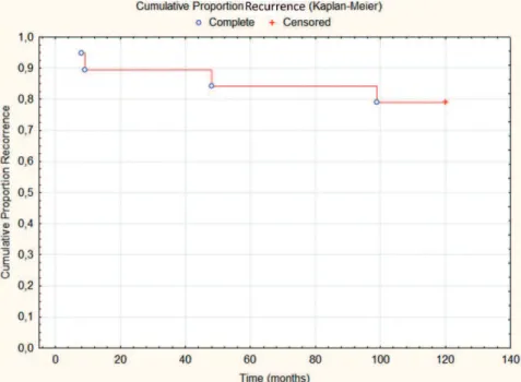
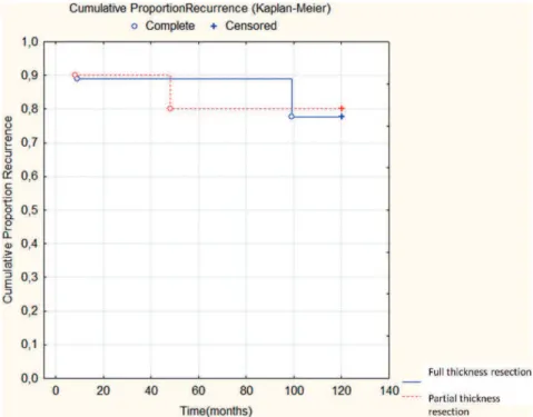
The full results of the statistical analysis are indicated in Table 2. Male sex was a risk factor (p = 0.05). None of the other variables were found to be recurrence-associated factors (Table 2). The overall cumulative proportion and the cumulative proportion of recurrence depending on the margin status and resection thickness were calculated using a Kaplan-Meier curve (Figures 2, 3, and 4). Positive margins and the resection thickness were not significant risk factors for a later recurrence, both with an odds ratio (OR) of 1.1428 (95% Confidence Interval [CI] = 0.3392 to 28.0204).
DISCUSSION
Achieving negative margins should be the goal; however, given the propensity of the tumor to invade vital structures, obtaining negative margins for desmoid tumors involving some chest regions often represents a challenge for the surgeon.5 Moreover, the prognostic significance of micro-scopically positive margins remains controversial. In our series, microscopically positive margins were present in 9 patients (47%), but only 2 (22%) of these patients experi-enced recurrence. The other 7 patients had a mean disease-free survival time of 31.2 months (12 to 78 months). The patients with positive margins had large tumors (mean, 12 cm) in regions where it is difficult to obtain margins, such as the cervicothoracic transition, the axillae (with axillary vessel involvement) and the thoracic spine. Thus, we believe that the incidence of positive margins was high.
Abbas et al. reported 53 cases of desmoid tumors of the chest wall that were surgically treated and concluded that microscopically positive margins are a risk factor for recurrence (p,0.0001).5 However, Lev et al. described the outcomes of 146 patients who underwent operations for desmoid tumors at the M.D. Anderson Cancer Center and concluded that patients with microscopically positive or negative margins had no differences in their long-term recurrence rates (19% vs. 16%).6
This topic is particularly important when dealing with tumors that require disfiguring or extensive operations to obtain histologically negative margins. In these particular situations, the focus should perhaps be on the preservation of organ function and the patients’ quality of life rather than on striving for microscopically negative margins (at least in the first resection). Moreover, in contrast to most malignant tumors, the local recurrence of a desmoid tumor does not seem to portend an increased likelihood of uncontrolled future tumor growth.7
Although conducted with few patients, this study assesses one of the largest series of patients with desmoid tumors located exclusively on the chest wall over a more recent retrospective period (11 years). This study suggests that microscopically positive margins alone may not predict recurrence, without the bias of radiotherapy. Male sex was a risk factor for recurrence, but similar
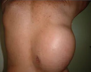
Figure 1 - Patient presenting a large desmoid tumor on the posterior thoracic wall.
Table 1- Descriptive characteristics of the patients.
Total patients 19 (100%)
Younger than 40 years 12/19 (63.1%)
Male 10/19 (52.6%)
Positive margins 9/19 (47.3%)
Full-thickness resection 10/19 (52.6%)
Previous biopsy 13/19 (68.4%)
Recurrence 4/19 (21%)
Table 2- Variables tested for the evaluation of recurrence.
Variable P value
95% confidence interval
younger than 40 years 0.52 -24.95 to 46.38
Male 0.05 9.63 to 70.36
Positive margins 0.66 -34.55 to 38.99
Full-thickness resection 0.24 -59.31 to 12.64
Previous biopsy 0.18 5.68 to 55.86
Desmoid tumors of the chest wall
Fernando CA et al. CLINICS 2011;66(4):705-708
results were not observed in the literature. One hypothesis is that male patients, who have less estrogen than do female patients, do not benefit from hormonal depriva-tion. However, in our sample, hormonal deprivation was not used. This result needs to be confirmed, but it may be important for thoracic surgeons in the cases of large tumors in areas where negative surgical margins are difficult to achieve.
We conclude that male patients seem to have an increased risk of recurrence and that the status of the margins and other variables should be studied further. However, in the cases where the first resection results in substantial functional impairment in the form of a scapulothoracic disarticulation or a significant increase in morbidity or mortality, a resection with negative macroscopic margins should not be a priority in selected cases.
Figure 2- Cumulative proportion of recurrence events during follow-up (months).
Figure 3- Cumulative proportion of recurrence events during follow-up (months).
CLINICS 2011;66(4):705-708 Desmoid tumors of the chest wall
Fernando CA et al.
REFERENCES
1. Aaron AD, O’Mara JW, Legendre KE, Evans SR, Attinger A, Montgomery EA. Chest wall fibromatosis associated with silicone breast implants. Surg Oncol.1996;5:93-9, doi: 10.1016/S0960-7404(96)80006-5.
2. Kaplan J, Davidson T. Intrathoracic desmoids: report of two cases. Thorax. 1986;41:894-5, doi: 10.1136/thx.41.11.894.
3. Abbas EA, Deschamps C, Cassivi SD, Nichols III FC, Allen MS, Scleck CD, et al. Chest wall desmoids tumors: results of surgical intervention. Ann Thorac Surg. 2004;78:1219-23, doi: 10.1016/j.athoracsur.2004. 03.015.
4. Mendez-Fernandez M, Gard DA. The desmoid tumors: benign neo-plasm, not a benigne disease. Plastic Reconstr Surg. 1991;87:956-60, doi: 10.1097/00006534-199105000-00025.
5. Ballo MT, Zagars GK, Pollack A, Pisters PW, Pollock RA. Desmoids tumor: prognostic factors and outcome after surgery, radiation therapy or combined surgery and radiation therapy. J Clin Oncol. 1999;17:158-67. 6. Lev D, Kotilingan D, Wei C, Ballo MT, Zagars GK, Pisters PW, et al. Optimizing treatment of desmoids tumors. J Clin Oncol. 2007;25:1785-91, doi: 10.1200/JCO.2006.10.5015.
7. Melis M, Zagerr JS, Sondak VK. Multimodality management of desmoid tumors: how important is a negative surgical margin? J Surg Oncol. 2008;98:594-602, doi: 10.1002/jso.21033.
Figure 4- Cumulative proportion of recurrence events during follow-up (months), depending on the margin status. OR = 1.1428 (95% CI, 0.3392 to 28.0204).
Desmoid tumors of the chest wall
Fernando CA et al. CLINICS 2011;66(4):705-708