DOI: 10.14260/jemds/2014/2012
CASE REPORT
Journal of Evolution of Medical and Dental Sciences/ Volume 3/ Issue 06/February 10, 2014 Page 1489
A RARE PRESENTATION OF FOREIGN BODY IN THE EAR WITH TRAUMATIC
DISLOCATION OF THE INCUS IN A FEMALE CHILD: A CASE REPORT
Santhana Krishnan K1, Poornima S. Bhat2
HOW TO CITE THIS ARTICLE:
Santhana Krishnan K, Poornima S. Bhat. A Rare Presentation of Foreign Body in the Ear with Traumatic Dislocation of the Incus in a Female Child: A Case Report . Journal of Evolution of Medical and Dental Sciences 2014; Vol. 3, Issue 06, February 10; Page: 1489-1491, DOI: 10.14260/jemds/2014/2012
ABSTRACT: Foreign bodies in the middle ear are a rare occurrence. This is a case report of a 5 year old female, with plastic button in the left ear, with history of attempted removal using a metal hook, leading to subtotal perforation of the tympanic membrane and traumatic dislocation of the incus. On surgery a yellow colored plastic button, measuring about 1x 1 cm size was removed from middle ear by retroauricular approach. Myringostapediopexy using autologous incus and temporalis fascia under general anesthesia was done. Patient improved clinically post operatively. Foreign bodies in the ear should be removed with caution. Blind removal by instrumentation can lead to complications like tympanic membrane perforation and ossicular disruption. Early surgical intervention with tympanoplasty under anesthesia with tympanoplasty at the same sitting can prevent further complications and correct the hearing loss.
KEYWORDS: Foreign body, incus, myringostapediopexy
INTRODUCTION: Foreign bodies in the ear are a common presentation in children. Most commonly foreign bodies are present in the external auditory canal, which can be removed on outpatient basis or sometimes under general anesthesia. A foreign body in the middle ear with ossicular disruption is a rare occurrence, which has to be treated by removal of foreign body and ossiculoplasty. Here is a rare case of plastic foreign body in the left ear of a 5 year old female, with tympanic membrane perforation and incus dislocation during attempted removal elsewhere, which was treated surgically.
DOI: 10.14260/jemds/2014/2012
CASE REPORT
Journal of Evolution of Medical and Dental Sciences/ Volume 3/ Issue 06/February 10, 2014 Page 1490
DISCUSSION: Foreign bodies in the middle ear are a rare occurrence. External auditory canal foreign bodies are most commonly removed by rinsing by a syringe and water.1, 2 Foreign bodies in the sulcus tympanicus are difficult to remove by syringing and they can be removed only by instrumentation.3 Syms and Nelson4 presented 4 cases of impression-material foreign bodies in middle ear induced chronic otitis media. A case of silicone foreign body in the middle ear caused by auditory canal impression in hearing aid fitting was reported by Shimanski.5 Metal spark perforation of the tympanic membrane causing facial paralysis and hearing loss is presented by Stage and Vinding.6 Hakuba et al studied 22 patients with traumatic ossicular disruption and found that most common cause was ear-pick injury and incudostapedial disarticulation was the most common finding7.
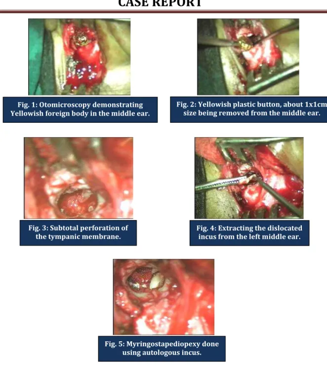
Fig. 1: Otomicroscopy demonstrating Yellowish foreign body in the middle ear.
Fig. 2: Yellowish plastic button, about 1x1cm size being removed from the middle ear.
Fig. 3: Subtotal perforation of the tympanic membrane.
Fig. 4: Extracting the dislocated incus from the left middle ear.
DOI: 10.14260/jemds/2014/2012
CASE REPORT
Journal of Evolution of Medical and Dental Sciences/ Volume 3/ Issue 06/February 10, 2014 Page 1491 In this case, there was traumatic dislocation of incus, secondary to attempted removal of a plastic foreign body, which was removed by retroauricular approach and hearing reconstruction was done at the same sitting.
CONCLUSION: Foreign bodies in the ear should be removed with caution. Blind removal by instrumentation can lead to complications like tympanic membrane perforation and ossicular disruption. Early surgical intervention under anesthesia with tympanoplasty at the same sitting can prevent further complications and correct the hearing loss.
REFERENCES:
1. Peltola TJ, Saarento R. Water used to visualize and remove hidden foreign bodies from the external ear canal. J Laryngol Otol 1992; 106:157–8.
2. Jones I, Moulton C. Use an electric ear syringe in the emergency department. J Accid Emerg Med 1998; 15:327- 8.
3. Heim SW, Maughan KL. Foreign bodies in the ear, nose, and throat. Am Fam Physician 2007; 76:1185-9.
4. Syms C, Nelson R: Impression-material foreign bodies of the middle ear and external auditory canal. Otolaryngology Head and Neck Surgery 1998, 119:406-407.
5. Schimanski G: Silicone foreign body in the middle ear caused by auditory canal impression in hearing aid fitting. HNO 1992, 40:67-68.
6. Stage J, Vinding T: Metal spark perforation of the tympanic membrane with deafness and facial paralysis. J Laryng Otol 1986, 100(6):699-700.
7.
Hakuba N et al, Ear-pick injury as a traumatic ossicular damage in Japan. Eur Arch Otorhinolaryngol. 2010 Jul;267(7):1035-9.AUTHORS:
1. Santhana Krishnan K. 2. Poornima S. Bhat
PARTICULARS OF CONTRIBUTORS:
1. Assistant Professor, Department of
Otorhinolaryngology, SMVMCH, Pondicherry. 2. Post Graduate, Department of ENT, VIMS,
Bellary.
NAME ADDRESS EMAIL ID OF THE CORRESPONDING AUTHOR:
Dr. Santhana Krishnan K, Assistant Professor, Department of E.N.T,
Sri Manakula Vinayagar Medical College and Hospital, Kalitheerthalkuppam, Madagadipet, Puducherry – 605107, India.
E-mail: santhanakrishnan2709@gmail.com