SpliceMet
Juliane Liepe1,2., Michele Mishto1,3., Kathrin Textoris-Taube1
, Katharina Janek1, Christin Keller1, Petra Henklein1, Peter Michael Kloetzel1*, Alexey Zaikin4
1Institut fu¨r Biochemie, Charite´, Universita¨tsmedizin Berlin, Berlin, Germany,2Centre for Bioinformatics, Division of Molecular Biosciences, Imperial College London, London, United Kingdom,3Interdepartmental Center for Studies on Biophysics, Bioinformatics and Biocomplexity ‘L. Galvani’ (CIG), University of Bologna, Bologna, Italy, 4Institute for Women’s Health and Department of Mathematics, University College London, London, United Kingdom
Abstract
The identification of proteasome-generated spliced peptides (PSP) revealed a new unpredicted activity of the major cellular protease. However, so far characterization of PSP was entirely dependent on the availability of patient-derived cytotoxic CD8+T lymphocytes (CTL) thus preventing a systematic investigation of proteasome-catalyzed peptide splicing (PCPS). For
an unrestricted PSP identification we here developed SpliceMet, combining the computer-based algorithm ProteaJ within vitro proteasomal degradation assays and mass spectrometry. By applying SpliceMet for the analysis of proteasomal processing products of four different substrate polypeptides, derived from human tumor as well as viral antigens, we identified fifteen new spliced peptides generated by PCPS either bycisor from two separate substrate molecules, i.e., by transsplicing. Our data suggest that 20S proteasomes represent a molecular machine that, due to its catalytic and structural properties, facilitates the generation of spliced peptides, thereby providing a pool of qualitatively new peptides from which functionally relevant products may be selected.
Citation:Liepe J, Mishto M, Textoris-Taube K, Janek K, Keller C, et al. (2010) The 20S Proteasome Splicing Activity Discovered by SpliceMet. PLoS Comput Biol 6(6): e1000830. doi:10.1371/journal.pcbi.1000830
Editor:Rob J. De Boer, Utrecht University, The Netherlands
ReceivedDecember 17, 2009;AcceptedMay 24, 2010;PublishedJune 24, 2010
Copyright:ß2010 Liepe et al. This is an open-access article distributed under the terms of the Creative Commons Attribution License, which permits unrestricted use, distribution, and reproduction in any medium, provided the original author and source are credited.
Funding:This work was financed in part by grants of the Deutsche Forschungsgemeinschaft Sonderforschungsbereich (Kl421/15, SFB 740, TR19) to PMK and by VW foundation to AZ and JL and the UCLH/UCL NIHR Comprehensive Biomedical Research Centre to AZ. MM received funding from the AV Humboldt PostDoc fellowship. The funders had no role in study design, data collection and analysis, decision to publish, or preparation of the manuscript.
Competing Interests:The authors have declared that no competing interests exist.
* E-mail: [email protected]
.These authors contributed equally to this work.
Introduction
The multiple subunit 20S proteasome is the central catalytic unit of the ubiquitin proteasome system (UPS) and catalytic core of the 26S proteasome that is built by the association of the two 19S regulator complexes with the catalytic 20S core [19S-20S-19S]. With its N-terminal threonine residues as the single active site of theb-subunitsb1,b2 andb5, the 20S proteasome is a N-terminal nucleophilic hydrolase responsible for the generation of the vast majority of virus- or tumor-derived peptides presented by MHC class I molecules at the cell surface for recognition by peptide-specific cytotoxic T lymphocytes (CTL) [1,2]. This function is generally aided by the interferon-c- (IFN-c)-induced synthesis of the alternative catalytic subunitsb1i,b2i,b5i, with concomitant formation of immunoproteasome subtypes possessing altered proteolytic properties as well as by the IFN-c-induced up-regulation of the proteasome activator subunits PA28-a and PA28-b[3,4]. Peptides generated by the 20S proteasome were so far thought to exhibit a linear sequence identical to that found in the unprocessed parental protein. This view was dramatically changed by the identification of three epitope peptides derived from the melanocyte protein gp100, the SP100 nuclear phospho-protein and fibroblast growth factor (FGF-5), which represented fusions of proteasomal cleavage products and were shown to be generated by proteasomes [5–8]. Proteasome-catalyzed peptide
proteasomal splicing reaction, as such, must be a considerably more frequent event than so far assumed. But even if peptide splicing is a rare event, PSP may still play a crucial role within the immune response. This is due to the sensitivity of CTL cells, which are able to detect very small numbers of MHC class I peptide complexes [11], and in the most extreme example even a single MHC class I complex [12].
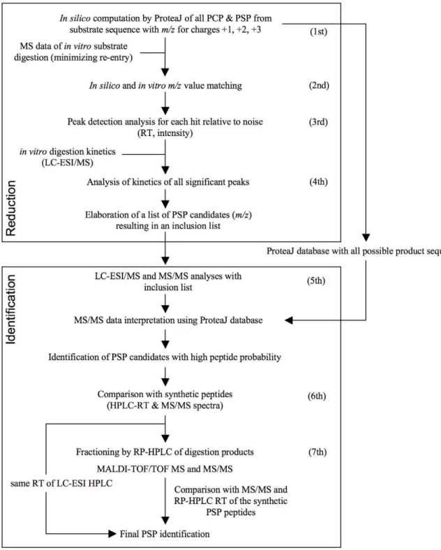
To allow a systematic, CTL-independent investigation of PSP we therefore developed SpliceMet: a method that combines combinatorial computations (ProteaJ) with mass spectrometric (MS) analyses of proteasome-generated peptides. Based on a given protein or peptide sequence, ProteaJ produces a data set with the
m/zvalue of all theoretically possible PSP that may be generated by the proteasome through the combination of any two fragments (greater than one amino acid in length) generated from the same substrate molecule (incis) or from separate substrate molecules (in
trans) and ligated in a normal or reverse order. This is followed by MS analysis ofin vitrodigests of the synthetic peptide substrate and by comparison of the MS signals obtained with the theoretical ProteaJ-computedm/zvalues. By matching the theoretical values with the experimentally obtained m/z values and verifying the peptide generation kinetics, a restricted list of candidate PSP is generated. Their presence in 20S proteasome digests of substrates is then investigated by LC-ESI-MS/MS and LC-MALDI-TOF/ TOF-MS/MS leading to the final identification of the PSP (Fig. 1).
Results
SpliceMet
The SpliceMet method is organized into two main experimental blocks characterized by 7 main steps (Figure 1). To reduce the number of possible proteasome generated spliced peptides (PSP) the first block utilizes the following 4 main steps that are subsequently investigated in the second block. The first experi-mental block combines the computational algorithm ProteaJ with proteasomein vitrodigests of a synthetic peptide of choice and mass spectrometric (MS) analyses as follows:
1) Calculation of all combinatorially possible PSP and setting of the ProteaJ database. The digestion of the substrate of length L with a sequence of amino acids ai, i = 1..Lmay result
in SCP~ 1
2ðL{Lextz1ÞðL{Lextz2Þ cleavage products (PCP) each of which can be denoted as PCPij, where the product starts at the
position i, i = 1…L-Lext+1 (C-terminus) and ends at the position j = i+Lext-1…L(N-terminus). Lextdescribes the minimal length of a
PCP that can produce a PSP (here Lext = 2). Any two PCPijand
PCPkn may be spliced into PSPi-j/k-n. For the total amount of
generated products Sall, including PCP and PSP, we have i = 1…
L-Lext+1, j = i+Lext-1…L, k = 1…L-Lext+1, n = k+Lext-1…L and we
can calculateSall~ P L{Lextz1
i~1 PL j~izLext{1
P L{Lextz1
k~1
PL n~kzLext{1
1. Note
that Sall~ 1
4ðL{Lextz1Þ
2
L{Lextz2
ð Þ2~S2PCP. PSP can be classified into two main groups: cis splicing (PSPcis) and trans
splicing (PSPtrans), wherebycissplicing occurs in the same order as
in the substrate (PSPcis,normal, where i+Lext#j+1#k+1#n-Lext) or in
reverse order (PSPcis,reverse, k+Lext#n+1#i#j-Lext+1). The total
number of all PSP is then SPSP~ 1
4ðL{Lextz3ÞðL{Lextz2Þ
L{Lextz1
ð ÞðL{LextÞ. The number of pure trans PSP can be calculated as PSPtrans= SPSP-PSPcis,normal- PSPcis,reverse. Table 1
summarizes the conditions for each product and their total amount.
Estimation of mass-to-charge ratios (m/z) of all possible PSP. To list all possible PSP, in which four indicesijkndefine the sequence, we computed the molecular weight (Mr, calc.) of each peptide and the correspondingm/zvalues for charge states z = 1, 2, 3 (m/z= (Mr+z)/z). Since them/zvalues of PSP can differ by less than the mass accuracy of 0.5 Da for the used ESI-ion trap mass spectrometer (LCQ-classic & DECA XP instruments), we clustered allm/zvalues into groups with am/zrange of 0.2 Da (accordingly to the MS instrument resolution). For each group we determined the average m/z value thereby obtaining a set of theoreticalm/zvalues that could be further analyzed.
2) Matching with the LC-ESI/MS full spectra. The presence of the theoretical m/z values was detected among MS signals of the digestion products of the investigated peptide of choice.
3) Peak detection of all the computed m/z values. In the LC-ESI mass chromatogram we identified the significant peaks for each theoreticalm/zvalue. For each theoreticalm/zvalue either no peak or several peaks could be detected and defined by theirm/ zand retention time (RT).
4) Analysis of m/z time-dependent kinetics and establishment of an inclusion list for the LC-ESI/MS measurements. In time-dependent processing experiments (signal intensity versus time of digestion) identified peaks that did not fulfill the following criteria were eliminated from the candidate list: i. initial intensity (t = 0) smaller than MAX (e.g.here = 107for measurements by DECA XP MAX instrument); ii. monotonously ascending signal intensity towards a maximum followed by a monotonous decline in case assay condition allowed re-entry of the PSP. It was assumed that the monotonous increase resulted from the continuous production of PSP and the decrease from the ‘‘re-entry’’ event.
Next, we defined tmaxas the digestion time when the highest
amount of generated PSP was observed and sorted all pairs (m/z, RT) with respect to tmaxinto groups indexed as g of the size Dg. If
Dg.Dmax (here 15 depending on MS resolution) then the
corresponding group was split into subgroups gi of size smaller
than Dmax. The number of groups determined the number of
additional up-scaled processing assays in which the absolute concentration of substrate and proteasome were increased keeping the relative substrate/proteasome ratio constant, whereas the total
Author Summary
number of subgroups represented the number of requested new MS runs. The resultingm/z, RT, tmaxestablished the inclusion list.
The second block consists of the following 3 steps:
5) LC-ESI-MS/MS analysis with inclusion list. Precursor ion selection for MS/MS analysis was performed using the
established inclusion list enabling the fragmentation analysis of even low-abundance peptides. MS/MS spectra were analyzed with Bioworks software version 3.3 (Thermo Fisher) using the ProteaJ database. Significant hits which were annotated as PSP showed a peptide probability p,0.00005.
Figure 1. SpliceMet.Applying the computer program ProteaJ on a peptide sequence of choice,m/zvalues of all theoretically possible proteasomal cleavage (PCP) and splicing (PSP) products are calculated (1ststep). This is followed by anin vitrodigest of the synthetic substrate and the comparison of the obtained MS signals with the theoreticalm/zvalues (2nd). Matching of the signals and verification of peptide generation kinetics results in an inclusion list for LC-ESI-MS/MS analysis required for identification of the PSP (3rd, 4th). For final confirmation, the MS/MS spectra (5th) and the HPLC-RT of proposed PSP (6th) are compared with those of the analogous synthetic peptides. For the identification of those PSP candidates that do not fully satisfy these requisites, the generation of PSP is up-scaled followed by HPLC fractionation with an extended gradient and the fractions are analyzed by nano-LC-MALDI-TOF/TOF-MS (7th).
6) Comparison with synthetic peptides. All identified PSP resulting from step 5 were manually confirmed by comparison with synthetic peptides of the same sequence. The candidate PSP and their synthetic analogues had to exhibit a similar RT (delta RT,0.5 min) and fragmentation pattern in the LC–ESI-MS/MS analysis.
7) Validation of PSP sequences by MALDI-TOF. In some experiments the requirements outlined in step 5 and 6 were not fully met requesting further MS identification. In this case, we proceeded by fractionating the digestion products by reverse phase (RP)-HPLC and by analyzing each fraction by LC-ESI-MS/MS using an inclusion list with them/zvalues of the PSP candidates. Their RT in the HPLC run was also compared with that of the corresponding synthetic peptides. Those fractions with MS/MS and RT that matched the PSP were lyophilized and fractionated again using a more focused HPLC method to decrease the number of peptides in each fraction. The up-scaled fractions were subsequently compared with the RT of the synthetic PSP and analyzed by nano-LC-MALDI-TOF/TOF-MS/MS.
Validation of SpliceMet
For proof of principle we initially investigated 20S proteasome catalyzed peptide splicing during proteasomal degradation of the synthetic 13mer peptide (gp100PMEL1740–52, RTKAWNRQLYPEW),
previously shown to serve as substrate for PSP generation [6]. For the experiments we used 20S proteasomes of Lymphoblastoid cell Lines (LcL), which possess splicing activity [7] and predominantly resemble
the immunoproteasome subtype [13,14]. Following each step of SpliceMet we obtained a progressive decrease of the number of candidate PSP leading to the identification of the previously described PSP gp100PMEL1740–42/47–52[6] by LC-ESI/MS/MS at the 6
th
step of SpliceMet (Figure 2). The substantial reduction of PSP in the candidate list (Table 2) and the final identification of the PSP gp100PMEL1740–42/47–52validated our analysis method.
To verify the hypothesis of the occurrence of a proteasome-dependenttranssplicing reaction we performedin vitrodigestions in which the unmodified 13mer gp10040–52 peptide was applied to
proteasomal processing in the presence of the same peptide but with the heavy amino acid residues 13C6-Lys and 15N-Leu
(RTK+6
AWNRQL+1
YPEW). As shown in Figure 3, we indeed detected PSP variants as being the results ofcis(variants2a&2d) or oftrans(variants2b&2c) splicing, demonstrating that PCPS can occur not only incisbut also intrans(see also Figure S1).
Identification of nine new PSP in the proteasomal
digestion of gp10035–57
By applying SpliceMet we investigated the generation of new PSP derived from the proteasomal degradation products of the 23mer peptide gp10035–57, which is a N- and C- terminally
extended version of gp10040–52 by LcL 20S proteasome
(Figure 4A). In these experiments we identified eight new PSPcis,
four of which were identified at step 6 (Figure 4) and four at step 7 of SpliceMet (Table 3 & Figure S2). We also identified a ninth PSP Table 1.Computation of cleavage and splicing products.
products conditions total amount
all fragments i = 1…L-Lext+1, j = i+Lext-1…L, k = 1…L-Lext+1, n = k+Lext-1…L 1
4ðL{Lextz1Þ
2
L{Lextz2
ð Þ2
PCP i = 1…L-Lext+1 j = i+Lext-1…L 1
2ðL{Lextz1ÞðL{Lextz2Þ cis - normal i = 1…L-2Lextj = i+Lext-1…L-Lext-1 k = j+2…L-Lext+1 n = k+Lext-1…L 1
24ðL{2Lextz1ÞðL{2LextÞðLz3{2LextÞðLz2{2LextÞ cis - reverse k = 1…L-2Lext+1 n = k+Lext-1…L-Lexti = n+1…L-Lextj = i+Lext-1…L 1
24 L{ 2Lextz1
ð ÞðLz4{2LextÞðLz3{2LextÞðLz2{2LextÞ
Described are the conditions to compute all products of a specific type (PCP, cis-normal PSP and –reverse PSP). The indices i, j, k and n are the amino acid positions of the product,e.g.PSPi-j,k-n, L is the length of the substrate, Lextis the minimal length of a PCP that can produce a PSP.
doi:10.1371/journal.pcbi.1000830.t001
Figure 2. By applying SpliceMet we identified the known PSP produced by digestion of the synthetic 13mer gp10040–52by 20S
proteasomes.Sequence of the substrate gp10040–52and of the PSP gp10040–42/47–52and its ESI-MS/MS spectrum (double protonated withm/z
610.8) are shown. In the spectra B- and Y-ions are reported. Ions’ loss of water is symbolized byu. In the experiments (100ml of reaction) 4 nmol of
gp10040–52were cleaved for 36 hours by 1mg 20S proteasome purified from LcL.
with the sequence [VSRQL][VSRQL] derived from splicing of two distinct molecules of the PCP gp10035–39 (Figure 5). The
identification of this PSP was of particular relevance because it was the first example of PSPtrans detected in in vitro proteasomal
digestion of a single peptide sequence.
PSP formation is a general phenomenon not restricted to
the gp10035–57sequence
Since the sequence requirement for PCPS are not yet known one might argue that the observed frequent PSP generation when gp100PMEL1735–57 was used as substrate was due certain
gp10035–57 sequence specificities. To test this we applied
SpliceMet for the analysis of PSP derived from another polypeptide sequence of the same protein, i.e. gp100201–229.
Among the proteasome-generated degradation products of this 29mer we identified three PSP (Table 4 and Figure S3). Since peptide fragments with overlapping sequences were spliced together these PSP were generated by atranssplicing event.
In order to exclude a peculiar and rare tendency of the entire gp100 sequence to be spliced by PCPS we investigated thein vitro
digestion products of two other peptides, i.e. the 30mer HIV-derived gag-pol29–58and the murine cytolomegalovirus
(MCMV)-derived 25mer polypeptide pp8916–40. Thein vitro processing of
gag-pol29–58by proteasomes produced at least one PSPtrans(Table 4
& Figure S4), whereas two PSPtrans were detected after the
digestion of the MCMV derived pp89 polypeptide peptide (Table 4 & Figure S5).
Discussion
SpliceMet
The aim of our study was to develop a method for the identification of spliced peptides which would allow the identifi-cation of any theoretically possible PSP and which was independent of adventitiously available CD8+ T cells and T-cell recognition assays permitting the detection of only a single spliced epitope peptide. The availability of such a method would greatly facilitate systematic studies required to elucidate the molecular mechanism of PCPS. Therefore we have developed and applied a method – SpliceMet – that, by combining computational and experimental methods, facilitates the identification of proteasome-generated spliced peptides.
Although in this investigation we have considered only polypeptide substrates up to a length of 30 amino acid residues, SpliceMet could also be applied to longer peptides or proteins to further our understanding of the mechanisms that govern PCPS and, in particular, trans-splicing. It has to be pointed out however that an increase in substrate length will lead to an
exponential expansion of the ProteaJ data base as well as the number of peaks detectable by MS and therefore will require the application of restricting parameters such as size or sequence quality to match this approach with the capacity of the presently available MS technologies.
In our experiments we observed a substantial number of peak spectra at the 5th step of SpliceMet, which could not be
identified with sufficient confidence due to the low MS/MS quality. The number of unidentified spectra depends on the size of the ProteaJ database and to technical difficulties of MS analysis. Therefore, to reduce the number of unidentifiable spectra we incorporated the 7thstep into our method. Indeed, up-scaling of the digestion products by two rounds of HPLC fractionation permitted a better separation of the digestion products thereby limiting the number of overlapping peptides with similarm/zand RT and increased product concentration in this manner facilitating the identification of PSP by MS. Furthermore, at step 7 we analyzed the sample with a second MS instrument, a MALDI-TOF/TOF mass spectrometer, which has a higher resolution and sensitivity than the used ESI-ion trap mass spectrometer. Its application in other studies allowed the identification of peptides not previously detected by ESI-MS/MS, not only because of the higher sensitivity but also due to the different method of ionization and detection, which led to the identification of a complementary pool of peptides [15,16]. Accordingly, we used both techniques to identify as many PSP as possible. LC-ESI/MS analysis was primarily adopted because it is a less time consuming technique and allowed the analyses of as large a number of samples as needed at SpliceMet step 4. Likely, a further minimization of unidentified spectra could be obtained by exploiting the high performance of the new generations of MS analyzers.
The computational algorithm ProteaJ is based on a combina-torial approach. Therefore the amount of calculated PSP strongly depends on parameters like substrate length L and the minimal length of a PCP Lext, as well as the kind of PSP allowed,i.e. cisor
transPSP. Thus ProteaJ parameter settings were used which in preliminary experiments seemed to be most reliable; for example, we limited the PCP Lextto a minimum of 2 and accordingly we
identified PSP such as gp10047–48/35–39 or gag-pol45–57/48–49. In
contrast, when we considered PCP Lext = 1 in a preliminary
experiment on gp10035–57 we were not able to identify any new
PSP (data not shown).
SpliceMet applications and PSP implications
By applying SpliceMet we here showed that 20S proteasomes possess a substantial in vitro splicing activity. Since in vitro
experiments for generation of spliced and non-spliced epitope Table 2.PSP candidate reduction by applying SpliceMet.
number of m/z number of sequences
SpliceMet steps 1 2 3 4 5 6 7
gp10040–52 2580 (100) 280 (10.8) 32 (1.2) 18 (0.7) 2 1
gp10035–57 7229 (100) 1288 (17.8) 1121 (15.5) 239 (3.3) 20 4 5
Reduction of number of PSP candidates during the progression of SpliceMet step by step. The number of possible PSP detectable in thein vitrodigestion of a peptide declines continuously during the consecutive steps of SpliceMet (Figure 1). Here the PSP number reduction observed for the 13mer gp10040–52and 23mer gp10035–57is reported both as total number and as a percentage compared to the theoretical PSP number (in brackets). The values are referring to the number of possible PSP at the end of the SpliceMet step. For example, although 5664 PSP could be generated from gp10040–52assuming 2 as the minimum length of the native PCP (Lext), only 2580 represent them/zvalue clusters (obtained with a cluster range of 0.2) that will be matched with the LC-ESI/MS full spectrum at the beginning of step 2. Moreover, up to step 4 the numbers are referred to as the number ofm/zvalues whereas from step 5 they are referred to as the possible sequence because they have been identified by MS/MS.
peptides are known to closely resemble thein vivosituation [3] our data reveal that 20S proteasomes represent a molecular machine that facilitates the generation of spliced peptides from its own cleavage products. Therefore, our data may have considerable biological implications in that they provide evidence that proteasome-dependent protein degradation results in the genera-tion of a second, so far undetected pool of spliced peptides, from which novel potentially functionally relevant peptides can be selected. Indeed, the two previously identified PSP were shown to be MHC class I epitopes recognized by CTL of human patients [6,7]. This and the relatively high number of PSP that we identified raises the possibility that peptide splicing in general may lead to an increase in the peptide pool available for epitope selection. For example, from the melanocytic gp100PMEL17tumor
antigen (661 amino acids) 1,786,862 9mers with a unique sequence could be theoretically produced. Of these, a maximum of 652 are unspliced proteasomal cleavage products while the rest (99.96%) represent theoretical PSP. At the moment we do not have any sufficient information to judge on how many of these PSP (as well as normal PCP) are really produced and which percentage of them may efficiently bind MHC class I molecules. Based on our preliminary data we are tempted to speculate that specific PCP are generated more efficiently than PSP even if the MS signal of some PSP (e.g.gp10047–55/35–39) was as high as that of
many PCP (data not shown). Nevertheless, if, for example PCP were produced 1000-fold more efficiently than any given PSP, spliced peptides generated from gp100PMEL17would still represent a significant peptide pool (i.e.the 73.26% of the 9mers derived Figure 3. Generation of PSP by proteasomaltranssplicing.(A) To demonstrate the generation of a PSPtransby the binding of two fragments originated from two distinct molecules of substrate, 5 nmol gp10040–52 and its heavy analogue with amino acids 13C6-Lys and 15N-Leu
(RTK+6AWNRQL+1YPEW) were digested together for 36 hours by 1.5
mg LcL 20S proteasomes in 100ml buffer. Theoretically four different PSP could
be generated from thecisortransligation of the proteasomal fragments [RTK] and [QLYPEW] with sequences [RTK][QLYPEW]: gp10040–42/47–52-a,
[M+H]+ = 1220.7; gp10040–42/47–52-b, [M+H]+= 1221.7; gp10040–42/47–52-c, [M+H]+= 1226.7; gp10040–42/47–52-d, [M+H]+ = 1227.7. (B) LC-MALDI-TOF/ TOF-MS spectra at RT = 41.3 min show peaks which can be assigned to all four possible PSP of gp10040–42/47–52(for MS/MS spectra see Figure S1).
from the digestion of gp100PMEL17) from which antigenic spliced peptides could be selected.
This basic computational analysis assumes that the splicing of proteasomal cleavage products can occur alsoin vivo. Our observation that thein vitrosplicing reaction not only occurs incisbut also intrans
indirectly supports such an assumption. The existence of thetransPSP implies the likely situation that two or more substrate molecules are present at the same time within the proteasomal cavity as suggested by some excellent previous studies [17–19] or that the cleavage products of a first substrate molecule remain within the catalytic chamber while a second molecule of substrate is cleaved. Very recently, Dalet and co-workers investigatedtransproteasome splicing
in vivo, providing some very interesting albeit not final insights. They showed that PSPtrans were generated in vivo when the precursor
peptides of FGF-5 and gp100 were electroporated into COS cells, whereas only the FGF-5-derived PSPtrans(and in a very small amount)
could be detected by CTL assay when COS cells were transfected with FGF-5 or gp100 plasmid [8]. Taking into account the high number of PSPtranswe identified withinin vitrodigestion products of
four peptides, we are led to conclude that further studiesin vitroandin vivo on different cellular and proteasome models are required to clarify this phenomenon.
An extensive application of SpliceMet on a wide range of polypeptides substrates would also help to identify putative peptide sequence motifs that facilitate the proteasomal splicing reaction. For example, in seven of the nine gp10035–57-derived PSP, the
sequence VSR represents the N-terminus of those PCP, which according to the transpeptidation model [6,20] perform a nucleophilic attack on the acyl-enzyme intermediate, thereby forming the detected PSP. Likewise, for four PSP the sequence YPEW represents the C-terminus, which forms the acyl-enzyme intermediate that is subsequently attacked by the second PCP generating the new PSP. From these observations one might infer a higher affinity of these two peptide sequences for a
transpep-tidation reaction. However, only a more extensive investigation of this specific issue with SpliceMet, covering a large number of different polypeptides would allow to validate such a hypothesis.
For this and other aims, studies performed with the help of SpliceMet could be powered if coupled with algorithms for the prediction of proteasomal cleavages, mathematical modeling of degradation kinetics as well as of the MHC class I antigen presentation [21–26]. Such an approach would also facilitate the reduction of the theoretical PSP number, which might represent a limitation of SpliceMet application to very long proteins such as gp100PMEL17. By combining the SpliceMet results with the estimation of these and other algorithms it would be theoretically possible to restrict the PSP identification to a group of PSP possessing features of interest (e.g.epitope-specific for a defined HLA I haplotype) and to predict their altered expression upon proteasome modification [24].
Methods
I. Peptides and peptide synthesis
All peptides were synthesized using Fmoc solid phase chemistry as previously described [27]. Exception had to be made for heavy analogues of gp10040–52. The isotope-labeled amino acids 15
N-Fmoc-L-Leucine (3eq. amino acid, 3eq. HBTU, 6eq. DIEA in DMF) and L-Lysine-a-N-Fmoc, e-N-T-Boc, 13C6 (1.92eq. amino acid,
1.92eq. HBTU, 3.84eq. DIEA in DMF) were coupled over night. The sequence enumeration for the peptides gp10040–52, gp10035–57
and gp100201–229 is referred to the human gp100PMEL17sequence
described by Adema and colleagues [28], for the peptide pp8916–40is
referred to the murine cytomegalovirus pp89 protein described by Lyonset al.[29]. The peptide sequence here named gag-pol29–58is a
modified version of the sequence 29–57 of the HIV gap-pol protein as described by Reitzet al.[30], where a Valin was inserted before the Threonin 53. All peptide sequences were extrapolated on the web site http://www.uniprot.org/.
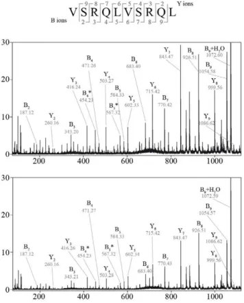
Figure 4. Identification of PSP in gp10035–57digestion by SpliceMet [step 6].(A) Sequence of gp10035–57. The bracket indicates the
previously described substrate gp10040–52. Arrows indicate the cleavage positions that are necessary to generate the newly identified PSP. The colors
correspond to the identified PSP sequences as reported in (B) to (E). (B–E) LC-ESI/MS/MS spectra (upper panels) and extracted ion chromatograms (middle and lower panels) of the double-protonated PSP. (B) [QLYPEWTEA][VSRQL] (gp10047–55/35–39) -m/z860.4, (C) [QLYPEWTEA][RTK] (gp10047–55/40–42) -m/z761.6, (D) [YPEW][VSRQL] (gp10049–52/35–39) –m/z589.5, (E) [YPEW][VSR] (gp10049–52/35–37) –m/z469.0. The RT of the detected peaks in the digestion is
consistent (with a maximum difference of 0.5 min) with those of synthetic peptide of same sequences (lower panel of extracted ion chromatograms). 40mM gp10035–57were digested for 24 hours in 100ml reaction by 0.5mg 20S proteasomes purified from LcLs.
doi:10.1371/journal.pcbi.1000830.g004
Table 3.PSP identified in the proteasomal digestion of the polypeptide gp10035–57.
Peptide (gp100) Sequence Mr, calc PSP type
Identification step of SpliceMet
49–52/35–39 [YPEW][VSRQL] 1176.59 cis,reverse 6
49–50/35–37 [YPEW][VSR] 935.45 cis,reverse 6
47–55/40–42 [QLYPEWTEA][RTK] 1520.76 cis,reverse 6
47–55/35–39 [QLYPEWTEA][VSRQL] 1718.86 cis,reverse 6
47–52/35–37 [QLYPEW][VSR] 1176.59 cis,reverse 7
47–48/35–39 [QL][VSRQL] 842.50 cis,reverse 7
45–52/35–37 [NRQLYPEW][VSR] 1446.74 cis,reverse 7
37–38/49–57 [RQ][YPEWTEAQR] 1462.70 cis,normal 7
35–39/35–39 [VSRQL] [VSRQL] 1184.70 trans 7
The PSP identified by the application of SpliceMet on the proteasome-mediated digestion of the substrate gp10035–57are here described. PSPnormalor PSPreverseresult from splicing in the same order as the substrate or in reverse order to the substrate, respectively. PSPcisare derived from the splicing of two non-overlapping sequences
of the original substrate. In contrast, PSPtransnecessarily originate from two distinct substrate molecules because of the overlapping sequences of the two peptides
spliced together.
II. Cell cultures
Lymphoblastoid cell lines (LcLs) are human B lymphocytes immortalized with Epstein Barr virus (EBV) which mainly express active immunoproteasomes [13,14]. LcLs were cultured in RPMI1640 medium supplemented with 10% FCS.
III. 20S proteasome purification
20S proteasomes were purified from 3*E+09 LcLs as previously reported [31]. The purity of 20S proteasome preparation was verified by SDS-PAGE electrophoresis (12, 5% poly-acrylamide gel stained with Coomassie dye) (Figure S6). Furthermore, a non-proteasome proteolytic activity of the preparation was tested and excluded (data not shown) by the digestion of 40mM gp10040–52
for 24 hours by 1mg of LcL 20S proteasomes in presence of
400mM Lactacystin (previously incubated with 20S proteasomes at room temperature for 10 min).
IV.In vitrodigestion of synthetic peptide substrates
Synthetic peptides at different concentrations (from 40 to 100mM) were digested by 0.25–1.5mg 20S proteasomes in 50–
100ml Hepes buffer (Hepes 20 mM, KCl 1 mM, MgCl 0.5 mM,
DTT 1 mM, NaN3 1 mM, pH 7.3) for different time periods
(from 20 min to 48 hours) at 37uC. Digestions were stopped by acidic inactivation and frozen. Digestions were performed also in TEAD buffer (Tris 20 mM, EDTA 1 mM, NaN3 1 mM, DTT
1 mM, pH 7.2) and no remarkable differences compared to Hepes buffer emerged (data not shown). In contrast, for SpliceMet step 7, 1.1mmol of the peptides (at the final concentration of 100mM) were digested for 24 hours by 62mg of LcL 20S proteasomes in 10 ml Hepes buffer and the products up-scaled by RP-HPLC separation. All experiments reported in this study were repeated at least twice and each set of experiments was measured by each MS instrument at least twice.
V. LC-ESI MS
In LC-runs the peptide separation was carried out on a 2.1 mm (mRPC C2/C18, 100 mm62.1 mm, 3mm, 120 A˚ , Amersham) and a 1 mm RP column (Beta Basic-18, 100 mm61 mm, 3mm, 150 A˚ , ThermoFisher) using a Surveyor system (ThermoFisher Scientific, USA). The mobile phase (A) was 100% water containing 0.05% (v/v) TFA and (B) was 70:30 (v/v) acetoni-trile/water containing 0.045% (v/v) TFA or 0.1% acetic acid for the PSP identifications reported in Figure 3. Online MS analysis was performed by DECA XP MAX iontrap instrument (Thermo-Fisher Scientific, USA) and by LCQ-classic iontrap (Thermo(Thermo-Fisher Scientific, USA) after HPLC separation (HP1100, Agilent). MS data were acquired with a triple scan method in positive ion mode (MS - mass range 250–2000m/z, zoom scan, MS/MS). Analysis Figure 5. MS/MS identification of the PSPtransgp10035–39/35–39
[VSRQL][VSRQL] within the gp10035–57 digestion products. MALDI-TOF/TOF-MS/MS spectrum of precursor ion [M+H]+ = 1185.7
observed in the up-scaled digestion (see ‘‘Methods’’ section) of the 23mer gp10035–57by 20S LcL proteasomes (upper panel) is shown in
comparison with the synthetic peptide [VSRQL][VSRQL] (lower panel). In the spectra B-, Y-ions, the loss of ammonia symbolized by *, the relative abundance (%) in y-axis andm/zin the x-axis. are reported.
doi:10.1371/journal.pcbi.1000830.g005
Table 4.PSP identified in proteasomal digestions of three additional polypeptides.
PSP Sequence Mr, calc PSP type
Identification step of SpliceMet
substrategp100201–230- AHSSSAFTITDQVPFSVSVSQLRALDGGNK
201–204/201–209 [AHSS][AHSSSAFTI] 1301.60 trans 6
201–209/201–207 [AHSSSAFTI][AHSSSAF] 1607.70 Trans 7
201–207/201–207 [AHSSSAF][AHSSSAF] 1393.61 trans 7
substrategag-pol29–58-YKLKHIVWASRELERFAVNPGLLEVTSEGC
45–57/48–49 [AVNPGLLEVTSEG][PG] 1439.58 trans 6
substratepp8916–40–RLMYDMYPHFMPTNLGPSEKRVWMS
27–30/23–30 [PTNL][PHFMPTNL] 1381.61 trans 6
27–32/20–30 [PTNLGP][DMYPHFMPTNL] 1943.89 trans 6
To verify that the relatively high PSP number was not peculiar to the sequence gp10035–57we extended our investigation to three additional peptides. Six new PSP were identified within their products ofin vitroproteasomal digest by applying SpliceMet. Three of them derived from the digestion of the sequence gp100201–230, one from HIV gag-pol29–58and two from MCMV pp8916–40. All of them were produced by atranssplicing reaction.
of ESI/MS data was accomplished using Bioworks version 3.3 (ThermoFisher Scientific, USA). Database searching was per-formed using the ProteaJ database and the following parameters: no enzyme, mass tolerance for fragment ions 1amu. In time-dependent processing experiments (signal intensity versus time of digestion) we analyzed the kinetics of the identified peaks by using LCQuan software version 2.5 (Thermo Fisher). At step 3 of SpliceMet the significant peaks for each theoreticalm/z value in the LC-ESI mass chromatogram were identified by Bioworks peak detection algorithm with a signal-to-noise ratio larger thand (here = 2).
VI. Digestion product up-scaling by RP-HPLC
Further identification of the PSP at step 7 of SpliceMet was performed by MALDI-TOF/TOF-MS analysis of the gp10035–57
digestion products separated by two distinct rounds of RP-HPLC. In the first round 57 fractions were collected, lyophilized and analyzed by LC-ESI/MS to identify PSP candidates. The fractions containing the PSP candidates were then separated with more focused gradients (different for each selected fraction of the first round of HPLC separation) on the same column obtaining 47 fractions, which were lyophilized and investigated by MALDI-TOF/TOF-MS analysis. Each round was obtained by collecting the eluted fractions of the 5-15 runs (5–20ml each) to maintain a good separation of the digestion products on the chromatogram. The runs were carried out on the column C18 (3364.6 mm; ODS1 1.5mm) by the HPLC Beckman SytemGold and different gradients of acetonitrile.
VII. Nano-LC-MALDI-TOF/TOF-MS
Peptide separation was carried out using an Ultimate HPLC system (Dionex, Idstein, Germany). Samples were concentrated on a trap column (PepMap C18, 5 mm6300mm65mm, 100 A˚ ,
Dionex) and eluted onto an analytical column (PepMap C18, 150 mm675mm63mm, 100 A˚ , Dionex). The mobile phase (A) was 2:98 (v/v) acetonitrile/water containing 0.05% (v/v) TFA and (B) was 80:20 (v/v) acetonitrile/water containing 0.045% (v/v) TFA. Runs were performed at a flow rate of 200 nL/min using a binary gradient 0–15% B in 4 min, 15–60% B in 45 min, 60– 100% B in 5 min. Column effluent was mixed with MALDI matrix (5 mg/mla-cyano-4-hydroxy-cinnamic acid in 70:30 (v/v) acetontrile/water containing 0.1% (v/v) TFA, 1ml/min) and spotted at ten second intervals on MALDI steel targets using a Probot fractionation device (Dionex). MS analysis was performed on a 4700 Proteomics Analyzer (Applied Biosystems,
Framing-ham, MA, USA). MS data were acquired in positive ion mode in the mass range 800–4000m/zby accumulation of 1200 laser shots per spot and processed with default calibration. MS/MS spectra were generated by 1 keV collisions and accumulation of 2500 to 10000 laser shots. Analysis of MALDI MS data was accomplished using MASCOT version 2.1 (Matrixscince, London, UK). Database search was performed using ProteaJ database and the following parameters: no enzyme, mass tolerance for precursors, +/2 80 ppm and for MS/MS fragment ions, +/2 0.3 Da. Spectral images for manual validation were prepared with Data Explorer Software version 4.8 (Applied Biosystems).
Supporting Information
Figure S1 Verification of the PSP gp10040–42/47–52 with
sequence RTKQLYPEW generated bycisandtranssplicing. Found at: doi:10.1371/journal.pcbi.1000830.s001 (0.97 MB TIF)
Figure S2 MS/MS identification of four gp10035–57PSP at step
7 of SpliceMet.
Found at: doi:10.1371/journal.pcbi.1000830.s002 (0.55 MB TIF)
Figure S3 Identification of three PSP originated from the synthetic substrate gp100201–230.
Found at: doi:10.1371/journal.pcbi.1000830.s003 (0.90 MB TIF)
Figure S4 Identification of the PSP gag-pol45–57/48–49.
Found at: doi:10.1371/journal.pcbi.1000830.s004 (0.34 MB TIF)
Figure S5 Identification of two PSP originated from the synthetic substrate pp8916–40.
Found at: doi:10.1371/journal.pcbi.1000830.s005 (0.63 MB TIF)
Figure S6 SDS-PAGE Electrophoresis with 20S proteasome purified from LcLs.
Found at: doi:10.1371/journal.pcbi.1000830.s006 (0.95 MB TIF)
Acknowledgments
We thank Agathe Niewienda, Elena Bellavista, Eberhard Krause and Heike Stephanowitz for their excellent technical assistance and supervision, Sascha Bulik for the estimation of the PSP number and Hermann-Georg Holzhu¨tter for inspiring discussions.
Author Contributions
Conceived and designed the experiments: JL MM PMK AZ. Performed the experiments: JL MM. Analyzed the data: JL MM KTT KJ CK. Contributed reagents/materials/analysis tools: PH. Wrote the paper: JL MM PMK AZ.
References
1. Kloetzel PM, Ossendorp F (2004) Proteasome and peptidase function in MHC-class-I-mediated antigen presentation. Curr Opin Immunol 16: 76–81. 2. Groll M, Ditzel L, Lowe J, Stock D, Bochtler M, et al. (1997) Structure of 20S
proteasome from yeast at 2.4 A resolution. Nature 386: 463–471.
3. Kloetzel PM (2001) Antigen processing by the proteasome. Nat Rev Mol Cell Biol 2: 179–187.
4. Kloetzel PM (2004) Generation of major histocompatibility complex class I antigens: functional interplay between proteasomes and TPPII. Nat Immunol 5: 661–669.
5. Hanada K, Yewdell JW, Yang JC (2004) Immune recognition of a human renal cancer antigen through post-translational protein splicing. Nature 427: 252–256. 6. Vigneron N, Stroobant V, Chapiro J, Ooms A, Degiovanni G, et al. (2004) An antigenic peptide produced by peptide splicing in the proteasome. Science 304: 587–590.
7. Warren EH, Vigneron NJ, Gavin MA, Coulie PG, Stroobant V, et al. (2006) An antigen produced by splicing of noncontiguous peptides in the reverse order. Science 313: 1444–1447.
8. Dalet A, Vigneron N, Stroobant V, Hanada K, Van den Eynde BJ (2010) Splicing of distant Peptide fragments occurs in the proteasome by transpepti-dation and produces the spliced antigenic peptide derived from fibroblast growth factor-5. J Immunol 184: 3016–3024.
9. Borissenko L, Groll M (2007) Diversity of proteasomal missions: fine tuning of the immune response. Biol Chem 388: 947–955.
10. Schaefer H, Chamrad DC, Marcus K, Reidegeld KA, Bluggel M, et al. (2005) Tryptic transpeptidation products observed in proteome analysis by liquid chromatography-tandem mass spectrometry. Proteomics 5: 846–852. 11. Cresswell P (2004) Cell biology. Cutting and pasting antigenic peptides. Science
304: 525–527.
12. Sykulev Y, Joo M, Vturina I, Tsomides TJ, Eisen HN (1996) Evidence that a single peptide-MHC complex on a target cell can elicit a cytolytic T cell response. Immunity 4: 565–571.
13. Mishto M, Santoro A, Bellavista E, Sessions R, Textoris-Taube K, et al. (2006) A structural model of 20S immunoproteasomes: effect of LMP2 codon 60 polymorphism on expression, activity, intracellular localisation and insight into the regulatory mechanisms. Biol Chem 387: 417–429.
14. Mishto M, Bellavista E, Ligorio C, Textoris-Taube K, Santoro A, et al. (2010) Immunoproteasome LMP2 60HH variant alters MBP epitope generation and reduces the risk to develop multiple sclerosis in Italian female population. PLoS One 5: e9287.
16. Hofmann S, Gluckmann M, Kausche S, Schmidt A, Corvey C, et al. (2005) Rapid and sensitive identification of major histocompatibility complex class I-associated tumor peptides by Nano-LC MALDI MS/MS. Mol Cell Proteomics 4: 1888–1897.
17. Hutschenreiter S, Tinazli A, Model K, Tampe R (2004) Two-substrate association with the 20S proteasome at single-molecule level. Embo J 23: 2488–2497.
18. Lee C, Prakash S, Matouschek A (2002) Concurrent translocation of multiple polypeptide chains through the proteasomal degradation channel. J Biol Chem 277: 34760–34765.
19. Sharon M, Witt S, Felderer K, Rockel B, Baumeister W, et al. (2006) 20S proteasomes have the potential to keep substrates in store for continual degradation. J Biol Chem 281: 9569–9575.
20. Berkers CR, de Jong A, Ovaa H, Rodenko B (2009) Transpeptidation and reverse proteolysis and their consequences for immunity. Int J Biochem Cell Biol 41: 66–71.
21. Kesmir C, Nussbaum AK, Schild H, Detours V, Brunak S (2002) Prediction of proteasome cleavage motifs by neural networks. Protein Eng 15: 287–296. 22. Tenzer S, Peters B, Bulik S, Schoor O, Lemmel C, et al. (2005) Modeling the
MHC class I pathway by combining predictions of proteasomal cleavage, TAP transport and MHC class I binding. Cell Mol Life Sci 62: 1025–1037. 23. Luciani F, Kesmir C, Mishto M, Or-Guil M, de Boer RJ (2005) A mathematical
model of protein degradation by the proteasome. Biophys J 88: 2422–2432. 24. Mishto M, Luciani F, Holzhutter HG, Bellavista E, Santoro A, et al. (2008)
Modeling the in vitro 20S proteasome activity: the effect of PA28-alphabeta and
of the sequence and length of polypeptides on the degradation kinetics. J Mol Biol 377: 1607–1617.
25. Peters B, Sette A (2007) Integrating epitope data into the emerging web of biomedical knowledge resources. Nat Rev Immunol 7: 485–490.
26. Salimi N, Fleri W, Peters B, Sette A (2010) Design and utilization of epitope-based databases and predictive tools. Immunogenetics 62: 185–196. 27. Textoris-Taube K, Henklein P, Pollmann S, Bergann T, Weisshoff H, et al.
(2007) The N-terminal flanking region of the TRP2360-368 melanoma antigen determines proteasome activator PA28 requirement for epitope liberation. J Biol Chem 282: 12749–12754.
28. Adema GJ, de Boer AJ, Vogel AM, Loenen WA, Figdor CG (1994) Molecular characterization of the melanocyte lineage-specific antigen gp100. J Biol Chem 269: 20126–20133.
29. Lyons PA, Allan JE, Carrello C, Shellam GR, Scalzo AA (1996) Effect of natural sequence variation at the H-2Ld-restricted CD8+T cell epitope of the murine cytomegalovirus ie1-encoded pp89 on T cell recognition. J Gen Virol 77 (Pt10): 2615–2623.
30. Reitz MS, Jr., Hall L, Robert-Guroff M, Lautenberger J, Hahn BM, et al. (1994) Viral variability and serum antibody response in a laboratory worker infected with HIV type 1 (HTLV type IIIB). AIDS Res Hum Retroviruses 10: 1143–1155.

![Figure 4. Identification of PSP in gp100 35–57 digestion by SpliceMet [step 6]. (A) Sequence of gp100 35–57](https://thumb-eu.123doks.com/thumbv2/123dok_br/18397691.358301/8.918.92.825.798.1031/figure-identification-psp-gp-digestion-splicemet-step-sequence.webp)