J of Evolution of Med and Dent Sci/ eISSN- 2278-4802, pISSN- 2278-4748/ Vol. 3/ Issue 40/Sept. 01, 2014 Page 10076
PALATOSCOPY / RUGOSCOPY: A POTENTIAL TOOL IN HUMAN
IDENTIFICATION
Roopali Mahajan1, Mohd. Arif Dar2, Sanjeet Singh Risam3
HOW TO CITE THIS ARTICLE:
R. Mahajan, Mohd. Arif Dar, Sanjeet Singh Risam. Palatoscopy/Rugoscopy: A Potential Tool in Human Identification. Journal of Evolution of Medical and Dental Sciences 2014; Vol. 3, Issue 40, September 01; Page: 10076-10088, DOI: 10.14260/jemds/2014/3307
ABSTRACT: Identification of individuals is a challenging task in forensic odontology. In circumstances where identification of an individual by fingerprint or dental record comparison is difficult, the palatal rugae may be considered as an alternative source. Palatal rugae have been shown to be highly individualistic and unique and it maintains consistency in shape throughout life. In forensic dentistry, palatal rugae patterns can lead us to important information and help in a person's identification This paper reviews different classifications, techniques, advantages, limitations and clinical implications of palatoscopy.
KEYWORDS: Palatal rugae; Palate; Rugoscopy; Forensic Odontology; Human identification
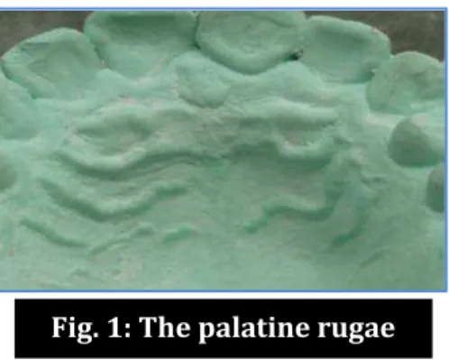
INTRODUCTION: Palatoscopy, or palatal rugoscopy, is the name given to the study of palatal rugae in order to establish a person's identity.1,2 The palatal rugae are located on the anterior portion of the maxilla (Figure 1). Anatomically, in hard mucosal palate, one can identify an antero-posterior thin central groove, bordered, on each side, by a crest: the palatal raphae. From this crest, laterally, three to seven smaller crests emerge. These crests are called palatal rugae. Palatal rugae are irregular, asymmetric ridges of mucous membrane extending lateral from the incisive papilla and the anterior part of the median palatal raphe3,4. Palatal rugae are formed in the 3rd month in utero from the hard connective tissue covering the bone.5 Lund(1924)6 observed that a connective tissue core is
embedded deeply between the sub mucosal fatty tissue and the stratum reticulum of the palate.
This core represents a foundation over which the substance of the rugae builds to become a fold like projection in the roof of the mouth. Hausser E. Zur Bedeutung (1951)7 suggested with the
increase in size of the anterior part of the palate in the early years of life, the length of the rugae and the distance between them increases. The pattern of orientation of the rugae remains unchanged throughout life. The number of rugae on each side of the palate varies between three and five. The palatine rugae do not extend posteriorly beyond the anterior half of the hard palate and never cross the midline. The anterior rugae usually are more prominent than the posterior rugae.
J of Evolution of Med and Dent Sci/ eISSN- 2278-4802, pISSN- 2278-4748/ Vol. 3/ Issue 40/Sept. 01, 2014 Page 10077
Two thirds of the rugae are curved, and the rest are angular. The last rugae frequently are divided into the medial and lateral parts are not connected and do not continue in their axial orientation. Fragmentary rugae frequently are present, particularly in the posterior half of the rugae territory. The shape, length, width, prominence, number and orientation of palatine rugae vary considerably among people.
The inclination of the rugae to the sagittal plane can differ markedly between both sides. In general, no bilateral symmetry exists in the rugae pattern. Friel (1949)8 demonstrated in a study that
the teeth move forward in relation to the rugae in conjunction with growth of the jaws. He showed that the posterior boundary of the rugae in relation to the teeth tends to extend backward until age 20 years.
Once formed, they do not undergo any changes except in length, due to normal growth, remaining in the same position throughout an entire person's life. Not even diseases, chemical aggression or trauma seem to be able to change palatal rugae for1 The ability of palatal rugae to resist decomposition changes for up to seven days after death was also noted. However, some events can contribute to changes in rugae pattern, including extreme finger sucking in infancy and persistent pressure due to orthodontic treatment.9 Camargo et al.10 referred that, in gingival graft surgery, the selection of the palatal donor site should avoid the rugae areas because they may persist in the grafted tissue. However, extractions may produce a local effect on the direction of the rugae.11
The occurrence, number and arrangement of palatal rugae in mammals are species-specific12. In humans they are asymmetrical, which is an exclusive feature of human beings.3 According to English's studies, 13 palatal rugae patterns are sufficiently characteristic to discriminate between individuals. In fact, these authors found it legitimate to base identification upon their comparison1 allowing for human identification even in extreme circumstances14. The relationship between hard palate measurements and dental arches has also been used to determine group ethnicity.
FUNCTIONS OF PALATAL RUGAE:
1. To facilitate food transportation through the oral cavity, prevent loss of food from the mouth and participate in the chewing process12.
2. Due to the presence of gustatory and tactile receptors, they contribute to the perception of taste, the texture of food qualities and tongue position.12
J of Evolution of Med and Dent Sci/ eISSN- 2278-4802, pISSN- 2278-4748/ Vol. 3/ Issue 40/Sept. 01, 2014 Page 10078 only of soft tissue, they are not present in skeletons. Fiene18 (1958) discovered that the palatal rugae could be helpful in anthropological paternity investigations.
CLINICAL IMPLICATIONS OF PALATINE RUGAE:
1. Landmark during orthodontic treatment: Hausser7 observed orthodontically treated patients
who underwent extraction of four premolars and concluded that the lateral edges of the rugae moved forward about one-half the distance of the migration of the adjacent teeth, while the medial rugae were not affected.
Peavy and Kendrick19said "the closer the rugae are to the teeth, the more prone they are to
stretch in the direction that their associated teeth move." In addition to these findings of the importance of using medial points, it has been said that the more posterior rugae are less susceptible to changes with tooth movement, being the third palatal rugae pair in particular the most stable reference reported that the lateral ends of the rugae that terminated close to the teeth followed the movement of the teeth in the sagittal plane, but not in the transverse plane.
Van der Linden20 evaluated changes in the position of posterior teeth in relation to palatine
rugae in 65 normally growing children (aged 6 to 16 years) and in six orthodontically treated patients. The authors noted larger movements at both the medial and lateral rugae points in the orthodontically treated patients.
2. Tooth movement: Hoggan and Sadowsky21 investigated the use of the palatine rugae as reference
points for measuring tooth movement in a manner comparable with cephalometric superimpositions. The authors evaluated the anteroposterior movement of the maxillary first molars and central incisors with the use of two cephalometric variables and six study model variables, and they combined the right and left sides of the palate. The results showed no statistical differences between the mean incisor and molar movement measured cephalometrically and the tooth movement measured relative to the medial and lateral end of the third palatine ruga. Thus, the authors concluded that palatine rugae could be used reliably to assess anteroposterior tooth movements.
Simmons and colleagues22 used the longitudinal database of the Child Research Council of
Denve to examine the anteroposterior stability of the medial rugal region. Their analysis of the data indicated that the medial ruga region increased significantly in anteroposterior length but not uniformly between the sexes. The authors concluded that such changes were characteristic of general craniofacial growth and suggest that the rugae region is responding to the differential growth of the underlying bone. Thus, the authors concluded that the medial rugal landmarks did not appear to be a stable reference point for tooth migration research.
3. Palatine rugae in cleft palate: Park and colleagues23 studied the pattern of palatine rugae in
submucosal clefts. The palatal mucosa had a unique feature in 87.5 percent of the submucosal clefts and in 100 percent of the isolated clefts: one or more of the palatine rugae curved toward the region of the bony notch in the posterior border of the hard palate.
Kratzsch and Opitz24 investigated the characteristics of the palatal rugal zone by means of
J of Evolution of Med and Dent Sci/ eISSN- 2278-4802, pISSN- 2278-4748/ Vol. 3/ Issue 40/Sept. 01, 2014 Page 10079
rugae counts per segment decreased significantly, but the third ruga was never lost after surgery. The primary rugae in unilateral and bilateral cleft lip and palate were the same as those in isolated cleft palates, and they did not differ from those in people who did not have cleft lip or palate.
The linear distance from the tuberosity line to the rugal zone increased in the unilateral and bilateral cleft segments before palatal cleft repair, indicating sagittal maxillary development in the posterior area of the palate. Surgical repair of the cleft palate resulted in a significant lessening of the distance in both segments of unilateral cleft, most likely due to the displacement of mucosa and periosteum required to cover the palatal cleft.
In a second study, Kratzsch and Opitz25 investigated the relationship of palatine rugae to
points (landmarks) and distances on the cleft palate during the period from birth to the time of early mixed dentition. The authors identified changes in the distances from the lateral palatine rugae points of the first and third rugae to the incisal point, the canine point and the tuberosity line. The results of their study indicated that a comparison of distances from the palatine rugae with distances between equivalent points revealed the changes that occurred in the anterior palate during various stages of orthodontic therapy and growth.
4. Palatal vault: Landa26 reported that rugae in dentures are ineffectual or sometimes detrimental to
speech if they add unnecessary thickness to the anterior palatal region
5. Variation of rugae pattern in different ethnic groups: There seems to be a significant
association between rugae forms and ethnicity. Kapali and colleagues (1997)9 studied the palatal
rugae pattern in Australian Aborigines and whites. They observed the number, length, shape, direction and unification of rugae. The authors concluded that the mean number of primary rugae in Australian Aborigines was higher than that in whites, although whites had more primary rugae that exceeded 10 mm in length. The most common shapes in both ethnic groups were wavy and curved forms, while straight and circular forms were least common.
Kashima (1990)27 compared the palatine rugae and shape of the hard palate in Japanese and
Indian children. They found the following:
Japanese children had more primary rugae than Indian children, but both groups had the same number of transverse palatine rugae.
The two groups differed with regard to primary rugae shapes, the posterior boundary of the rugal zone, and the number and position of the secondary and fragmentary rugae.
The palatal raphae of the Japanese child were wider than those Indian children.
Both groups had many transverse palatine rugae on the left side of the palate. The posterior border of the rugal zone on the left side was shifted farther back than it was on the right side. There were no significant differences between the two sexes in either group.
Shetty and colleagues (2005)28 compared the palatine rugae patterns in Indians with those in a
J of Evolution of Med and Dent Sci/ eISSN- 2278-4802, pISSN- 2278-4748/ Vol. 3/ Issue 40/Sept. 01, 2014 Page 10080
6. Forensic identification: It is a well-established fact that the rugae pattern is as unique to a human
as are his or her fingerprints and it retains its shape throughout life16. The anatomical position of the
rugae inside the mouth—surrounded by cheeks, lips, tongue, buccal pad of fat, teeth and bone— keeps them well-protected from trauma and high temperatures. Thus, they can be used reliably as a reference landmark during forensic identification.
Thomas and Van Wyk (1987)29 described the identification of a severely charred edentulous
body with the help of dentures in the victim’s mouth that were compared with another set found in the person’s home. Plaster casts of the tissue surface of both sets of maxillary dentures were made. The investigators delineated and photographed the rugae and midpalatal raphae. They made tracings of each set of rugae on acetate paper and superimposed them on the photograph of the other cast. The tracings established a concordance between the two sets of dentures.
Jacob and Shalla (1987)30 evaluated the use of dental stone casts derived from maxillary
tissues and from the internal aspects of maxillary dentures for postmortem identification of edentulous people. They reported results of 100 percent accuracy when they evaluated the entire cast and results of 79 percent accuracy when they evaluated only the rugae tracings from the casts. Thus, their investigation supported the use of stone casts derived from the internal aspects of maxillary dentures for forensic science identification when the entire cast topography is considered.
Limson and Julian (2004)31 used a computer software program to evaluate the use of palatine
rugae patterns for forensic identification. The authors obtained 250 casts by using irreversible hydrocolloid. They used a sharp pencil to delineate rugae and photographed the rugae pattern with a digital camera; they then transferred the image to a computer. They compared the digitized casts with the stored records. The digitized rugae pattern samples matched the patterns in the stored records.
Muthusubramanian and colleagues (2005)32examined the extent of palatine rugae
preservation for use as an identification tool in burn victims and cadavers, thus simulating forensic cases of incineration and decomposition. Patients with panfacial third-degree burns were examined within 72 hours after their accident. In addition, human cadavers stored in a mortuary at 5°C with 30 to 40 percent relative humidity and kept for a minimum of seven days were assessed for the condition of the palatine rugae. The authors took photographs of the palatine rugae by using a palatal mirror. The study results showed that among the subjects with third-degree panfacial burns, 93 percent of the palatine rugae were normal. The authors observed no changes in the color or surface anatomy of the palatine rugae in 77 percent of the human cadavers. They concluded that the palatine rugae could be used reliably as a reference landmark during forensic identification.
CLASSIFICATIONS OF PALATAL RUGAE:
1. Goria (1911)33: Developed the first system of classification and was rudimentary. The rugae
pattern was categorized in two ways: specifying the number of rugae and specifying the extent of the rugal zone relative to the teeth. In this system, compound rugae of two or more branches were counted as one, whether they were V or Y shaped. Goria further distinguished two types: simple or primitive and more developed.
J of Evolution of Med and Dent Sci/ eISSN- 2278-4802, pISSN- 2278-4748/ Vol. 3/ Issue 40/Sept. 01, 2014 Page 10081 morphology. In this manner, there were four known types of palatal rugae: B-bilious personality rugae; N-nervous personality rugae; S-sanguinary personality rugae; L-lymphatic personality rugae. The letters B, N, L, and S, stand for the different personalities. The letters l and r stand for the left and right side of the palate, and are followed by a number, which specifies the palatal rugae number on each side.
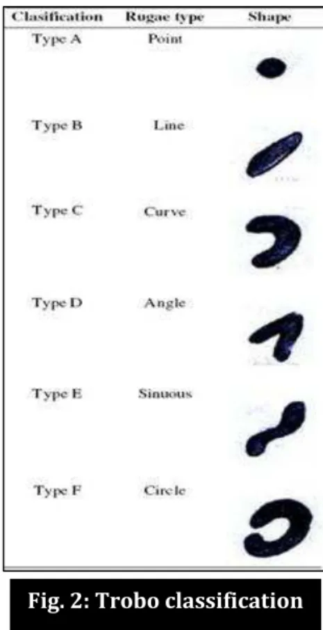
3. Trobo classification (1932)34: This classification also divides rugae into two groups: Simple rugae, classified from A to F(Table 1 & Fig 2) and composed rugae, classified with the letter X. Composed rugae result from two or more simple rugae unions.
Classification Rugae type
Type A Point
Type B Line
Type C Curve
Type D Angle
Type E Sinuous
Type F Circle
Table 1: Trobo classification
4. Carrea classification (1937)15: This author divides palatal rugae into four different types, as shown in Table. 2. Palatal rugae are classified only according to their form and no formula (rugogram) is developed.
J of Evolution of Med and Dent Sci/ eISSN- 2278-4802, pISSN- 2278-4748/ Vol. 3/ Issue 40/Sept. 01, 2014 Page 10082 Classification Rugae type
Type I Posterior-anterior directed rugae Type II Rugae perpendicular to the raphae Type III Anterior-posterior directed rugae Type IV Rugae directed in several directions
Table 2: Carrea classification
,
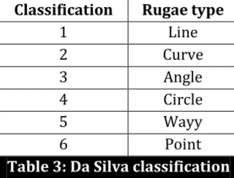
5. Da Silva classification (1938)34 : In this classification, palatal rugae are divided into two groups: simple, from 1 to 6( Table 3) and composed, resulting from two or more simple rugae. They are named according to each rugae number. It is possible to classify each rugae individually (describing its form), but also to describe all the palatal rugae system (describing each ruga type number), making this a difficult classification to use.
Classification Rugae type
1 Line
2 Curve
3 Angle
4 Circle
5 Wayy
6 Point
Table 3: Da Silva classification
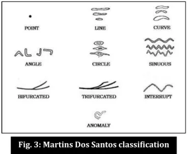
6. Martins Dos Santos classification (1946)15: Based on the form and position of each palatal rugae, this classification indicates and characterizes the following:
One initial rugae; the most anterior one on the right side is represented by a capital letter. Several complementary rugae; the other right rugae are represented by numbers.
One sub initial rugae; the most anterior one on the left side is represented by a capital letter. Several sub complementary rugae; the other left rugae are represented by numbers.
The numbers and letters given to each rugae, relate to its form and can be seen in Table 4; Fig 3.
Ruage type Anterior position Other positions
Point P 0
Line L 1
Curve C 2
Angle A 3
Circle C 4
Sinuous S 5
Bifurcated B 6
Trifurcated T 7
Interrupt I 8
Anomaly An 9
J of Evolution of Med and Dent Sci/ eISSN- 2278-4802, pISSN- 2278-4748/ Vol. 3/ Issue 40/Sept. 01, 2014 Page 10083
7. Lysell’s classification (1955)16: This is the most important classification and has been used
widely in research involving rugae. It is comprehensive and includes the IP. Rugae are measured in a straight line between the origin and termination and are grouped into three categories:
Primary: 5 millimeters or more. Secondary: 3-5 mm.
Fragmentary: 2-3 mm.
Rugae smaller than 2 mm are disregarded. The rugae on both sides of the palate are numbered separately from anterior to posterior and classified according to shape, position or origin in relation to the median palatal raphae. Three categories of unification are recognized in this system:
Common origin diverging laterally; Separate origins converging laterally;
Separate origins converging laterally but involving one primary and one secondary ruga.
Branching, breaks, papillations, annular formations and spirals are counted, while the rugae directions are measured in degrees relative to the distribution of secondary and fragmentary rugae by noting their proximity to the nearest primary rugae while observing the posterior border relationship with the teeth. The clinician measures the IP and classifies it according to one of seven shapes.
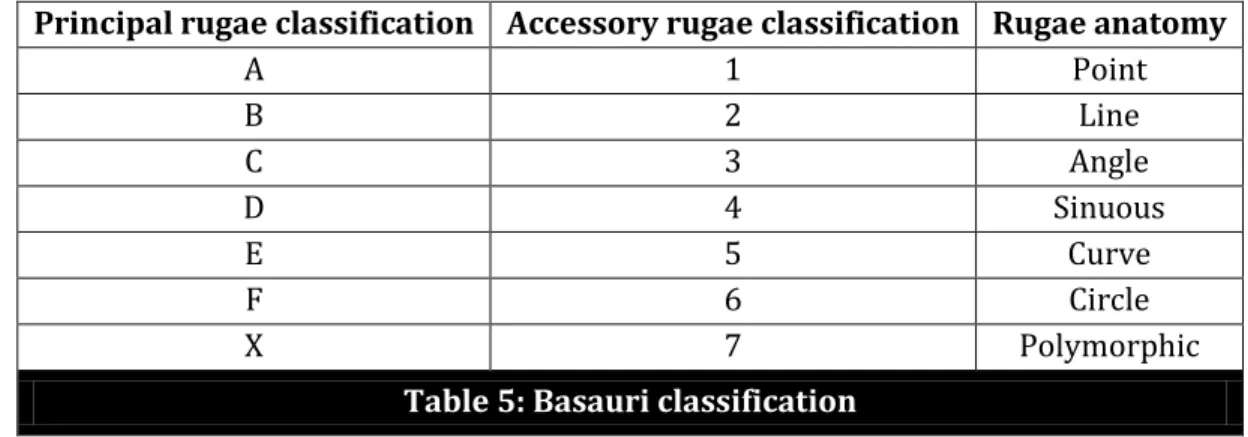
8. Basauri classification (1961)34:Like the Trobo classification, this is a very easy classification to use. It distinguishes between the principal rugae, which is the more anterior one (labelled with letters) and the accessory rugae, which concerns all the remaining rugae(labelled with numbers), as seen in Table 5.
J of Evolution of Med and Dent Sci/ eISSN- 2278-4802, pISSN- 2278-4748/ Vol. 3/ Issue 40/Sept. 01, 2014 Page 10084 Principal rugae classification Accessory rugae classification Rugae anatomy
A 1 Point
B 2 Line
C 3 Angle
D 4 Sinuous
E 5 Curve
F 6 Circle
X 7 Polymorphic
Table 5: Basauri classification
9. Lima (1968)35: Consists of four main types: punctate, straight, curved and composite.
10.Tzatscheva and Jordanov(1970)36: Classified rugae according to their direction, branching,
symmetry and radiality.
11.Cormoy system classification34: This system classifies palatal rugae according to their size, in: (1)principal rugae(over 5 mm).
(2)accessory rugae(ranging from 3 to 4 mm). (3)fragmental rugae(with less than 3 mm length).
The form(line, curve, and angle), origin(medial extremity) and direction of each ruga are also described. Possible ramifications are also pointed out. Rugae that share the same origin, interrupted rugae and the incisive papilla are described as well. It is a very complete system. However, its use does not lead to rugogram elaboration, which makes the managing and processing of data difficult.
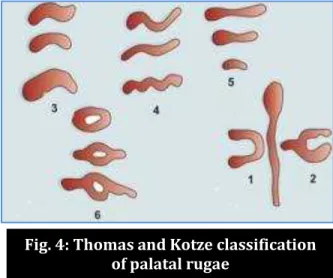
12. Thomas and Kotze (1983) classification37:The rugae pattern is classified based on their length, shape, direction and unification, proposed by Lysell (1955) and later modified by Thomas and Kotze(1983)(Table 6) and(Fig 4).
Criteria Length
Primary rugae A-5 to 10mm B-10 mm or more
Secondary rugae: 3-5mm Shape
Fragmentary rugae: less than 3mm Curved
Wayy Straight Circular
J of Evolution of Med and Dent Sci/ eISSN- 2278-4802, pISSN- 2278-4748/ Vol. 3/ Issue 40/Sept. 01, 2014 Page 10085 ANALYSING AND RECORDING PALATAL RUGAE: There are several ways to analyze palatal rugae. Intraoral inspection is probably the most usedand also the easiest and the cheapest. However, it can create difficulties if a future comparative exam is required.34 A more detailed and exact study, as well as the need to preserve evidence may justify oral photography or oral impressions.34 Calcorrugoscopy, (Fig 5) or the overlay print of palatal rugae in a maxillary cast, can be used in order to perform comparative analysis.34 By using stereoscopy, one can obtain a three dimensional image of palatal rugae anatomy.
It is based on the analysis of two pictures taken with the same camera, from two different points, using special equipment. Another technique is the sterophotogrammery which, by using a special device called Traste Marker, allows for an accurate determination of the length and position of every single palatal ruga.34 However, due to its simplicity, price and reliability, the study of maxillary dental casts is the most used technique.34
ADVANTAGES OF PALATOSCOPY:
1. Palatal rugae are used in human identification due to their singularity and unchangeable nature. Changes that occur from orthodontic movement, extraction, aging, and palatal expansion do not modify the rugae enough to hamper identification29.
Fig. 4: Thomas and Kotze classification of palatal rugae
J of Evolution of Med and Dent Sci/ eISSN- 2278-4802, pISSN- 2278-4748/ Vol. 3/ Issue 40/Sept. 01, 2014 Page 10086 2. Low utilization costs.15
3. It is possible to have antemortem data established such as records found in dental practice in different forms (dental casts, old prosthetic maxillary devices and intraoral photographs) to compare with post mortem data.29
4. Rugoscopy is rather simple technique not requiring any complex instrumentation29.
PROBLEMS WITH PALATOSCOPY:
1. Palatoscopy might not be so useful in crime scene investigations in the linking of suspects to crime scenes. In fact, this kind of evidence is not expected to be found in such circumstances.38 2. Possibility of rugae pattern forgery. In a case report, Gitto et al.39 described a method where
palatal rugae were added to a complete denture in order to improve speech patterns in some patients. This process can lead to false identity exclusion due to misleading ante-mortem data.
CONCLUSION: Identification of living or dead people is often a difficult, challenging and time-consuming process. Palatal rugae have been shown to be highly individualistic and consistent in shape throughout the life. It is well-established fact that the palatal rugae pattern is unique to human being, as his fingerprints, thus its use in forensic identification is fairly justified. Analysis of palatal rugae pattern combined with other methods is an important alternative and complementary technique for human identification, providing a significant contribution in cases of criminal investigation. Rugoscopy is rather simple technique not requiring any complex instrumentation. Palatoscopy has been successfully used in necro identification. Thus, palatal rugae hold potential as a supplementary tool, along with the dentition, to establish the identity of an individual. Nevertheless, we believe that larger samples should be examined in detail to further validate the findings of this study and come to definitive conclusions.
REFERENCES:
1. Carbajo C, Identificación de cadáveres y aspectos forenses de los desastres, Publicaciones de la Unidad de Investigación en Emergencia y Desastres, http://www.desastres.org.
2. Gilbert J.A. Calabuig, Medicina Legal y Toxicologia (5th ed.)
3. J.D. Simmons, R.N. Moore and L.C. Erickson, A longitudinal study of anteroposterior growth changes in the palatine rugae, J. Dent. Res. 66(1987) (9), pp. 1512-1515.
4. Abdel-Aziz H.M. and Sabet N.E., Palatal rugae area: a landmark for analysis of pre- and post-orthodontically treated adult Egyptian patients, East Mediterr. Health J. 2001. (1/2), p. 60-66.
5. T.W. Sadler, Langman's Medical Embryology(6th ed.), Williams & Wilkins, Baltimore (1990) p. 316-320]
6. Lund O. Histologische beitrage zur anatomie des munddachs und paradentiums. Vrtlzschr F Zahnh 1924; 40: 1-20.
7. Hausser E. Zur Bedeutung und Veranderung der Gaumenfalten des menschen [The palatal ridges in man: their significances and their modifications]. Stoma (Heidelb) 1951; 4(1) :3-26.
8. Friel S. Migration of teeth. Dent Rec (London) 1949; 69(3): 74-84.
J of Evolution of Med and Dent Sci/ eISSN- 2278-4802, pISSN- 2278-4748/ Vol. 3/ Issue 40/Sept. 01, 2014 Page 10087 10.Camargo P.M., Melnick P.R. and Kenney E.B., The use of free gingival grafts for aesthetic
purposes, Periodontology 27(2001), p. 72-96.
11.Bowles R.G., First premolar extraction decisions and effects, Thesis presented for the Graduate Studies Council, The University of Tennessee, Health Science Center, 2005.]
12.Buchtová M, Tichy F, Putnová I. and Míšek I. The development of palatal rugae in the European pine vole, Microtus subterraneus(Arvicolidae, Rodentia), Folia Zoo 52(2003) (2), pp. 127-136. 13.English W.R., Robinson S.F., Summitt J.B., Oesterle L.J., Brannon R.B. and Morlang W.M.,
Individuality of human palatal rugae, J. Forensic Sci. 33(1988) (3), pp. 718-726.
14.Ermenc B. and Rener K., Possibilities for dental identification in the case of mass disaster in Slovenia, Forensic Sci. Int. 103(1999), pp. 867-875.
15.Campos ML, Rugoscopia palatina, Available from URL : http://www.pericias-forenses.com.br. 16.Lysell, Plica palatinae transverse and papilla incisive in man. A morphological and genetic
study, Acta Odont. Scand. 13(1955) (Suppl. 18), p. 5-137.
17.Sassouni V. Palato print, physioprint and roentgenographic cephalometry as new methods in
human identification (preliminaryreport). J Forensic Sciences 1957; 2: 428-42.
18.Fiene M. Vergleichend-morphologische studien über das Gaumen faltenrelief von Zwillingen.
Fortschr Kieferorthop1958; 19: 229-36.
19.Peavy DC Jr, Kendrick GS. The effects of tooth movement on the palatine rugae. J Prosthet Dent 1967; 18 (6): 536-542.
20.Van der Linden FP. Changes in the position of posterior teeth in relation to ruga points. Am J Orthod 1978; 74(2): 142-161.
21.Hoggan BR, Sadowsky C. The use of palatal rugae for the assessment of anteroposterior tooth movements. Am J Orthod Dentofacial Orthop 2001; 119(5): 482-488.
22.Simmons JD, Moore RN, Erickson LC. A longitudinal study of anteroposterior growth changes in the palatine rugae. J Dent Res 1987; 66(9): 1512-1515.
23.Park S, Eguti T, Kato K, Nitta N, Kitano I. The pattern of palatal rugae in submucous cleft palates and isolated cleft palates. Br J Plast Surg 1994; 47(6): 395-399.
24.Kratzsch H, Opitz C. Investigations on the palatal rugae pattern in cleft patients, part I: a morphological analysis. J Orofac Orthop 2000;61 (5): 305-317.
25.Kratzsch H, Opitz C. Investigations on the palatal rugae pattern in cleft patients, part II: changes in the distances from the palatal rugae to maxillary points. J Orofac Orthop 2000; 61(6): 421-431.
26.Landa J. The importance of phonetics in full denture prosthetics. Dent Dig 1935; 41: 154-160. 27.Kashima K. Comparative study of the palatal rugae and shape of the hard palatal in Japanese
and Indian children [in Japanese]. Aichi Gakuin Daigaku Shigakkai Shi 1990; 28 (1 part 2): 295-320.
28.Shetty SK, Kalia S, Patil K, Mahima VG. Palatal rugae pattern in Mysorean and Tibetan populations. Indian J Dent Res 2005; 16 (2): 51-55.
29.Thomas CJ, Van Wyk CW. Elastic fibre and hyaluronic acid in the core of human palatal rugae. J Biol Buccale 1987; 15 (3): 171-174.
J of Evolution of Med and Dent Sci/ eISSN- 2278-4802, pISSN- 2278-4748/ Vol. 3/ Issue 40/Sept. 01, 2014 Page 10088
31.Limson KS, Julian R. Computerized recording of the palatal rugae pattern and an evaluation of its application in forensic identification. J Forensic Odontostomatol 2004; 22(1): 1-4.
32.Muthusubramanian M, Limson KS, Julian R. Analysis of rugae in burn victims and cadavers to simulate rugae identification in cases of incineration and decomposition. J Forensic Odontostomatol 2005; 23 (1): 26-29.
33..Goria C. Le rughe del palato in speciale rapporto coll anthropologia criminale e la psichiatria. 1911. Cited by: Lysell L. Plicae palatinae transversae and papilla incisiva in man. Acta Odontol Scand.
34.Pueyo V.M., Garrido B.R. and Sánchez J.A.S., Odontología Legal y Forense, Masson, Barcelona (1994) pp. 277-292
35. Lima OC. Rugoscopia [Rugoscopy Correia Lima’s process ]. Rev Bras Med ; (12): 806-807.1955;13: (suppl 18): 5-13
36.Tzatscheva L, Jordanov J. Plicae palatinae transversae undpapilla incisive bei den Bulgaren [Plica palatinae transversae and papilla incisiva in Bulgarians]. Z Morphol Anthropol 1970; 62(3): 276-284.
37.Thomas CJ, Kotze TJ. The palatal ruga pattern: a new classification.J Dent Assoc S Afr 1983; 38 (3): 153-157.
38.Caldas IM, Magalhães T, Afonso A. Establishing identity using cheiloscopy and palatoscopy. Forensic Sci Int 2007; 165: 1-9.
39.Gitto C.A., Exposito S.J. and Draper J.M., A simple method of adding palatal rugae to a complete denture, J. Prosthet. Dent. 81 (1999), pp. 237-239.
AUTHORS: 1. R. Mahajan 2. Mohd. Arif Dar 3. Sanjeet Singh Risam
PARTICULARS OF CONTRIBUTORS:
1. Registrar, Department of Oral Medicine and Radiology, Govt. Dental College and Hospital Srinagar, Jammu and Kashmir.
2. Registrar, Department of Oral Medicine and Radiology, Govt. Dental College and Hospital Srinagar, Jammu and Kashmir.
3. Senior Lecturer, Department of Oral Medicine and Radiology, Govt. Dental College and Hospital Srinagar, Jammu and Kashmir.
NAME ADDRESS EMAIL ID OF THE CORRESPONDING AUTHOR: Dr. Mohd Arif Dar,
Registrar,
Dept. of Oral Medicine and Radiology, Govt. Dental College and Hospital, Shreen Begh, Srinagar,
Jammu and Kashmir.