Type 2 by Bortezomib Sensitizes Jurkat Leukemia T Cells
against Glucocorticoid-Induced Apoptosis
Yi Tao1., Lu Gao2., Xiaosong Wu1, Hongmei Wang1, Guang Yang1, Fenghuang Zhan3, Jumei Shi1*
1Department of Hematology, Shanghai Tenth People’s Hospital, Tongji University School of Medicine, Shanghai, People’s Republic of China,2Department of Physiology, Second Military Medical University, Shanghai, People’s Republic of China,3Department of Internal Medicine, University of Iowa Carver College of Medicine, Iowa City, Iowa, United States of America
Abstract
11b-hydroxysteroid dehydrogenases type 2 (11b-HSD2), a key regulator for pre-receptor metabolism of glucocorticoids (GCs) by converting active GC, cortisol, to inactive cortisone, has been shown to be present in a variety of tumors. But its expression and roles have rarely been discussed in hematological malignancies. Proteasome inhibitor bortezomib has been shown to not only possess antitumor effects but also potentiate the activity of other chemotherapeutics. In this study, we demonstrated that 11b-HSD2 was highly expressed in two GC-resistant T-cell leukemic cell lines Jurkat and Molt4. In contrast, no 11b-HSD2 expression was found in two GC-sensitive non-hodgkin lymphoma cell lines Daudi and Raji as well as normal peripheral blood T cells. Inhibition of 11b-HSD2 by 11b-HSD inhibitor 18b-glycyrrhetinic acid or 11b-HSD2 shRNA significantly increased cortisol-induced apoptosis in Jurkat cells. Additionally, pretreatment of Jurkat cells with low-dose bortezomib resulted in increased cellular sensitivity to GC as shown by elevated induction of apoptosis, more cells arrested at G1 stage and up-regulation of GC-induced leucine zipper which is an important mediator of GC action. Furthermore, we clarified that bortezomib could dose-dependently inhibit 11b-HSD2 messenger RNA and protein levels as well as activity (cortisol-cortisone conversion) through p38 mitogen-activated protein kinase signaling pathway. Therefore, we suggest 11b-HSD2 is, at least partially if not all, responsible for impaired GC suppression in Jurkat cells and also indicate a novel mechanism by which proteasome inhibitor bortezomib may influence GC action.
Citation:Tao Y, Gao L, Wu X, Wang H, Yang G, et al. (2013) Down-Regulation of 11b-Hydroxysteroid Dehydrogenase Type 2 by Bortezomib Sensitizes Jurkat Leukemia T Cells against Glucocorticoid-Induced Apoptosis. PLoS ONE 8(6): e67067. doi:10.1371/journal.pone.0067067
Editor:Zhengqi Wang, Emory University, United States of America
ReceivedDecember 21, 2012;AcceptedMay 13, 2013;PublishedJune 24, 2013
Copyright:ß2013 Tao et al. This is an open-access article distributed under the terms of the Creative Commons Attribution License, which permits unrestricted use, distribution, and reproduction in any medium, provided the original author and source are credited.
Funding:1. National Natural Science Foundation of China (No. 81071856 and 30973450 to JS), (http://www.nsfc.gov.cn/). 2. Joint Research Fund for Overseas, Hong Kong and Macao Scholars(No. 81228016 to FZ and JS), (http://www.nsfc.gov.cn/). 3. Shanghai Pujiang Program (No. 11PJ1407900 to JS), (http://www.stcsm. gov.cn). 4. Shanghai Tenth People’s Hospital Funds (No. 12RQ112 to YT, No. 10RD103 and 11SC103 to JS), (http://www.shdsyy.com.cn). The funders had no role in study design, data collection and analysis, decision to publish, or preparation of the manuscript.
Competing Interests:The authors have declared that no competing interests exist.
* E-mail: shijumei@hotmail.com
.These authors contributed equally to this work.
Introduction
Glucocorticoids (GCs), stress hormones secreted from the adrenal gland, are physiologically involved in metabolism, cell differentiation, and several aspects of the maintenance of homeostasis. They play their parts by combining with cognate intracellular glucocorticoid receptor (GR) and translocating to the nucleus afterwards [1]. Pharmacologically, GCs have pro-apoptotic effects and are administered for the treatment of lymphoproliferative disorders [2]. In childhood acute lymphoblas-tic leukaemia (ALL) treatment protocols, an introductory mono-therapy GC has been used to reduce leukemic blasts in GC sensitive patients in the initial therapy. However, GC sensitivity is different from person to person and GC-resistance is a therapeutic problem with an unclear molecular mechanism. Some studies have suggested that the GC receptor is underexpressed or mutated in GC-resistant cells, but others have reached contradictory results [3,4,5], indicating the possibility of multiple diverse mechanisms involved in GC resistance.
Bortezomib (Velcade, PS-341) is the first proteasome inhibitor that was clinically tested in patients and becomes a therapeutic modality for multiple myeloma [12]. Moreover, bortezomib is also highly cytotoxic to a variety of malignancies. The antitumor mechanism of bortezomib not only promotes apoptosis in cancer cells, but also sensitizes these cells to chemotherapy [13]. In addition, bortezomib has been demonstrated to overcome GC resistance at the hypoxic blood-brain barrier to reduce brain edema in acute ischemic stroke [14]. But it is unclear if bortezomib could increase cell susceptibility to GC-induced cytotoxicity.
In this study, we investigated the 11b-HSD2 expression in GC-resistant T-cell lymphoblastic lymphoma/leukemia lines and further determined its contribution to GC resistance by using 11b-HSD inhibitor or 11b-HSD2 gene silencing. To clarify whether bortezomib could improve GC sensitivity, we treated Jurkat T-cell lymphoblastic lymphoma/leukemia cells with cortisol following bortezomib pretreatment. Herein we reported 11b -HSD2 presence was partly responsible for GC resistance in leukemia T cells and bortezomib enhanced GC sensitivity in Jurkat cells by P38 mitogen-activated protein kinase (MAPK)-mediated down-regulation of 11b-HSD2, suggesting that 11b -HSD2 could be used as a potential therapeutic target in GC-resistant lymphoproliferative disorders and also a novel down-stream target of bortezomib action.
Materials and Methods
Cell Lines and Cell Treatment
Jurkat and Molt4, two T-ALL cell lines, were kindly provided by Dr. J. Hou (Second Military Medical University, Shanghai, China). Daudi and Raji, two Non-hodgkin lymphoma (NHL) cell lines, were derived from ATCC (Manassas, VA, USA). Cells were grown in RPMI 1640 (Bio Whitaker, Rockland, ME, USA) at 37uC, 5% carbon-dioxide and saturated humidity. The media were supplemented with 10% fetal calf serum (Sigma), 100 U/ml penicillin, 100 mg/ml streptomycin and 2 mM L-glutamine (GibcoBRL, Paisley, UK). Peripheral blood samples were obtained from healthy volunteers after written informed consent in accordance with Declaration of Helsinki. Approval was obtained by Shanghai Tenth People’s Hospital for Medical Sciences Institutional Review Board for these studies. T cells were isolated from peripheral blood using CD3 positive magnetic bead selection (Stem Cell Technologies). Jurkat cells in log-phase growth were treated with increasing concentrations of bortezomib (Millennium Pharmaceuticals, USA) for 12 or 24 hours before the detection of 11b-HSD2 mRNA and protein expression. After 24 hours of indicated concentrations of bortezomib exposure, Jurkat cells were collected and washed once with medium and treated with indicated concentrations of cortisol (Sigma) for 48 hours. Cells unexposed to bortezomib were used as a control. For analysis of 11b-HSD2 inhibition, Jurkat cells were treated with 5mM 11b -HSD inhibitor 18b-glycyrrhetinic acid (Sigma) and 10mg/ml cortisol in the presence or absence of GC receptor antagonist RU486 (1mM) for 48 hours. For analysis of p38 MAPK signal pathway, Jurkat cells were pretreated with 5 nM bortezomib in the presence or absence of 10mM p38 MAPK inhibitor SB203580 (Cell Signaling Technology, MA, USA) for 24 hours. Then cells were exposed to 10mg/ml cortisol for 48 hours.
Reverse Transcriptase–polymerase Chain Reaction (RT-PCR) and Quantitative Real-time PCR (qRT-(RT-PCR)
Total RNA was prepared from individual samples by using TRIzol reagent (Invitrogen, USA), and 5mg total RNA was used for the RT reaction using the PrimeScriptTM RT reagent kit
(Takara, Tokyo, Japan) and then stored at 220uC. The cDNA samples obtained was then amplified by PCR with Taq DNA polymerase (Takara, Tokyo, Japan). The PCR condition used was as follows: 96uC, 3 min, for initial denaturation; 95uC (15 sec), 64uC (15 sec) and 72uC (15 sec) in a total 40 cycles with a final extension step at 72uC for 10 min. The nucleotide sequences of the primers were as follows: 11b-HSD2, sense: 59-GCTGCTGCGCTCAGACCT-39, antisense: 59-GGCTGTTCAACTCCAATACG-39 (accession number NM_000196). b-actin, sense: 59-TGTGTTGGCGTA-CAGGTCTTTG-39, antisense: 59-GGGAAATCGTGCGTGA-CATTAAG-39 (accession number NM_001101). All primer sets were designed to span at least one intron to avoid false-positive signals derived from genomic DNA. 10ml of the reaction mixture were subsequently electrophoresed on a 1.5% agarose gel and visualized by ethidium bromide, using a 100 bp DNA ladder (Invitrogen) to estimate the band sizes. Placenta cDNA (from our own laboratory) was used as a positive control for 11b-HSD2.
To measure 11b-HSD2 and Glucocorticoid receptora(GRa) mRNA levels, quantitative real-time PCR (qRT-PCR) was carried out using Rotor-Gene 3000 (Corbett Research, Sydney, Australia) in a total of volume of 25ml reaction mixture following the manufacturer’s protocol using the 26Taq PCR master mix (QIAGEN) and 0.2mM of each paired primer. qRT-PCR conditions were optimized according to preliminary experiments to achieve linear relationships between initial RNA concentration and PCR product. The annealing temperature was set at 64uC and amplification cycles were set at 40 cycles. The nucleotide sequences of the primers were as follows: 11b-HSD2, sense: 59-GTGCCATCGAGCTGCGTACCT-39, antisense: 59 -TCCGCATCAGCAACTACTTCA-39 (accession number NM_000196). GRa, sense: 59-AACTGGCAGCGGTTTTAT-39, antisens: 59-ATTTTGGTATCTGATTGGTGAT-39 (acces-sion number NM_000176). GILZ, sense: 59 -AGCGTGGTGGC-CATAGACAAC-39, antisense: 59 -AGCCACTTACACCGCA-GAA-39 (accession number NM_198057). The specificity of the primers was verified by examining the melting curve as well as subsequent sequencing of the qRT-PCR products. Each sample was normalized on the basis of itsb-actin mRNA content. The relative expression of the genes of interest was determined by using comparative threshold cycle (Ct) method. Briefly, DCt in each group was yielded by subtracting the Ct of theb-actin gene from the Ct of the target gene yields theDCt in each group (control and experimental groups). Then subtractingDCt of control group from the experimental group obtains theDDCt, which was entered into the equation 2 - DDCt and calculated for the exponential amplification of PCR.
Western Blot Analysis
Infrared Imaging System (LI-COR), as previously described [15]. The expression levels ofb-actin were used as the loading control for the immunoblots.
Specific 11b-HSD2 Gene Silencing by Lentivirus Expression Vector System
Two synthetic double-stranded oligonucleotide sequences spe-cific for 11b-HSD2 silencing were purchased from Sigma. A nonsense scrambled oligonucleotide was used as a control. 11b -HSD2 short hairpin RNA (shRNA) double-stranded oligonucle-otides were cloned into lentiviral pGMLV-SC1 vectors. Recom-binant lentiviruses were produced by transient transfection of 293T cells. Efficiency of viral transfection was determined by counting the number of green fluorescent protein (GFP)– expressing cells using flow cytometry, and 95% transduction efficiency of Jurkat cells was achieved.
11b-HSD2 Activity Assay
11b-HSD2 oxidase activity was assayed using thin-layer chromatography (TLC)/liquid scintillation (LS). Briefly, cells were cultured in 12-well plates in the presence or absence of indicated concentrations of bortezomib for 24 hours, washed with PBS three times after the treatments. Then the cells were incubated with 1mM cortisol (Sigma) containing 200,000 cpm [3H]cortisol (Amersham Life Science, Little Chalfont, UK) in FCS-free medium for 12 h at 37uC. Cells were then removed from the medium by centrifugation (10,0006g, 20 min, 4uC) and cell numbers per well were counted. To measure conversion, a mixture of cortisol and cortisone (40mg each) was added to the collected medium to allow visual localization of the steroids when subjected to TLC. Steroids in the media were extracted with ethyl acetate. The extract was dried, reconstituted with ethyl acetate (100ml), and applied to a TLC plate (Fisher Scientific, Pittsburgh, PA). Cortisol and cortisone were separated in the solvent system chloroform/ethanol (95:5, vol/vol), steroids were visualized under UV light, scraped off, and extracted with ethyl acetate. The solvent was dried, scintillation fluid added, and the radioactivity counted in a liquid scintillation counter. 11b-HSD2 activity was expressed as a percentage: pmol cortisone produced per 106cells of the experimental sample/pmol cortisone produced per 106cells of the control sample.
Cell Viability, Apoptosis and Cell Cycle Analyses
The live cells were counted by trypan blue dye exclusion assay for 5 days. Cell viability is expressed as the percent relative to control cells. For analysis of induction of apoptosis, cells were stained with Annexin V/PI (BD Pharmingen) according to the product instructions. Cells were then analyzed for apoptosis by flow cytometry using the Cell Quest software. For analysis of cell cycle, cells were washed with PBS, fixed in 70% ethanol overnight at 220uC, treated with RNaseA (Sigma, USA), stained with propidium iodide (Sigma), and assayed for DNA content by flow cytometry. The acquired histograms were analyzed by ModFit LT software to determine the cell cycle phase distribution.
Statistical Analysis
All data are reported as mean6SD. Paired Student’s t test or two-way ANOVA followed by the Student-Newman-Keuls test was used to assess significant differences between groups. Significance was set at p,0.05. The values for n refer to the number of experiments performed with different cell preparation.
Results
Jurkat and Molt4 Cells show Resistance to Cortisol-induced Apoptosis and Antiproliferation
Firstly, we tested the cytotoxic effect of cortisol on Jurkat and Molt4 cells (two T-cell lymphoblastic lymphoma/leukemia cell lines) as well as Daudi and Raji cells (two non-hodgkin lymphoma cell lines). For this, we incubated each cell line with 100 nM cortisol for 24 and 48 hours and measured drug-induced apoptosis by Annexin V/PI staining. We observed that the drug was potent in inducing apoptosis in Daudi and Raji cells by 24 and 48 hours. In contrast, Jurkat and Molt4 cells showed resistance to cortisol-induced apoptosis (Fig. 1A and B).
We then proceeded to study the ability of cortisol to inhibit proliferation of each cell line in vitro. When the same cell lines were incubated with indicated concentrations of drug for 5 days, we observed a clear dose-dependent inhibition of proliferation of in Daudi and Raji cells tested (Fig. 1C). The inhibitory effect of cortisol on proliferation was evident even at a lower dose than was observed in the apoptosis assay. On the other hand, the drug imposed few effects on proliferation of Jurkat and Molt4 cells, a reflection of cell insensitivity to GC (Fig. 1C).
High Expression of 11b-HSD2 and Failure in Auto-induction of GRain Cortisol-resistant Cells
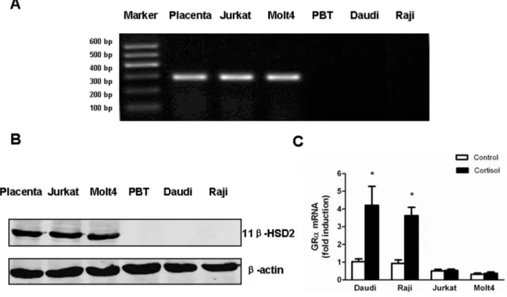
The relative resistance to cortisol suggested the likely presence of 11b-HSD2 in Jurkat and Molt4 cells. Taking placental cells as a positive control, RT-PCR showed high levels of 11b-HSD2 mRNA expression in Jurkat and Molt4 cells. Whereas, 11b-HSD2 mRNA was not detectable in Daudi and Raji cells as well as peripheral blood T (PBT) cells from healthy volunteers (Fig. 2A). Consistent with 11b-HSD2 mRNA levels, western blotting showed high levels of 11b-HSD2 protein in Jurkat and Molt4 cells, but no detections in Daudi, Raji and normal PBT cells (Fig. 2B).
Due to a close relationship between GC sensitivity and GRa
expression, qRT-PCR was applied to detect GRalevel before and after cortisol treatment. The results showed that the basal level of GRamRNA was higher in Daudi and Raji cells than Jurkat and Molt4 cells. In Daudi and Raji cells, GRamRNA was significantly increased by cortisol treatment, whereas almost no induction of GRaoccurred in Jurkat and Molt4 cells (Fig. 2C).
Inhibition of 11b-HSD2 Increases Sensitivity to Cortisol-induced Apoptosis
which is another evidence indicative of elevated cell apoptosis (Fig. 3B).
To further clarify the definite role of 11b-HSD2 in cell resistance to cortisol, specific shRNA (using the lentivirus shRNA expression vector system) was used to knockdown 11b-HSD2 in Jurkat cells. 11b-HSD2 protein expression was confirmed by Western blots (Fig. 3C). Similar to the effect of 18b-GA, 11b -HSD2 knockdown made Jurkat cells become highly sensitive to cortisol-induced apoptosis as compared to wild-type and scramble-transfected control cells (Fig. 3D), strongly suggesting that GC resistance in Jurkat cells was partly related to the presence of 11b -HSD2.
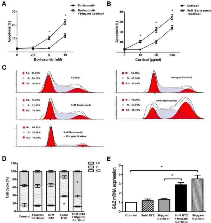
Bortezomib Pretreatment Sensitizes Jurkat Cells to Cortisol-induced Apoptosis and G1 Cell Cycle Arrest
Treatment of Jurkat cells with increasing doses of bortezomib resulted in direct cytotoxicty associated with enhanced apoptotsis (Fig. 4A). Different studies demonstrated that bortezomib
poten-tiates the activity of other chemotherapeutics, in part by down-regulating chemoresistance pathways [13]. As shown in Fig. 1, Jurkat cells were resistant to cortisol-induced cytotoxicty. Howev-er, we observed that Jurkat cells underwent increased cortisol-induced apoptosis when pretreated with increasing doses of bortezomib (Fig. 4A). Particularly, pretreatment with low-dose bortezomib (5 nM) for 24 hours that did not exert any direct cytotoxicty significantly enhanced the potency of cortisol-induced apoptosis (Fig. 4B).
It is known that GC induces G1 cell cycle arrest in GC-induced T-cell apoptosis [16]. Whereas, bortezomib has been demonstrat-ed to greatly arrest Jurkat cells at G2 phase in our study (Fig. 4C) and others [17]. We observed more cells were arrested by cortisol at G1 phase when pretreated with low-dose bortezomib (Fig. 4C and D), suggesting that bortezomib pretreatment may enhance the effects of cortisol on Jurkat cells.
GC-induced leucine zipper (GILZ), an important mediator of GC action, is up-regulated by GC [18]. Consistently, our results Figure 1. Resistance to cortisol-induced apoptosis and antiproliferation in Jurkat and Molt4 cells compared to Daudi and Raji cells. (A) Jurkat, Molt4, Daudi and Raji cells treated with 100 nM cortisol for 24 hours (middle panels) or 48 hours (bottom panels) were stained with Annexin V/PI and analyzed by flow cytometry. Annexin V single-positive cells were considered apoptotic. (B) Graphical representation of the data displayed in panel A as mean6SD of 3 independent experiments. *p,0.05vs.untreated cells. (C) Jurkat, Molt4, Daudi and Raji cells treated with
indicated concentrations of cortisol for 5 days. Percentages of viable cells after treatment were determined by using trypan blue dye exclusion. Data are shown as mean6SD of triplicate samples. *p,0.05vs.untreated cells.
also showed that cortisol dose-dependently enhanced GILZ mRNA level in Jurkat cells (Fig. 4E). To further clarify the effects of botezomib pretreatment on cell response to cortisol, we investigated the level of GILZ mRNA in Jurkat cells. As expected, cortisol increased GILZ mRNA in bortezomib-pretreated Jurkat cells (Fig. 4E), confirming the role of low-dose bortezomib in sensitizing Jurkat cells to GC.
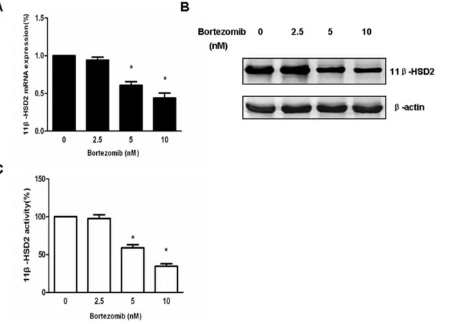
Bortezomib Decreases 11b-HSD2 mRNA and Protein Expression as well as Enzyme Activity in a Dose-dependent Manner
As mentioned above, high expression of 11b-HSD2 has been demonstrated in Jurkat cells and inhibition of 11b-HSD2 could restore cell susceptibility to cortisol. Therefore, we evaluated if bortezomib pretreatment improved GC cell sensitivity by regulat-ing 11b-HSD2 expression. Jurkat cells were treated with increasing amounts of bortezomib (2.5–10 nM), and the mRNA expression levels of 11b-HSD2 were examined 12 hours posttreatment. As shown in Fig. 5A, 11b-HSD2 levels in Jukat cells were significantly reduced by 5 nM bortezomib and further decreased in a dose-dependent manner. The control of 11b-HSD2 by bortezomib was then examined by quantifying 11b-HSD2 protein levels and enzyme activity 24 hours posttreatment using immunoblotting and enzyme activity assay respectively. Consistent with the reduced mRNA levels, bortezomib also dose-dependently
resulted in inhibition of both 11b-HSD2 protein level and cotisol-cortisone converting activity (Fig. 5B and C), indicating 11b -HSD2 was exactly a downstream target of bortezomib action.
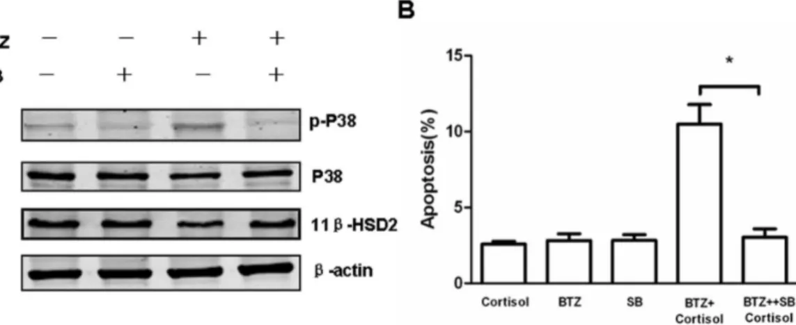
Bortezomib Decreases 11b-HSD2 Expression through p38 MAPK Signaling Pathway
It has been reported that the activation of p38 MAPK enhances GC sensitivity in GC-resistant T lymphoblastic cells [16]. Bortezomib has been demonstrated to exert numerous biological effects that include activating p38 MAPK [19]. Therefore, we hypothesize if bortezomib regulates 11b-HSD2 expression through p38 MAPK signaling pathway. Jurkat cells were treated with bortezomib in the presence or absence of SB203580, a selective p38 inhibitor, to examine 11b-HSD2 expression. Immunoblotting showed low-dose bortezomib resulted in phos-phorylation of p38 MAPK while decreasing 11b-HSD2 (Fig. 6A). Furthermore, co-treatment of SB203580 reversed bortezomib-induced inhibition of 11b-HSD2 (Fig. 6A), indicating p38 MAPK may play an important role in decreasing 11b-HSD2 by bortezomib. As expected, we further demonstrated p38 inhibitor SB203580 reversed cortisol-induced apoptosis in Jurkat cell pretreated with bortezomib (Fig. 6B).
Figure 2. Jurkat and Molt4 cells express high expression of 11b-HSD2 and fail to auto-induce GRaafter cortisol treatment.(A) 11b -HSD2 mRNA expression of each sample was detected using RT-PCR. These samples include (from left to right): Placenta, a 11b-HSD2 positive control; Jurkat, a T-ALL cell line; Molt4, a T-ALL cell line; PBT, peripheral blood T cells from healthy volunteers; Daudi, a B-NHL cell line; Raji, a B-NHL cell line. (B) Representative western blots showed 11b-HSD2 protein expression in each sample. 50mg protein extracts from each sample were probed with 11b -HSD2 antibody. The expression levels ofb-actin in the lysates served as the loading control. An immunoreactive protein of,42 kDa was found in Jurkat and Molt4 cells, but not in PBT, Daudi and Raji cells. (C) Jurkat, Molt4, Daudi and Raji cells were treated with 1mM cortisol for 24 hours. GRa levels of cortisol-treated cells relative to those of untreated cells (control) in each sample were analyzed by quantitative real-time PCR. Data are shown as mean6SD of triplicate samples. *p,0.05vs.untreated cells. There was no induction of GRaafter cortisol treatment in Jurkat and Molt4 cells in
Discussion
GC action is reported to be dependent on 11 beta-HSD activities contained in tissues, the amount of GC receptors in target cells as well as the concentration of circulating GC [20]. Many studies owe GC resistance to the GR within unresponsive cells. A deficient up-regulation of the GR and its downstream target, induced leucine zipper (GILZ) was found in GC-resistant subclones generated from a GC-sensitive cell line, which was attributed to mutational events [3]. Conversely, Beesley et al. examined 15 T-ALL cell lines and showed that GR resistance could not be explained by mutations in GR or variations in levels of its expression [4]. Additionally, the GR protein expression showed no difference between GR-responsive and GR-resistant patients in a case-control study [5].
Our data showed the basal level of GRa mRNA was lower in Jurkat and Molt4 cells compared to Daudi and Raji cells. Treatment with cortisol resulted in no auto-induction of GRain Jurkat and Molt4 cells thus giving an approximately 8-fold difference in GRa mRNA between Jurkat and Daudi cells.
However, even a 10-fold higher dose of cortisol could not induce apoptosis in Jurkat cells to the levels achieved in Daudi cells, suggesting other mechanisms apart from the low numbers of GR were involved in GC resistance. Jurkat cells have been demon-strated to express one wild-type and one mutated (R477H) GR allele. However, GC-sensitive cells transduced with the same mutated (R477H) GR underwent GC-induced apoptosis to the same degree and with similar kinetics as those transduced with wide type GR, thus indicating the mutation alone did not sufficiently account for the GC-resistant phenotype of Jurkat cells [21]. Here we have shown that high levels of 11b-HSD2 are present in Jurkat cells and inhibition of 11b-HSD2 by 18b-GA sensitizes Jurkat cells to GC. Similar results were also observed in Molt4 cells consistent with findings from Sai et al [2]. We further observed that 11b-HSD2 knockdown by specific shRNA sensitized Jurkat cells to cortisol-induced apoptosis, confirming 11b-HSD2 contribution to GC resistance in Jurkat cells. However, we demonstrated there was no 11b-HSD2 expression in normal peripheral blood T cells, in accord with pervious findings of 11b -Figure 3. Inhibition of 11b-HSD2 activity enhances susceptibility to cortisol-induced apoptosis.(A) Jurkat and Molt4 cells were treated with 11b-HSD inhibitor 18b-GA (5mM) plus 10mg/ml cortisol in the presence or absence of GC receptor antagonist RU486 (1mM) for 48 hours. Cells
were then analyzed for apoptosis by flow cytometry using Annexin V/PI binding. Data are shown as mean6SD of 3 independent experiments. * indicates p,0.05. (B) Immunoblot analysis of Jurkat cells with different treatments as indicated was performed using anti–caspase-8, caspase-9, caspase-3 and PARP antibodies. The expression levels ofb-actin in the lysates served as the loading control. (C) Western blots were used to examine 11b-HSD2 expression in 11b-HSD2 knockdown Jurkat cells. Wild-type (WT) and nonsense scramble shRNA (SCR)-transfected cells were used as controls. (D) WT, SCR-transfected and 11b-HSD2 knockdown Jurkat cells were treated with 10mg/ml cortisol for 48 hours. Cells were then analyzed for
apoptosis by flow cytometry using Annexin V/PI binding. Data are shown as mean6SD of 3 independent experiments. * p,0.05vs.control cells.
Figure 4. Pretreatment with bortezomib sensitizes Jurkat cells to cortisol-induced apoptosis and G1 cell cycle arrest.(A) Jurkat cells were pretreated with indicated concentrations of bortezomib (2.5–10 nM) for 24 hours. Cells were collected and washed once with medium. Afterwards, cells were treated with 10mg/ml cortisol for 48 hours. Cells were then analyzed for apoptosis by flow cytometry using Annexin V/PI
binding. Data are shown as mean6SD of triplicate samples. *p,0.05vs.the cells treated with bortezomib alone. (B) Jurkat cells were pretreated with
5 nM bortezomib for 24 hours. Cells were collected and washed once with medium. Afterwards, cells were treated with indicated concentrations of cortisol for 48 hours. Cells were then analyzed for apoptosis by flow cytometry using Annexin V/PI binding. Data are shown as mean6SD of triplicate samples. *p,0.05vs.cells without bortezomib pretreatment. (C) Jurkat cells were pretreated with 5 nM bortezomib (BTZ) for 24 hours. The cells were
collected and washed once with medium. Afterwards, cells were treated with 10mg/ml cortisol for 48 hours and analyzed for cell cycle by flow
cytometry. (D) Graphical representation of the data displayed in panel C as mean6SD of 3 independent experiments. * in G2 phase indicates p,0.05
vs.control cells. * in G1 phase indicates p,0.05vs.cells treated with cortisol alone. (E) Jurkat cells were pretreated with 5 nM bortezomib for 24
hours. The cells were collected and washed once with medium. Afterwards, cells were treated with 10mg/ml cortisol for 48 hours and analyzed for
HSD2 absent in normal tissues but present in malignant counterparts [10,22]. Moreover, Rabbitt et al. reported that cells transfected with cDNA encoding 11b-HSD2 significantly in-creased cell proliferation [23], which may explain the possible role 11b-HSD2 played in the formation of T-lymphoblastic leukemias. Although no 11b-HSD2 expression was found in B-NHL Daudi and Raji cells, maybe it is because T cells developed in a way different from B cells. As is known, T cells undergo positive and negative selection in thymus before they mature and transfer to the periphery. Specifically, it is thought that negative selection induces the death of thymocytes that bind strongly to self-peptide [24]. It has long been known that the apoptosis in thymocytes could be induced by GC, whose levels are able to be controlled within lymphoid organs through oxidative inactivation by 11b-HSD2 [20]. It is possible that the presence of 11b-HSD2 makes these T-lymphoblastic leukemic cells escape negative selection resulted from GC-induced apoptosis, leaving them to survive and finally transform into lymphoblastic cells. It has been shown that caspase-8, -9 and -3 activity is essential for GC-induced apoptosis in thymocytes as well as T-ALL cells [25]. Consistently, we observed processing of caspase-8,-9 and -3 and cleaved PARP in cortisol-treated Jurkat cells after 11b-HSD2 inhibition, indicating that
both death receptor–dependent (caspase 8) and –independent (caspase 9) apoptotic pathways were involved.
Bortezomib, a new selective proteasome inhibitor drug, not only directly inducesapoptosis in cancer cells, but also sensitizes these cells to chemotherapy [13,26,27]. In our study, we demonstrated that bortezomib also possessed the ability to enhance cell sensitivity to cortisol even at a non-cytotoxic low dose (5 nM). The improved sensitivity to cortisol by bortezomib pretreatment could be confirmed in 3 aspects. First, elevated Annexin V-binding indicated elevated cellular apoptosis in cortisol-treated Jurkat cells following bortezomib pretreatment. Second, cortisol arrested increased cells at G1 stage after bortezomib intervention, which is characteristic of enhanced cortisol effects considering cells were arrested at G2 stage with bortezomib treatment alone. Third, GILZ has a distinguished reputation as a critical mediator of GC effects and also participates in the regulation of cell apoptosis by GC [28]. Therefore, up-regulated GILZ mRNA is another solid evidence for the definite effects of bortezomib on sensitizing Jurkat cells to cortisol action. Although the same effects need to be confirmed by further studies in animal models which we plan to conduct in the near further, the in vitro results strongly indicate the synergistic advantage of combining proteasome inhibitor with cortisol clinically.
Figure 5. Bortezomib dose-dependently inhibits 11b-HSD2 mRNA and protein expression as well as enzyme activity.(A) Jurkat cells were exposed to the indicated concentrations of bortezomib for 12 hours and 11b-HSD2 mRNA levels were measured by quantitative real-time PCR and normalized tob-actin levels. Data are shown as mean6SD of triplicate samples. *p,0.05vs.untreated cells. (B) Jurkat cells were exposed to the
indicated concentrations of bortezomib for 24 hours and 11b-HSD2 protein levels were measured by immunoblot analysis using anti–11b-HSD2 antibody. The expression levels ofb-actin in the lysates served as the loading control. One representative experiment is presented. (C) Jurkat cells were exposed to the indicated concentrations of bortezomib for 24 hours and 11b-HSD2 enzyme activities were determined by measuring production of cortisone from 100 nM cortisol added to the medium for 24 hours. The 11b-HSD2 activity was expressed as a percentage: pmol cortisone produced per 106cells of the treated cells/pmol cortisone produced per 106cells of the untreated cells. Data are shown as mean6SD of triplicate samples. *p,0.05vs.untreated cells.
The mechanism by which bortezomib affects cell sensitivity to cortisol is elusive. It is well known that most GC actions are mediated by GR. Methotrexate has been reported to be a steroid sparing agent by means of increasing the expression of the active receptor (GRa) and/or decreasing the expression of the dominant negative receptor (GRb) [29]. Whereas, we observed neither the number of GR nor the ratio of GRa to GRb was changed in Jurkat cells after 5 nM bortezomib treatment (data not shown). Taking into account the potential role 11b-HSD2 plays in GC resistance, further we testified whether bortezomib had any effects on 11b-HSD2, a cortisol-inactivating enzyme in Jurkat cells. Interestingly, bortezomib decreased 11b-HSD2 mRNA and protein expression as well as enzyme activity in a dose-dependent manner, which could explain its effects in improving GC susceptibility. Further, the ability of low dose (5 nM) bortezomib to inhibit 11b-HSD2 activity and enhance GC sensitivity prompted us to clarify the intracellular mechanism. It has been demonstrated that bortezomib exerts numerous biological effects through NF-kB, AKT or MAPKs signal pathways [30,31]. In this study, we observed 5 nM bortezomib did not have any direct cytotoxic effects and NF-kB inhibitory activity (data not shown), which was in line with findings from Kashkar el al. [13]. Numerous reports have indicated that 11b-HSD2 can be regulated by MAPKs signalling [32,33]. Furthermore, MAPKs have been proven to modify GC sensitivity, namely that the activation of extracellular signal-regulated protein kinase (ERK) and p38 MAPK reduce and enhance GC sensitivity, respectively [16]. We showed that blockade of p38 MAPK using SB203580, a selective p38 inhibitor abrogated the effects of bortezomib on inhibiting 11b-HSD2 and failed to sensitize cell to cortisol-induced apoptosis, implying p38 MAPK pathway activation could be an excellent potential therapeutic strategy in overcoming GC resistance.
Although it has been reported that non-metabolisable GC dexamethasone is poorly inactivated by 11b-HSD2 [2], possibly accounting for why dexamethasone is widely applied in clinical fields, cortisol is still superior to dexamethasone in some respects. For example, cortisol has a shorter half-life of 8 hours compared to dexamethasone of 54 hours, which can reduce the adverse effects and complications of GC such as immunodeficiency, hyperglyce-mia and steroid-induced osteoporosis [34]. In addition, GCs also represent the mainstay strategy for diseases caused by an overactive immune system, such as allergies, asthma, autoimmune diseases and sepsis, which are suitable for being treated by short-acting cortisol.
Taken together, here we demonstrate at least two mechanisms responsible for GC resistance in leukemia T cells: failure of GR auto-induction and presence of cortisol-inactivating 11b-HSD2 enzyme. We also show that upon bortezomib pretreatment, Jurkat cells exhibit an increased sensitivity to cortisol, suggesting proteasome inhibitors could be potential GC sparing agents, specifically for diseases with GC resistance resulted from 11b -HSD2 presence.
Acknowledgments
All authors would like to thank Weiwei Zhang from Second Military Medical University for technical assistance.
Author Contributions
Conceived and designed the experiments: YT LG XW FZ JS. Performed the experiments: YT LG HW GY. Analyzed the data: YT LG. Contributed reagents/materials/analysis tools: XW FZ JS. Wrote the paper: YT LG JS.
References
1. Rabbitt EH, Gittoes NJ, Stewart PM, Hewison M (2003) 11beta-hydroxysteroid dehydrogenases, cell proliferation and malignancy. J Steroid Biochem Mol Biol 85: 415–421.
2. Sai S, Nakagawa Y, Yamaguchi R, Suzuki M, Sakaguchi K, et al. (2011) Expression of 11beta-hydroxysteroid dehydrogenase 2 contributes to glucocor-ticoid resistance in lymphoblastic leukemia cells. Leuk Res 35: 1644–1648. Figure 6. The effects of the p38 MAPK inhibitor on increased sensitivity to cortisol in Jurkat cells pretreated with bortezomib.(A) Jurkat cells were pretreated with 5 nM bortezomib (BTZ) in the presence or absence of 10mM SB203580 (SB), a selective p38 inhibitor for 24 hours.
Immunoblot analysis of Jurkat cells after different treatments as indicated in the figure was probed with p-p38 MAPK, p38 MAPK and anti-11b-HSD2, to determine the activation of p38 MAPK and the expression of 11b-HSD2. The expression levels ofb-actin in the lysates served as the loading control. (B) Jurkat cells were pretreated with 5 nM bortezomib (BTZ) in the presence or absence of 10mM SB203580 (SB) for 24 hours. Cells
were collected and washed once with medium. Afterwards, cells were treated with 10mg/ml cortisol for 48 hours. Cells were then analyzed for
apoptosis by flow cytometry using Annexin V/PI binding. Data are shown as mean6SD of triplicate samples. *p,0.05vs. cortisol-treated cells
3. Schmidt S, Irving JA, Minto L, Matheson E, Nicholson L, et al. (2006) Glucocorticoid resistance in two key models of acute lymphoblastic leukemia occurs at the level of the glucocorticoid receptor. Faseb J 20: 2600–2602. 4. Beesley AH, Weller RE, Senanayake S, Welch M, Kees UR (2009) Receptor
mutation is not a common mechanism of naturally occurring glucocorticoid resistance in leukaemia cell lines. Leuk Res 33: 321–325.
5. Lauten M, Cario G, Asgedom G, Welte K, Schrappe M (2003) Protein expression of the glucocorticoid receptor in childhood acute lymphoblastic leukemia. Haematologica 88: 1253–1258.
6. Stewart PM, Krozowski ZS (1999) 11 beta-Hydroxysteroid dehydrogenase. Vitam Horm 57: 249–324.
7. Koyama K, Myles K, Smith R, Krozowski Z (2001) Expression of the 11beta-hydroxysteroid dehydrogenase type II enzyme in breast tumors and modulation of activity and cell growth in PMC42 cells. J Steroid Biochem Mol Biol 76: 153– 159.
8. Pacha J, Lisa V, Miksik I (2002) Effect of cellular differentiation on 11beta-hydroxysteroid dehydrogenase activity in the intestine. Steroids 67: 119–126. 9. Rabbitt EH, Ayuk J, Boelaert K, Sheppard MC, Hewison M, et al. (2003)
Abnormal expression of 11 beta-hydroxysteroid dehydrogenase type 2 in human pituitary adenomas: a prereceptor determinant of pituitary cell proliferation. Oncogene 22: 1663–1667.
10. Eyre LJ, Rabbitt EH, Bland R, Hughes SV, Cooper MS, et al. (2001) Expression of 11 beta-hydroxysteroid dehydrogenase in rat osteoblastic cells: pre-receptor regulation of glucocorticoid responses in bone. J Cell Biochem 81: 453–462. 11. Nigawara T, Iwasaki Y, Asai M, Yoshida M, Kambayashi M, et al. (2006)
Inhibition of 11beta-hydroxysteroid dehydrogenase eliminates impaired gluco-corticoid suppression and induces apoptosis in corticotroph tumor cells. Endocrinology 147: 769–772.
12. Richardson PG, Barlogie B, Berenson J, Singhal S, Jagannath S, et al. (2003) A phase 2 study of bortezomib in relapsed, refractory myeloma. N Engl J Med 348: 2609–2617.
13. Kashkar H, Deggerich A, Seeger JM, Yazdanpanah B, Wiegmann K, et al. (2007) NF-kappaB-independent down-regulation of XIAP by bortezomib sensitizes HL B cells against cytotoxic drugs. Blood 109: 3982–3988. 14. Kleinschnitz C, Blecharz K, Kahles T, Schwarz T, Kraft P, et al. (2011)
Glucocorticoid insensitivity at the hypoxic blood-brain barrier can be reversed by inhibition of the proteasome. Stroke 42: 1081–1089.
15. Fiskus W, Verstovsek S, Manshouri T, Rao R, Balusu R, et al. (2011) Heat shock protein 90 inhibitor is synergistic with JAK2 inhibitor and overcomes resistance to JAK2-TKI in human myeloproliferative neoplasm cells. Clin Cancer Res 17: 7347–7358.
16. Tanaka T, Okabe T, Gondo S, Fukuda M, Yamamoto M, et al. (2006) Modification of glucocorticoid sensitivity by MAP kinase signaling pathways in glucocorticoid-induced T-cell apoptosis. Exp Hematol 34: 1542–1552. 17. Lu S, Yang J, Song X, Gong S, Zhou H, et al. (2008) Point mutation of the
proteasome beta5 subunit gene is an important mechanism of bortezomib resistance in bortezomib-selected variants of Jurkat T cell lymphoblastic lymphoma/leukemia line. J Pharmacol Exp Ther 326: 423–431.
18. Ayroldi E, Riccardi C (2009) Glucocorticoid-induced leucine zipper (GILZ): a new important mediator of glucocorticoid action. Faseb J.
19. Lioni M, Noma K, Snyder A, Klein-Szanto A, Diehl JA, et al. (2008) Bortezomib induces apoptosis in esophageal squamous cell carcinoma cells through activation of the p38 mitogen-activated protein kinase pathway. Mol Cancer Ther 7: 2866–2875.
20. Hennebold JD, Ryu SY, Mu HH, Galbraith A, Daynes RA (1996) 11 beta-hydroxysteroid dehydrogenase modulation of glucocorticoid activities in lymphoid organs. Am J Physiol (43) 270: R1296–1306.
21. Riml S, Schmidt S, Ausserlechner MJ, Geley S, Kofler R (2004) Glucocorticoid receptor heterozygosity combined with lack of receptor auto-induction causes glucocorticoid resistance in Jurkat acute lymphoblastic leukemia cells. Cell Death Differ 11 Suppl 1: S65–72.
22. Korbonits M, Bujalska I, Shimojo M, Nobes J, Jordan S, et al. (2001) Expression of 11 beta-hydroxysteroid dehydrogenase isoenzymes in the human pituitary: induction of the type 2 enzyme in corticotropinomas and other pituitary tumors. J Clin Endocrinol Metab 86: 2728–2733.
23. Rabbitt EH, Lavery GG, Walker EA, Cooper MS, Stewart PM, et al. (2002) Prereceptor regulation of glucocorticoid action by 11beta-hydroxysteroid dehydrogenase: a novel determinant of cell proliferation. Faseb J 16: 36–44. 24. Davey GM, Schober SL, Endrizzi BT, Dutcher AK, Jameson SC, et al. (1998)
Preselection thymocytes are more sensitive to T cell receptor stimulation than mature T cells. J Exp Med 188: 1867–1874.
25. Wang D, Muller N, McPherson KG, Reichardt HM (2006) Glucocorticoids engage different signal transduction pathways to induce apoptosis in thymocytes and mature T cells. J Immunol 176: 1695–1702.
26. Dasmahapatra G, Nguyen TK, Dent P, Grant S (2006) Adaphostin and bortezomib induce oxidative injury and apoptosis in imatinib mesylate-resistant hematopoietic cells expressing mutant forms of Bcr/Abl. Leuk Res 30: 1263– 1272.
27. Fribley AM, Evenchik B, Zeng Q, Park BK, Guan JY, et al. (2006) Proteasome inhibitor PS-341 induces apoptosis in cisplatin-resistant squamous cell carcinoma cells by induction of Noxa. J Biol Chem 281: 31440–31447. 28. Delfino DV, Agostini M, Spinicelli S, Vito P, Riccardi C (2004) Decrease of
Bcl-xL and augmentation of thymocyte apoptosis in GILZ overexpressing transgenic mice. Blood 104: 4134–4141.
29. Goecke IA, Alvarez C, Henriquez J, Salas K, Molina ML, et al. (2007) Methotrexate regulates the expression of glucocorticoid receptor alpha and beta isoforms in normal human peripheral mononuclear cells and human lymphocyte cell lines in vitro. Mol Immunol (4768) 44: 2115–2123.
30. Yang F, Jove V, Chang S, Hedvat M, Liu L, et al. (2012) Bortezomib induces apoptosis and growth suppression in human medulloblastoma cells, associated with inhibition of AKT and NF-kB signaling, and synergizes with an ERK inhibitor. Cancer Biol Ther 13: 349–357.
31. Zhou X, Fang Y, Jing H, Zhong L, Luo P, et al. (2011) Involvement of mitogen-activated protein kinase in signal transducer and activator of transcription-1 mediated differentiation induced by bortezomib in acute myeloid leukemia cells. Mol Carcinog.
32. Shang Y, Yang X, Zhang R, Zou H, Zhao R (2012) Low amino acids affect expression of 11beta-HSD2 in BeWo cells through leptin-activated JAK-STAT and MAPK pathways. Amino Acids 42: 1879–1887.
33. Sharma A, Guan H, Yang K (2009) The p38 mitogen-activated protein kinase regulates 11beta-hydroxysteroid dehydrogenase type 2 (11beta-HSD2) expres-sion in human trophoblast cells through modulation of 11beta-HSD2 messenger ribonucleic acid stability. Endocrinology 150: 4278–4286.