Therapeutically Relevant Drug Targets in FGFR and EGFR
Pathways in Sporadic Intrahepatic Cholangiocarcinoma
Mitesh J. Borad1,2,3.*, Mia D. Champion3,4., Jan B. Egan3., Winnie S. Liang5
, Rafael Fonseca1,2,3, Alan H. Bryce1,2,3, Ann E. McCullough6, Michael T. Barrett2,5, Katherine Hunt1, Maitray D. Patel7, Scott W. Young7, Joseph M. Collins7, Alvin C. Silva7, Rachel M. Condjella2, Matthew Block3,8, Robert R. McWilliams3,8, Konstantinos N. Lazaridis3, Eric W. Klee3,4, Keith C. Bible8, Pamela Harris9, Gavin R. Oliver3,4, Jaysheel D. Bhavsar3,4, Asha A. Nair3,4, Sumit Middha3,4, Yan Asmann3,4,
Jean-Pierre Kocher3,4, Kimberly Schahl3, Benjamin R. Kipp10, Emily G. Barr Fritcher10, Angela Baker5, Jessica Aldrich5, Ahmet Kurdoglu5, Tyler Izatt5, Alexis Christoforides5, Irene Cherni5, Sara Nasser5, Rebecca Reiman5, Lori Phillips5, Jackie McDonald5, Jonathan Adkins5, Stephen D. Mastrian5,
Pamela Placek5, Aprill T. Watanabe5, Janine LoBello5, Haiyong Han5, Daniel Von Hoff2,5, David W. Craig5", A. Keith Stewart1,2,3, John D. Carpten5"*
1Division of Hematology/Oncology Mayo Clinic, Scottsdale, Arizona, United States of America,2Mayo Clinic Cancer Center, Scottsdale, Arizona, United States of America, 3Center for Individualized Medicine, Mayo Clinic, Rochester, Minnesota, United States of America,4Department of Biomedical Statistics and Informatics, Mayo Clinic, Scottsdale, Arizona, United States of America,5Translational Genomics Research Institute, Phoenix, Arizona, United States of America,6Department of Pathology, Mayo Clinic, Scottsdale, Arizona, United States of America,7Department of Radiology, Mayo Clinic, Scottsdale, Arizona, United States of America,8Mayo Clinic Cancer Center, Rochester, Minnesota, United States of America,9Investigational Drug Branch, National Cancer Institute, Rockville, Maryland, United States of America,10Department of Laboratory Medicine and Pathology, Mayo Clinic, Rochester, Minnesota, United States of America
Abstract
Advanced cholangiocarcinoma continues to harbor a difficult prognosis and therapeutic options have been limited. During the course of a clinical trial of whole genomic sequencing seeking druggable targets, we examined six patients with advanced cholangiocarcinoma. Integrated genome-wide and whole transcriptome sequence analyses were performed on tumors from six patients with advanced, sporadic intrahepatic cholangiocarcinoma (SIC) to identify potential therapeutically actionable events. Among the somatic events captured in our analysis, we uncovered two novel therapeutically relevant genomic contexts that when acted upon, resulted in preliminary evidence of anti-tumor activity. Genome-wide structural analysis of sequence data revealed recurrent translocation events involving theFGFR2locus in three of six assessed patients. These observations and supporting evidence triggered the use of FGFR inhibitors in these patients. In one example, preliminary anti-tumor activity of pazopanib (in vitro FGFR2 IC50<350 nM) was noted in a patient with anFGFR2-TACC3 fusion. After progression on pazopanib, the same patient also had stable disease on ponatinib, a pan-FGFR inhibitor (in vitro, FGFR2 IC50<8 nM). In an independent non-FGFR2 translocation patient, exome and transcriptome analysis revealed an allele specific somatic nonsense mutation (E384X) inERRFI1, a direct negative regulator ofEGFRactivation. Rapid and robust disease regression was noted in thisERRFI1inactivated tumor when treated with erlotinib, an EGFR kinase inhibitor.FGFR2 fusions andERRFImutations may represent novel targets in sporadic intrahepatic cholangiocarcinoma and trials should be characterized in larger cohorts of patients with these aberrations.
Citation:Borad MJ, Champion MD, Egan JB, Liang WS, Fonseca R, et al. (2014) Integrated Genomic Characterization Reveals Novel, Therapeutically Relevant Drug Targets in FGFR and EGFR Pathways in Sporadic Intrahepatic Cholangiocarcinoma. PLoS Genet 10(2): e1004135. doi:10.1371/journal.pgen.1004135
Editor:Marshall S. Horwitz, University of Washington, United States of America ReceivedJuly 26, 2013;AcceptedDecember 6, 2013;PublishedFebruary 13, 2014
This is an open-access article, free of all copyright, and may be freely reproduced, distributed, transmitted, modified, built upon, or otherwise used by anyone for any lawful purpose. The work is made available under the Creative Commons CC0 public domain dedication.
Funding:This study was supported by the NIH Grant Number K12 CA90628 and the Center for Individualized Medicine. The funders had no role in study design, data collection and analysis, decision to publish or preparation of the manuscript.
Competing Interests:Dr. Kipp received grant funding from Abbott Molecular Inc, the suppliers of the FGFR2 FISH probe used in this study. The other authors have no conflicts to declare.
* E-mail: borad.mitesh@mayo.edu (MJB); jcarpten@tgen.org (JDC)
.These authors contributed equally to this work.
"DWC and JDC also contributed equally.
Introduction
Biliary tract cancers (BTC) comprise malignant tumors of the intrahepatic and extrahepatic bile ducts. Known risk factors for
B and hepatitis C [9–14], obesity [13], hepatolithiasis [15,16] and thorotrast contrast exposure [17,18]. Surgical approaches such as resection and liver transplantation represent the only curative treatment approaches for BTC [19]. Unfortunately, most patients present with surgically unresectable and/or metastatic disease at diagnosis. Systemic therapy with gemcitabine and cisplatin has been established as the standard of care for patients with advanced disease, but is only palliative [20], emphasizing the imminent need for novel therapies.
Multiple studies have reported the presence of mutations/allelic loss of known cancer genes in BTC [21–39] and recently, a prevalence set of 46 patients was used to validate 15 of these genes including:TP53, KRAS, CDKN2Aand SMAD4as well asMLL3, ROBO2, RNF43, GNAS, PEG3, XIRP2, PTEN, RADIL, NCD80, LAMA2 and PCDHA13. Recent studies have also identified recurrent mutations inIDH1(codon 132) andIDH2(codons 140 and 172) with a prevalence of 22–23% associated with clear cell/ poorly differentiated histology and intrahepatic primary [40,41]. Fusions with oncogenic potential involving the kinase geneROS1 have been identified in patients with BTC with a prevalence of 8.7% in a recent study [42]. Less frequently, mutations in sporadic BTC have been reported in EGFR [43,44], BRAF [45], NRAS [40,46],PIK3CA[40,46,47],APC[40],CTNNB1[40],AKT1[40], PTEN[40],ABCB4[48],ABCB11[49,50], andCDH1[51] as well as amplifications inERRB2[52].
Recently, two independent studies reported the presence of FGFR fusions in cholangiocarcinoma; a single case with FGFR2-AHCYL1 [53] as well as several cases identifying FGFR2-BICC1 fusions [53,54]. Arai et al. evaluated the presence ofFGFR2fusions in a cohort of 102 cholangiocarcinoma patients observing that the fusions occurred exclusively in the intrahepatic cases with a prevalence of 13.6% [53]. Due to the presence of known dimerization motifs in the fusion partners, Wu et al. conducted mechanistic studies that demonstrated the in vitro interaction of FGFR2-BICC1 and other fusions that was not observed in the presence of wildtypeFGFR2[54]. Furthermore, overexpression of theFGFR2-BICC1and other selected fusions resulted in altered cell
morphology and increased cell proliferation [54]. These data led to the conclusion that the fusion partners are facilitating oligomeriza-tion, resulting in FGFR kinase activation in tumors possessing FGFR fusions. In addition,in vitro and in vivo assessment of the sensitivity of cell lines containing an FGFR2 fusion to an FGFR inhibitor demonstrated sensitivity to treatment only in the fusion containing cells [53,54], suggesting the presence of FGFR fusions may be a useful predictor of tumor response to FGFR inhibitors.
To comprehensively explore the genetic basis of sporadic intrahepatic cholangiocarcinoma (SIC), with emphasis on eluci-dation of therapeutically relevant targets, we performed integrated whole genome and whole transcriptome analyses on tumors from 6 patients with advanced, sporadic intrahepatic cholangiocarci-noma (SIC). Notably, recurrent fusions involving the oncogene FGFR2(n = 3) were identified. A patient whose tumor presented with an FGFR2-MGEA5 fusion has demonstrated preliminary evidence of anti-tumor activity manifest as stable disease accompanied by CA19-9 reduction and tumor necrosis to ponatinib, a pan-FGFR inhibitor (in vitroFGFR1 IC50<24 nM,
FGFR2 IC50<8 nM, FGFR3 IC50<8 nM and FGFR4
IC50<34 nM). In another patient whose tumor possessed an FGFR2-TACC3fusion, preliminary anti-tumor activity of pazopa-nib (in vitro FGFR2 IC50<350 nM) was also noted. After
progression on pazopanib, the same patient also responded to ponatinib and again demonstrated tumor shrinkage. Additionally, a non-FGFR fusion patient was found to have allele-specific preferential expression of a loss of function mutation inERRFI1, a direct negative regulator of EGFR activation. Similarly, rapid and robust disease regression was noted in the patient with anERRFI1 mutant tumor when treated with erlotinib, an EGFR kinase inhibitor. Results suggest that these novel targets in the EGFR and FGFR pathways may be therapeutically relevant in patients with sporadic cholangiocarcinoma.
Results
Genomic landscape
We identified 327 somatic coding mutations, with an average of 55 mutations/tumor (range 34–112), within our cohort (Table 1, Figure 1). Nonsynonymous single nucleotide variations were the predominant class in all of the patients. Patients 1 and 2 accumulated a high number of synonymous mutations in comparison to the other patients. Patient 5 carried the most stops gained likely contributing to a higher number of pseudogenes in comparison to the others and was also the only patient to carry several predicted high impact mutations affecting splice site acceptor regions (Figure 1, light green, percentage ,5%). In addition, patient 6 also carried a codon change plus insertion variation. Sequencing statistics are provided inTable 2. Genes with mutations in more than one case included CSPG4 (n = 2), GRIN3A(n = 2) andPLXBN3(n = 2) (Table S1); with half of these predicted to be potentially damaging by SIFT [55], Polyphen [56], Mutation Assessor [57] and Mutation Taster [58]. While there was overlap in the somatic landscape of SIC with liver-fluke associated cholangiocarcinoma, hepatocellular cancer and pancreatic cancer, most of the aberrations detected in our study were distinct (Table 3). More importantly, using previously published methods [59], we identified molecular fusions involvingFGFR2that were felt to be therapeutically relevant in 3 patients. Additionally, these fusions were validated with a break apart Fluorescent In situ Hybridization (FISH) assay (Figure 2). Notably, the patients who did not harbor theFGFR2 fusions were negative using the same assay. Two of the three patients withFGFR2 fusions (Patients 4 and 6) were treated with FGFR inhibitors while the third patient Author Summary
(Patient 5), experienced clinical decline prior to the availability of results and as such did not receive any further therapy. Furthermore, overexpression of an SNV in ERRFI1(E384X), a negative regulator of EGFR, was detected in a non-FGFR2 translocation patient’s tumor. Taken together, our results consti-tute important therapeutically actionable alterations in patients with advanced SIC (Text S1).
Pathway analysis
Comparative pathway analysis of genes carrying small scale nucleotide variations (SsNVs) has implicated several major pathways, possibly interacting as a network, that are predicted to underlie disease in all of our studied biliary carcinoma patients. These shared pathways include EGFR, EPHB, PDGFR-beta, Netrin-mediated and Beta1 integrin mediated signaling pathways (Figure 3 and Tables S2 and S3). Interestingly, most of these pathways have known roles in mediating epithelial-to-mesenchy-mal cell transitions, which occur frequently during development as well as tumorigenesis. Cell growth and motility is inherent to the
successful progression of both biological processes. Studies of the nervous system and lung development have shown that Netrins act to inhibit FGF7 and FGF10 mediated growth or cell guidance [60]. In addition, Netrin-1 has a known role in mediating cell migration during pancreatic organogenesis [60]. Furthermore, Netrin-1 acts as a ligand fora3b1 anda6b4 integrins, both of which are involved in supporting adhesion of developing pancreatic epithelial cells with Netrin-1 although it is thought that a6b4 plays the principle role during this process [60]. Interestingly,a3b1 has been hypothesized to play a role during the process of angiogenesis, when chemoattractants and chemorepel-lents act to guide filopodia during migration [60]. The a3b1 integrin receptor may act together with additional pathways proposed to play a role during angiogenesis such as VEGF, PDGFR-beta [61], and EphrinB [62] as well as tumorigenesis [60]. Patients 3 and 4 also shared several genes acting in cadherin signaling pathways (Tables S3, S4), which are important for maintaining cell-cell adhesion and are known to be intimately integrated with EGFR and FGFR signaling pathways [63].
Table 1.Summary of mutation type by patient.
Patient 1 Patient 2 Patient 3 Patient 4 Patient 5 Patient 6
Nonsynonymous coding 20 30 31 44 101 34
Synonymous coding 13 12 0 0 0 0
Insertions/deletions 1 4 0 6 0 2
Stop gained 0 3 3 2 6 2
Start gained 0 1 0 0 0 0
Codon insertion 0 1 0 0 0 1
Codon deletion 0 0 0 0 0 1
Splice site donor 0 0 1 0 1 2
Splice site acceptor 0 0 0 0 4 0
Total 34 51 35 52 112 42
doi:10.1371/journal.pgen.1004135.t001
Figure 1. Sequence variation effects.Functional effects of high confidence sequence variations for all of the patients were identified as
described in the Methods. The abundance of variations in each functional category is provided as percentages relative to the total number of high confidence variations and raw counts are provided inTable 1. For categories where the percentage was less than 5%, values are not shown. Summaries by individual patients are shown as follows: A) Patient 1, B) Patient 2, C) Patient 3,D) Patient 4, E) Patient 5, andF) Patient 6. Nonsynonymous single nucleotide variations were the predominant class in all of the patients. Two patients, Patients 1 and 2 also accumulated a high number of synonymous mutations in comparison to the other patients; Patient 5 carries the most stops gained likely contributing to a higher number of pseudogenes in comparison to the others; Patient 5 was also the only patient to carry several predicted high impact mutations that affect the splice site acceptor regions (light green, percentage,5%). In addition to the major functional classes summarized, Patient 6 also carried a codon change plus insertion variation.
Exome Whole Genome RNA Seq
Patient Tissue
Aligned Reads (Millions)
Mean Target Coverage
% Target Bases 106
#of Functional Coding Variants
Aligned Reads (Millions)
Aligned Bases (Billions)
Physical Coverage
Aligned Reads (Millions)
Aligned Bases (Billions)
1 N 161 100 94% - 266 22 37 -
-T 156 112 94% 21 228 18 35 100 8.1
2 N 176 74 94% - 179 14 5 -
-T 202 81 94% 34 370 30 10 341 26
3 N 226 110 58% - 296 24 50 163 13
T 195 92 58% 52 321 26 50 101 8.1
4 N 167 80 95% - 317 26 42 -
-T 202 93 96% 52 163 13 12 264 20
5 N 257 146 96% - 335 27 51 -
-T 133 78 93% 250 349 28 39 401 31
6 N 350 243 92% - - -
-T 340 245 92% 43 - - - 713 31
Liver Control - - - 118 9.6
N = Normal, T = Tumor.
doi:10.1371/journal.pgen.1004135.t002
Novel
FGFR
and
EGFR
Targets
in
Cholangiocar
cinoma
Genetics
|
www.plosgen
etics.org
4
February
2014
|
Volume
10
|
Issue
2
|
In addition to the variations identified in genes acting in EGFR and/or FGFR signaling pathways, we also report multiple sSNVs and copy number variations (CNVs) (Figure 4) in genes such as HDAC1,TP53,MDM2andAKT1, acting in interaction networks or regulatory pathways involving the fusion partner genes in patients 5 (BICC1), and 6 (TACC3) (Table 4). Known mutations in BICC1have been shown to disrupt canonical Wnt signaling [64] and genes, such asBCL9, involved in this pathway are known to regulate a range of biological processes such as transcription and cell proliferation and carry variations in patient 5 (Table 4).
CSPG4, a target that is being investigated for antibody-based immunotherapy in preclinical studies of triple negative breast cancer [65], is involved in the Wnt signaling pathway, and carries variations in both patients 1 and 2, however, it is not mutated in patient 5. TACC3 is known to mediate central spindle assembly and multiple genes including CDCA8, BUB1, and TACC1, belonging to the TACC3 interaction network exhibit aberrant copy number in patient 6 (Table 4). A recent study has also implicated TACC3 in EGF-mediated EMT when overexpressed [64], and we find that thePLCG1,MAP2K1, andMAPK8genes,
Table 3.Comparison of mutation frequency in cholangiocarcinoma, pancreatic and liver cancers.
Gene
Non-liver fluke CCA (n = 6)
Liver fluke associated
CCA [111] (n = 54) CCA [40] (n = 62)
PDAC [112]
(n = 142) HCC [113] (n = 149)
AKT1 0% 0% 1.6% 0% 0%
APC 0% 0% 0% 0% 1.3%
ARID2 0% 0% NA 2.1% 6.0%
BAP1 16.7% 0% NA 0% 0%
BRAF 0% 0% 1.6% 0.7% 0%
CDKN2A 0% 5.6% NA 2.4% 7.4%
CSPG4 33.3% 0% NA 0% 0.7%
CTNNB1 0% 0% NA 0% 34.9%
DMXL1 0% 0% NA 0% 0%
EGFR 0% 0% 0% 0% 0%
ERRFI1 16.7% 0% NA 0% 0.7%
FLT3 0% 0% 0% 0% 0%
GNAS 0% 9.3% NA 0.7% 0%
GRIN3A 33.3% 0% NA 0% 0%
IDH1 0% 0% 13% 0% 0%
IDH2 16.7% 0% 2% 0% 0%
JAK2 0% 0% 0% 0% 0%
KIT 0% 0% 0% 0% 0%
KRAS 0% 16.7% NA 66.2% 1.3%
LAMA2 16.7% 3.7% NA 0% 0%
MLL3 16.7% 14.8% NA 4.9% 0%
NDC80 0% 3.7% NA 0% 0%
NLRP1 16.7% 0% NA 0% 0%
NOTCH1 16.7% 0% 0% 0% 0%
NRAS 16.7% 0% 3.2% 0% 0%
PCDHA13 16.7% 3.7% NA 0.7% 0%
PAK1 16.7% 0% NA 0% 0%
PEG3 0% 5.6% NA 1.4% 0%
PIK3CA 0% 0% 0% 0% 1.3%
PLXNB3 33.3% 0% NA 0% 0%
PTEN 0% 3.7% 2% 0% 0%
PTK2 16.7% 0% NA 0% 0%
RADIL 0% 3.7% NA 0% 0%
RNF43 0% 9.3% NA 0% 0%
ROBO2 0% 9.3% NA 1.4% 0%
SMAD4 0% 16.7% NA 11.3% 0%
TP53 33.3% 44.4% 8% 23.2% 19.5%
XIRP2 0% 5.6% NA 3.5% 0%
which act in both FGFR and EGFR regulatory pathways, exhibit CNV in patient 6. We also note that the DNAH5gene encoding a dynein protein which is part of the microtubule-associated motor protein complex carries two GRC missense mutations in patient 6 (Table S1). Several genes carrying more than one variation in either the same patient or different patients also included genes with known roles similar to genes in FGFR/EGFR pathways including axon guidance, invasive growth, or cell differentiation (NAV3, LAMC3, PLXNB3, and PTPRK) (Table S1). In the case of patient 4, our studies suggest that the primary effect of the FGFR2-MGEA5 fusion is on FGFR2 related signaling, since changes in expression were observed in FGF8(p,0.05) and the genome of this patient also carries a 4-bp insertion (‘GTGT) in the FGFR4 gene (Table
S1).
FGFR2-MGEA5 as a putative therapeutic target
Patient 4 is a 62 year-old white female found to have a left-sided intrahepatic mass with satellite lesions, with metastasis to regional lymph nodes (Table 5). A biopsy of the liver mass revealed the presence of a poorly differentiated adenocarcinoma that was consistent with intrahepatic cholangiocarcinoma (CK7+
, CEA+ , CK20+
, Hep-par 12, TTF-12) (Table 6). She received gemcita-bine and cisplatin and obtained clinical benefit in the form of stable disease for 6 months, followed by disease progression. She was re-treated with gemcitabine and capecitabine systemic therapy and attained stable disease for 6 months, followed by disease progression. A clinical trial of pegylated hyaluronidase (PEGPH20) produced only stable disease for 4 months, followed again by disease progression. At this juncture, she underwent a liver biopsy to obtain tissue for whole genome characterization of her tumor. She was found to have an FGFR2-MGEA5 fusion (Table 7, Figure 2) and ponatinib monotherapy was pursued as salvage treatment. Evaluation of pre-treatment immunohistochemistry demonstrated increased expression of FGFR2 and FGFR3 (Figure 5) and Clinical Laboratory Improvement Amendments (CLIA) validation by quantitative PCR revealed increased expression of FGFR3 (Table S5). In order to further validate the activation of the receptor, we conducted immunohistochem-istry (IHC) of pFRS2 Y436 and pERK(MAPK) that revealed strong expression of pFRS2 Y436 and pERK (Figure 6), thus confirming activation of the receptor.
Ponatinib was initiated at 45 mg given orally on a daily schedule. Approximately 6 weeks after initiation of therapy she was noted to have anti-tumor activity that was characterized by
central necrosis of a caudate liver lobe mass, shrinkage of metastatic lymph nodes involving the right cardiophrenic angle, central necrosis and shrinkage of a metastatic supraceliac axis lymph node (Figure 7) and reduction in CA 19-9 from 1408 U/ ml to 142 U/ml. Per RECIST criteria, she exhibited stable disease with a 14% decrease in the sum of largest diameters but with tumor necrosis and reduction in the CA19-9 tumor marker (89.8%). While the evidence is preliminary in nature, it was felt that the combination of tumor shrinkage not meeting the RECIST criteria definition of partial response, tumor necrosis and reduction in CA19-9 constituted preliminary evidence of anti-tumor activity. She has experienced no treatment related toxicities thus far and remains on therapy of approximately 3.5 months duration thus far.
TheFGFR2fusion partner observed in this patient,MGEA5, is an enzyme responsible for the removal of O-GlcNAc from proteins [66]. Interestingly, soft tissue tumors myxoinflammatory fibroblastic sarcoma (MIFS) and hemosiderotic fibrolipomatous tumor (HFLT) both share a translocation event resulting in rearrangements inTGFBR3andMGEA5[67,68]. Associated with this translocation event is the upregulation ofNPM3 and FGF8 [68], of which both genes are upregulated in this patient (fold change:NPM3= 6.17865,FGF8= 1.79769e+308). In breast can-cer, grade III tumors had significantly lowerMGEA5expression than grade I tumors with a trend of decreasing expression observed with increasing tumor grade [66]. In summary, MGEA5 may play an important role in carcinogenesis as an FGFR fusion partner.
FGFR2-TACC3 as a putative therapeutic target
Patient 6 is a 43 year-old white female who underwent a right salpingo-oophorectomy and endometrial ablation in the context of a ruptured ovarian cyst (Table 5). Postoperatively she developed dyspnea and was found to have pulmonary nodules as well as a 5 cm left sided liver mass. Pathological evaluation of the liver mass was consistent with a moderately differentiated intrahe-patic cholangiocarcinoma (CK7+
, CK202, TTF-12) in the absence of any known risk factors (Table 6). She was treated systemically with gemcitabine and cisplatin and had stable disease for approximately 6 months, but was subsequently found to have disease progression. She was treated with FOLFOX for 7 months and again attained stable disease as best response to therapy but eventually experienced disease progression. Upon disease progression, she was enrolled on a clinical study with the multi-kinase inhibitor pazopanib that is FDA-approved for the
Figure 2. Representative fluorescentin situhybridization (FISH) demonstrating the presence of FGFR2 fusion.A) Cholangiocarcinoma
with FGFR2 rearrangement (distinct orange and green signals are present in most of the cells). B) Cholangiocarcinoma negative for FGFR2 rearrangement (orange and green signals remain fused).
treatment of advanced renal cancer or sarcoma – and fortuitously has nanomolar activity against FGFR2 (in vitro IC50to FGFR2<350 nM) [69]. Transcriptome analysis revealed
the presence of anFGFR2-TACC3 fusion (Table 7, Figure 2). Evaluation of post-pazopanib tissue by immunohistochemistry revealed increased expression ofFGFR2 andFGFR3(Figure 5) Further evaluation of phosphorylation of downstream targets FRS2 Y436, and ERK(MAPK) revealed strong expression of pERK and moderate expression of pFRS2 Y436 (Figure 6), confirming activation of the receptor. She had been treated with pazopanib 800 mg orally daily for 4 months and demonstrated tumor shrinkage, which in retrospect, was postulated to be secondary to the FGFR2 inhibitory activity of pazopanib (Figure 8A). By RECIST criteria v1.1, the patient had a partial response to therapy as evidenced by a 71% decrease in the sum of diameters. Subsequently, the same patient was treated with a dedicated pan-FGFR inhibitor, ponatinib, (45 mg daily orally; in vitro IC50 : FGFR1<24 nM, FGFR2<8 nM,
FGFR3<8 nM and FGFR4<34 nM). She again attained minor tumor shrinkage (stable disease by RECIST criteria v1.1, decrease of 4% in sum of largest diameters) in multiple lesions after 2 months of therapy, despite undergoing a 50% dose reduction for abdominal pain felt to be related to drug (Figure 8B). She remains on therapy approximately 4 months since the initiation of ponatinib. As such, anti-tumor activity
was obtained with two distinct FGFR inhibitors in the same patient.
The FGFR2 fusion partner observed in this patient’s tumor, TACC3, is overexpressed in many tumor types with enhanced cell proliferation, migration, and transformation observed in cells overexpressingTACC3[70]. Furthermore regulation of ERK and PI3K/AKT by TACC3 may contribute in part to epithelial-mesenchymal transition (EMT) in cancer [70], a significant contributor to carcinogenesis. Interestingly, TACC3 has been identified as a fusion partner to FGFR3 in bladder cancer, squamous cell lung cancer, oral cancer, head and neck cancer and glioblastoma multiforme [54].
ERRFI1 as a putative therapeutic target
Patient 3 was a 50 year-old white male who presented with fevers and night sweats (Table 5). He was found to have a 4 cm tumor in his liver determined to be a poorly differentiated intrahepatic cholangiocarcinoma (CK7+, CK202, TTF12, CD562, synatophysin2, Hep-par 12) with sclerotic features (Table 6). No overt risk factors for cholangiocarcinoma were identified. A left hepatectomy was undertaken three months later. In addition to the primary tumor in segment 4, limited resections of segments 6 and 8 were undertaken to remove two tumor nodules. He was soon noted to have increased hyper-metabolism in the left lower cervical, upper mediastinal and
Figure 3. Gene Ontology pathway analysis.Genes carrying single nucleotide or frameshift variations, or aberrant in copy number were
annotated and clustered by GO term functional classes, some of which are known to play a role in Cancer (Tables S2 and S3). Major classes forA) SNVs andB) CNVs are labeled in the figure. Proteins predicted to be integral to the membrane and involved in transport, as well as transcriptional regulators were among the most abundant class in all of the patients affected by small scale sequence variations and copy number variations. Variations specifically affecting theEGFRorFGFRgene families were prevalent in Patients 4, 5, and 6 and are highlighted in the figure with the gene
name provided in parenthesis next to the pathway name. doi:10.1371/journal.pgen.1004135.g003
Figure 4. Copy number changes and structural rearrangements.Whole genome data was utilized to determine copy number alterations and
structural rearrangements in the genome for Patients 1–5. WGS was not conducted for patient 6. Red indicates copy number gain, green copy number loss and blue lines indicate structural rearrangements. Significant variability between samples was observed for both copy number changes and structural rearrangements. Patient 5 presented with numerous copy number changes and structural rearrangements contrasting with patient 4 who had minimal structural rearrangements and much smaller regions of copy number changes. Patient 3 is characterized by a large number of structural rearrangements with almost no copy number alterations; in contrast, Patient 1 has a moderate number of structural variations, but has large regions of copy number gain and loss. Patient 2 has a moderate number of structural rearrangements with multiple focal amplifications across the genome.
Table 4.Stable fusion partner gene pathways.
Patients
Gene in Interaction or Regulatory Network
Small-scale Variation (sSNV)/Copy Number
Variation (CNV) Associated Network Associated Pathway
4 FGFR4 ssNV FGFR glucose homeostasis
5 RAF1 CNV EGFR/FGFR axon guidance
5 RPS6KA5 CNV FGFR innate immune response
5 HGF CNV FGFR mitosis
5 FRS2 CNV FGFR ventricular septum development
5 FGFR2 CNV FGFR apoptotic process
5 FGFR4 CNV FGFR glucose homeostasis
5 FGFR1OP2 CNV FGFR response to wounding
5 FGFR1 CNV FGFR transcription, DNA-dependent
5 ANTXR1 CNV BICC1 actin cytoskeleton reorganization
5 ARL3 CNV BICC1 cell cycle
5 NKX3-1 CNV BICC1 multicellular organismal development
5 WIF1 CNV BICC1 multicellular organismal development
5 AXIN2 CNV BICC1 negative regulation of cell proliferation 5 SFRP1 CNV BICC1 negative regulation of cell proliferation 5 HDAC1 CNV BICC1 negative regulation of transcription from RNA
polymerase II promoter
5 HNF1A CNV BICC1 positive regulation of transcription, DNA-dependent 5 NR5A2 CNV BICC1 positive regulation of transcription, DNA-dependent
5 IPO13 CNV BICC1 protein import into nucleus
5 MAP3K7 CNV BICC1 transcription, DNA-dependent
5 SLC6A20 CNV BICC1 transmembrane transport
5 BTRC CNV BICC1 ubiquitin-dependent protein catabolic process
5 BCL9 CNV BICC1 Wnt receptor signaling pathway
5 TP53 ssNV BICC1 transcription, DNA-dependent
6 PLCG1 CNV EGFR/FGFR axon guidance
6 MAP2K1 CNV EGFR/FGFR innate immune response
6 MAPK8 CNV EGFR/FGFR peptidyl-threonine phosphorylation
6 GAB1 CNV FGFR heart development
6 ATF2 CNV FGFR innate immune response
6 MAPKAPK2 CNV FGFR innate immune response
6 RPS6KA5 CNV FGFR innate immune response
6 HGF CNV FGFR mitosis
6 FRS2 CNV FGFR ventricular septum development
6 FGF2 CNV FGFR apoptotic process
6 FGFR2 CNV FGFR apoptotic process
6 FGFR4 CNV FGFR glucose homeostasis
6 FGF17 CNV FGFR positive regulation of cell proliferation 6 FGF18 CNV FGFR positive regulation of cell proliferation 6 FGF20 CNV FGFR positive regulation of cell proliferation 6 FGFR1OP CNV FGFR positive regulation of cell proliferation
6 FGFR1 CNV FGFR transcription, DNA-dependent
6 MDM2 CNV TACC3 protein ubiquitination
6 E2F2 CNV TACC3 apoptotic process
6 GADD45A CNV TACC3 apoptotic process
6 HMGB2 CNV TACC3 apoptotic process
6 RHOA CNV TACC3 axon guidance
abdomino-retroperitoneal lymph nodes related to metastatic disease from his cholangiocarcinoma. He was treated with gemcitabine and cisplatin for 9 months and obtained stable disease as his best response, followed by eventual progression. He received treatment with pegylated hyaluronidase (PEGPH20) in the setting of an investigational study for one month and had no response to therapy. A biopsy of a left supraclavicular lymph node was obtained two months prior to the initiation of PEGPH20 in the context of a clinical study employing whole genome analysis for putative therapeutic target selection.
Since our study goal was to identify potential therapeutically relevant events, the novel loss of function mutation in ERRFI1 (E384X) detected in Patient 3’s metastatic, recurrent/refractory SIC (Table S1) warranted additional examination. Specifical-ly, the allelic fraction of the DNA mutation constituted only 11% of the sequencing reads, is consistent with tissue heterogeneity, and constituted 78% of the sequencing reads within the RNASeq data. Such allele specific expression of the mutated allele from the same tissue specimen suggests nearly complete loss of function of ERRFI1 in this patient’s tumor. Notably, the patient’s tumor did not harbor any mutations or amplifications in other EGFR signaling members such as EGFR and BRAF.
Upon availability of CLIA validated sequencing data (Table S5), the patient was treated with erlotinib 150 mg orally/daily. After 3 months, RECIST v1.1 partial response evidenced by a decrease of 58% in the sum of largest diameters was observed (Figure 9). Evaluation of pretreatment tumor tissue by immuno-histochemistry revealed increased expression of EGFR pathway members (Figure 10).
Discussion
Integrated analysis of sporadic intrahepatic cholangiocarcinoma (SIC) genomic and transcriptomic data led to the discovery of FGFR2fusion products in three of six assessed patients (Table 7, Figures 4 and 11). Members of the FGFR family (FGFR1-4) have been associated with mutations, amplifications and translo-cation events with oncogenic potential [71]. FGFR fusions with oncogenic activity have been previously identified in bladder cancer (FGFR3) [72], lymphoma (FGFR1 and FGFR3) [73,74], acute myeloid leukemia (FGFR1) [75], multiple myeloma [76], myeloproliferative neoplasms [77], and most recently glioblastoma multiforme (FGFR1andFGFR3) [78].FGFR2,FGFR3andFGFR4 have been found to be overexpressed in IDH1/IDH2 mutant biliary cancers [79], a context seen within Patient 1 in our study
Table 4.Cont.
Patients
Gene in Interaction or Regulatory Network
Small-scale Variation (sSNV)/Copy Number
Variation (CNV) Associated Network Associated Pathway
6 EVI5 CNV TACC3 cell cycle
6 CDCA8 CNV TACC3 cell division
6 CKAP5 CNV TACC3 cell division
6 PPP1CC CNV TACC3 cell division
6 BUB1 CNV TACC3 cell proliferation
6 GTSE1 CNV TACC3 DNA damage response, signal transduction by p53 class mediator resulting in cell cycle arrest
6 TACC1 CNV TACC3 microtubule cytoskeleton organization
6 KIF20A CNV TACC3 microtubule-based movement
6 KIF2C CNV TACC3 microtubule-based movement
6 NCAPH CNV TACC3 mitosis
6 NSUN2 CNV TACC3 mitosis
6 AKAP9 CNV TACC3 mitotic cell cycle
6 KIF23 CNV TACC3 mitotic cell cycle
6 MCM5 CNV TACC3 mitotic cell cycle
6 NPM1 CNV TACC3 negative regulation of cell proliferation
6 CBX5 CNV TACC3 negative regulation of transcription, DNA-dependent
6 MKI67 CNV TACC3 organ regeneration
6 AURKAIP1 CNV TACC3 positive regulation of proteolysis
6 AKT1 CNV TACC3 protein ubiquitination
6 BRCA1 CNV TACC3 protein ubiquitination
6 KLHL13 CNV TACC3 protein ubiquitination
6 KLHL9 CNV TACC3 protein ubiquitination
6 TTF2 CNV TACC3 regulation of transcription, DNA-dependent
6 RACGAP1 CNV TACC3 signal transduction
6 TDRD7 CNV TACC3 spermatogenesis
6 PRKACA CNV TACC3 transmembrane transport
Patient 1 Patient 2 Patient 3 Patient 4 Patient 5 Patient 6
Age (years) 64 66 50 62 50 43
Gender F M M F F F
Location of Primary Tumor
Intrahepatic Intrahepatic/ Gallbladder
Intrahepatic Intrahepatic Intrahepatic Intrahepatic
Stage III IV IV IV IV IV
CA19-9 (Units/ml) WNL 1008 WNL WNL* N/A 56
Sites of Metastasis N/A Abdominal Lymph Nodes
Cervical,Thoracic, Abdominal, Pelvic Lymph Nodes
Abdominal, Pelvic Lymph Nodes, Liver
Liver, Lungs, Peritoneum Lungs
Underlying Etiology Unknown Unknown Unknown Unknown Unknown Unknown
Liver fluke No No No No No No
Hepatitis B Unknown Unknown Negative Unknown Unknown Unknown
Hepatitis C Unknown Unknown Negative Unknown Unknown Unknown
Prior Surgical Resection
No Yes Yes No Yes No
Prior Radiation Therapy No No No No No No
Systemic Chemotherapy
Gem/Cis Gem/Cis, Capecitabine Gem/Cis Gem/Cis, Gem/Cape, PEGPH20
Gem/Cis, 5-FU/Carbo Gem/Cis, FOLFOX, Pazopanib
Survival Status Alive Dead Dead Alive Dead Alive
Survival Duration from biopsy (months)
14.5+ 8.8 9.0 9.3+ 4.1 5.5+
F = female; M = male; WNL = Within Normal Limits; Gem/Cis = Gemcitabine and Cisplatin; Gem/Cape = Gemcitabine and Capecitabine; PEGPH20 = pegylated hyaluronidase; FU/Carbo = Fluorouracil and Carboplatin; FOLFOX – 5-FU, Leucovorin and Oxaliplatin,
* = WNL at baseline but 1408 U/ml prior to therapy and N/A = Not Available.
doi:10.1371/journal.pgen.1004135.t005 Novel
FGFR
and
EGFR
Targets
in
Cholangiocar
cinoma
Genetics
|
www.plosgen
etics.org
11
February
2014
|
Volume
10
|
Issue
2
|
(Tables S1 and S6, Figure 5); although, no fusion events were depicted in these studies or in Patient 1.
Although the gene partner fused to FGFR2 was different for each patient (MGEA5, BICC1 and TACC3), the breakpoints in FGFR2all occurred within the last intron distal to the last coding exon and terminal protein tyrosine kinase domain (Figure 11). All three fusions were validated at the DNA and/or RNA level (Table 8). Amongst these fusions, theFGFR2-BICC1 fusion has recently been independently identified in SIC [53,54]. For this particular fusion product we observed, and validated, the presence of two fusion isoforms (FGFR2-BICC1 and BICC1-FGFR2). Interestingly, BICC1 is a negative regulator of Wnt signaling [80] and when comparing expression of tumor and normal tissue we observed differentially expressed Wnt signaling genes, APC (fold change -4.75027), GSK3B (fold change -3.35309), and CTNNB1 (fold change -1.73148), yet when the expression was compared to other cholangiocarincomas, no difference was observed.
The FGFR genes encode multiple structural variants through alternative splicing. Notably, RNASeq data revealed that the FGFR2-IIIb isoform was present in all fusions detected in our study and has been shown to have selectivity for epithelial cells as opposed to theFGFR2-IIIc isoform, which is found selectively in mesenchymal cells [81]. Paradoxically, wildtype FGFR2-IIIb has been described as a tumor suppressor in pre-clinical systems of bladder cancer and prostate cancer [82,83]. As such, FGFR signaling appears context-dependent and exhibits variability in disparate tumor types.
Importantly, one critical study has shown that FGFR2 carboxy-terminal deletion mutants induce ligand-independent
transformation and clonogenic growth [84]. This is important because all of the fusion events within our study would lead to loss of the carboxy-terminus of FGFR2. Furthermore, a very recent study that described FGFR fusions in solid tumors illustrated that FGFR fusion partners in SIC resulted in dimerization domains, and suggested that activation occurred through ligand indepen-dent dimerization and oligomerization [54]. It is likely that both loss of the carboxy terminus and the addition of dimerization domains leads to oncogenicFGFR2activity in these tumors.
Comparative pathway analysis of genes carrying mutations/ aberrant in copy number identified additional potential therapeu-tic targets belonging to, or intimately integrated with, the EGFR and FGFR signaling pathways (Figure 3, Tables S2, S3, S4). Interestingly, most of these pathways also have known roles in mediating epithelial-to-mesenchymal cell transitions, which occur frequently during development as well as during tumorigenesis [60]. Patients 3 and 4 harbored aberrations in several genes acting in cadherin signaling pathways (Tables S3, S4), which are important for maintaining cell-cell adhesion [63].
The preliminary anti-tumor activity noted in a patient with FGFR2-MGEA5(Patient 4) andFGFR2-TACC3 fusion (Patient 6) represent the first reports of application of FGFR inhibitors to the treatment of patients with cholangiocarcinoma harboring these alterations. These results suggest that oncogenic activation of FGFR2represent a potential therapeutically actionable event. The FGFR tyrosine kinase inhibitors (TKI) dovitinib [85] and NVP-BGJ398 [86] are currently in clinical development and the FGFR TKI ponatinib [75,87] was recently approved by the FDA for use in treating T315I mutant chronic myelogenous leukemia.FGF7 (keratinocyte growth factor) has been previously linked to poor
Table 7.Fusion events.
Gene1 Gene2 Gene1 break location Gene2 break location
Predicted Reciprocal
Translocation Patient
Fusions FGFR2 MGEA5 chr10:123243211 chr10:103552699 No 4
FGFR2 BICC1 chr10:123237843 chr10:60380614 Yes 5
BICC1 FGFR2 chr10:60272900 chr10:123237848 Yes 5
FGFR2 TACC3 chr10:123243211 chr4:1741428 No 6
doi:10.1371/journal.pgen.1004135.t007
Table 6.Pathological characteristics of 6 advanced, sporadic biliary tract cancer patients.
Patient 1 Patient 2 Patient 3 Patient 4 Patient 5 Patient 6
Grade/differentiation Moderate Moderate Undifferentiated** Poor Moderate Poor Biopsy Procedure U/S Guided Liver
Biopsy
U/S Guided Liver Biopsy
Excisional Biopsy Lymph Node
U/S Guided Liver Biopsy
U/S Guided Liver Biopsy
Excisional Lung Biopsy
%Necrosis (aliquots) 5 (1) 0 (2) 0–35 (7) 0 (3) 0–5 (3) 0
%Tumor 50 10–20 25–75 0–20 40–50 30
%Stroma and normal elements 50 80–90 25–75 80–100 50–60 70
Histological Type NST* NST NST NST NST NST
Clear Cell Histology (Yes/No) No No No No No No
U/S = Ultrasound. *NST: No special type.
**Rare gland formation with expression of cytokeratin, polyclonal CEA, and MOC-31.
All were adenocarcinomas of no special types and high grades as defined by the World Health Organization Classification of Tumors of the Digestive System (Lyon 2000). Degree of differentiation is based on the percentage of glands (defined as having visible lumens by visual estimate) as follow: 95% or more glands-well differentiated, 40–94% glands-moderately differentiated, 5–39% glands-poorly differentiated,,5% glands-undifferentiated.
Figure 5. Immunohistochemistry demonstrating FGFR2 and FGFR3 expression. A) Tumor stained with FGFR2 antibody. Patient 1 demonstrates moderate cytoplasmic positivity (solid arrows); background fibro-inflammatory tissue is negative (empty arrows). Patient 2 demonstrates moderate cytoplasmic expression for FGFR2; tumor nuclei are negative. Patient 3 demonstrates tumor cells with negative nuclear and weak cytoplasmic expression of FGFR2 (solid arrows) with cells demonstrating moderate basolateral or complete membranous staining as well. Patient 4 demonstrates weak/moderate cytoplasmic positivity with multi-focal weak/moderate membranous expression (solid arrows); background fibro-inflammatory tissue demonstrates negative/weak staining (empty arrows). Patient 5 demonstrates weak/moderate cytoplasmic positivity with multi-focal moderate/strong membranous expression (solid arrows); background fibro-inflammatory tissue is negative (empty arrows). Patient 6 demonstrates moderate/strong cytoplasmic positivity (solid arrows); background lymphocytes are negative (empty arrows).B) Tumor stained with FGFR3 antibody. Patient 1 demonstrates strong cytoplasmic positivity, variable nuclear expression and occasional moderate/strong membranous expression (solid arrows); background fibrous tissue is negative (empty arrows). Patient 2 demonstrates negatively staining background neutrophils (focally intraepithelial-far right) (empty arrows) and tumor cells with strong nuclear expression and moderate cytoplasmic positivity (solid arrows). Patient 3 demonstrates negatively staining background inflammation (empty arrows) and tumor cells with weak nuclear expression and moderate cytoplasmic positivity (solid arrows). Patient 4 demonstrates weak/moderate cytoplasmic positivity and variable nuclear expression; background fibro-inflammatory tissue demonstrates negative/weak positivity (empty arrows). Patient 5 demonstrates moderate cytoplasmic positivity, variable nuclear expression and strong multi-focal membranous expression (solid arrows); background fibrous tissue is negative. Patient 6 demonstrates diffuse/moderate/strong cytoplasmic and membranous positivity and variable nuclear expression (solid arrows); background lymphocytes are negative (empty arrows).
prognosis in patients with biliary tract cancer and a small molecule FGFR kinase inhibitor, Ki23057, has demonstrated efficacy in preclinical models [88]. It should be recognized that small molecule tyrosine inhibitors are almost universally promiscuous with regards to specificity and typically significant off-target effects are resultant. Off target efficacy resulting from inhibition of angiogenic kinases in addition to FGFR2 inhibition could explain the anti-tumor activity exhibited in patient 6, as pazopanib has been shown to have nanomolar range potency towardsVEGFR1-3, PDGFRA/BandCKITas well [89].
Larger trials, preferably of a randomized nature with a control arm, need to be conducted to truly define the role of FGFR inhibitors in the treatment of patients with cholangiocarcinoma, particularly those harboring FGFR2 fusions. While our results provide impetus and enthusiasm towards this end, at this stage they should be considered preliminary in nature.
The preliminary anti-tumor activity observed in patient 6 with both pazopanib, and subsequently ponatinib, is particularly intriguing, but also raises important questions. There was an initial response to pazopanib, followed by disease progression. This is a phenomenon observed with the clinical application of most targeted therapeutic approaches. Potential explanations include tumor heterogeneity resulting from clonal selection, transcriptional up-regulation of escape pathways, epigenetic mechanisms and other yet undefined mechanisms of resistance to therapy. The patient did not have additional known alterations in key oncogenic pathways in genes such asBRAF,KRAS,EGFRandPIK3CA, which if present, could provide a putative basis for eventual escape from FGFR pathway inhibition. It is unclear why patient 6 initially
responded to pazopanib followed by resistance and subsequently responded to ponatinib, another FGFR inhibitor. Putative explanations include the higher potency of ponatinib observedin vitro to FGFR2 (IC50<8 nM for ponatinib vs. 350 nM for pazopanib) and resistance being defined as .20% increase in sum of largest diameters per RECIST v1.1 standard criteria that triggered a discontinuation from pazopanib and recapturing of anti-tumor activity by subsequent inhibition of the FGFR pathway which still maintained therapeutic relevance in that patient at a later time point.
ERRFI1has a role as a negative regulator ofEGFRdependent skin morphogenesis [90,91], uterine steroid hormone responsive-ness [92] and as a tumor suppressor gene [90,93,94].ERRFI1is an endogenous inhibitor of EGFR, ERRB2, ERRB3 and ERRB4 through direct interaction with the kinase domains of these proteins [95,96] and endocytosis/lysosomal degradation of ERBB receptors [97].ERRFI1deletions have been found in glioblastoma multiforme and breast cancer [98–100]. Other mechanisms of ERRFI1loss include methylation, acetylation and loss of function mutations [98,101,102]. Consistent with a driver role of this mutation, previously germline homozygous disruption ofERRFI1 in mice induces hyperplasia and adenoma formation in the epithelium and development of spontaneous adenocarcinomas of the lung, gallbladder and biliary tract [103]. The tyrosine kinase inhibitor gefitinib has demonstrated anti-tumor activity in mice in spontaneous tumors driven byERRFI1germline loss [91].
Our results suggest immediate and actionable implications for SIC patients with tumors harboring ERFFI1 loss of func-tion mutafunc-tions orFGFR fusions, given the clinical availability of
Figure 6. Immunohistochemistry demonstrating pFRS2 Y436, and pERK expression in Patients 1, 4, 5 and 6. A) Tumor stained with
pFRS2 Y436 antibody. Patient 1 tumor cells demonstrating both strong cytoplasmic and nuclear expression of pFRS2 (solid arrows); background fibrous stroma is negative (empty arrows). Patient 4 tumor cells show strong nuclear expression and moderate to strong cytoplasmic positivity (solid arrows); occasional background fibrous stromal cells are negative for pFRS2 (empty arrows) and scattered tumor cells show basolateral/membranous staining as well (white arrows). Patient 5 tumor cells show intensely strong expression in both nuclei and cytoplasm (solid arrows); scattered background fibrous stromal cells are negative (empty arrows). Patient 6 tumor cells show negative nuclear expression of pFRS2, moderate cytoplasmic expression and basolateral or membranous expression of varying intensity (solid arrows); background fibrous stromal cells are negative (empty arrows).B) Tumor stained with pERK(MAPK) antibody. Patient 1 demonstrates negative/weak fibrous stroma (empty arrows) and tumor cells with negative nuclei and moderate to strong cytoplasmic expression (solid arrows). Patient 4 demonstrates negative inflammatory background (empty arrows) tumor cells with variable negative to strong nuclear expression and moderate to strong cytoplasmic positivity (solid arrows). Patient 5 demonstrates negative/weak fibrous stroma (empty arrows) and tumor cells with strong nuclear and cytoplasmic expression (solid arrows). Patient 6 demonstrates negative background lymphocytes/mononuclear inflammatory cells (empty arrows) and tumor cells with strong nuclear and cytoplasmic expression (solid arrows).
FDA-approved EGFR and FGFR tyrosine kinase inhibitors. Antibodies specific to FGFR2-IIIb have also shown preclinical efficacy and may serve as an additional platform for therapeutic development in this context [104]. Additional studies to characterize the prevalence of these aberrations in both sporadic and liver fluke associated BTC will need to be conducted. Nevertheless, our results suggest that prospective clinical studies designed to treat patient’s tumors harboring these novel genomic aberrations utilizing targeted agents on an individualized basis should be pursued more fully through larger clinical studies in order to explore the precise extent of clinical benefit that this tailored approach may have in patients with primary or advanced BTC.
Additionally, post-treatment biopsies to assess pathway down-regulation in patients 4 and 6 (treated with FGFR inhibitors) and patient 3 (treated with EGFR inhibitor) are not available, as the treatment was not conducted in the setting of a protocol that would allow for the collection of additional research biopsies. Incorporation of post-treatment biopsies in carefully designed prospective studies will be critical towards defining the association between the use of FGFR and EGFR inhibitors in appropriately selected patients with relevant genomic aberrations.
Materials and Methods
Ethics statement and sample collection
Clinical information was assimilated from patient records from the Mayo Clinic. Informed consent was obtained for each patient on two ongoing research protocols approved by the Mayo Clinic Institutional Review Board (10-006180 and 10-002879). Clinico-pathological features collected included age, gender, stage, histological grade, sites of metastasis, tumor sample assessment for overall cellularity/necrosis and percent tumor cellularity, prior therapies and risk factors (hepatitis B and C, Caroli’s disease, obesity, hepatolithiasis and cholelithiasis, primary sclerosing cholangitis, thorotrast exposure andH. pylori, H. bilis, S. typhiand S. paratyphiinfections). All patients were known to not have had prior exposure to liver flukes that have been implicated in biliary carcinogenesis (O. viverrini and C. sinensis). Tissue specimens were collected fresh frozen and maintained below280uC until nucleic acid extraction. A board certified pathologist who is experienced in biospecimen studies, evaluated a portion of each specimen to confirm the presence of tumor, the degree of necrosis and the percent cellularity.
Whole genome sequencing
Patients 1, 3, 4, and 5. 1.1mg genomic DNA was used to generate separate long insert whole genome libraries for each
Figure 7. Anti-tumor activity in Patient 4 harboring an
FGFR2-MGEA5fusion, to FGFR inhibitors. A) CT images of patient 4, whose tumor possessed an FGFR2-MGEA5 fusion, at baseline and 6 weeks
demonstrate central necrosis of a caudate liver lobe mass (left arrow), 2.6 cm at baseline and 6 weeks, and shrinkage of a metastatic supraceliac axis lymph node (right arrow), 3.1 cm and 2.9 cm at baseline and 6 weeks respectively.B) CT images of patient 4 showing shrinkage of metastatic lymph nodes involving the right cardiophrenic angle (red circles), 1.3 cm and 0.5 cm at baseline and 6 weeks respectively.
doi:10.1371/journal.pgen.1004135.g007
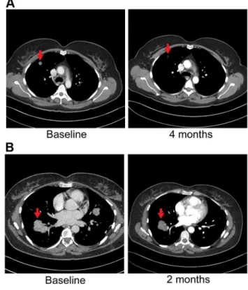
Figure 8. Anti-tumor activity in Patient 6, harboring an
FGFR2-TACC3fusion, to FGFR inhibitors. A) CT images of patient 6, whose tumor possessed anFGFR2-TACC3 fusion, at baseline and after four
months of pazopanib demonstrate significant tumor shrinkage (red arrows), 10.8 mm and 3.1 mm respectively.B) CT images of patient 6 at baseline and two months demonstrate significant tumor shrinkage (red arrows), 41.1 mm and 39.4 mm respectively after subsequent ponatinib treatment, 45 mg/daily, was begun.
sample using Illumina’s (San Diego, CA) TruSeq DNA Sample Prep Kit (catalog#FC-121-2001). In summary, genomic DNAs are fragmented to a target size of 900–1000 bp on the Covaris E210. 100 ng of the sample was run on a 1% TAE gel to verify fragmentation. Samples were end repaired and purified with Ampure XP beads using a 1:1 bead volume to sample volume ratio, and ligated with indexed adapters. Samples are size selected at approximately 1000 bp by running samples on a 1.5% TAE gel and purified using Bio-Rad Freeze ‘n Squeeze columns and Ampure XP beads. Size selected products are then amplified using PCR and products were cleaned using Ampure XP beads.
Patient 2. 300 ng genomic tumor and normal DNA was used to create whole genome libraries. Samples were fragmented on the Covaris E210 to a target size of 200–300 bp and 50 ng of the fragmented product was run on a 2% TAE gel to verify fragmentation. Whole genome libraries were prepared using Illumina’s TruSeq DNA Sample Prep Kit.
Exome sequencing
Patients 1 and 3. 1.1mg genomic DNA for each sample was
fragmented to a target size of 150–200 bp on the Covaris E210. 100 ng of fragmented product was run on TAE gel to verify fragmentation. The remaining 1mg of fragmented DNA was
prepared using Agilent’s SureSelectXTand SureSelectXTHuman All Exon 50 Mb kit (catalog#G7544C).
Patient 2. 50 ng genomic tumor and normal DNA was used to create exome libraries using Illumina’s Nextera Exome Enrichment kit (catalog# FC-121-1204) following the manufac-turer’s protocol.
Patients 4 and 5. 1mg of each tumor and germline DNA
sample was used to generate separate exome libraries. Libraries were prepared using Illumina’s TruSeq DNA Sample Prep Kit and Exome Enrichment Kit (catalog#FC-121-1008) following the manufacturer’s protocols.
Patient 6. 3mg of genomic tumor and normal DNA was fragmented on the Covaris E210 to a target size of 150–200 bp. Exome libraries were prepared with Agilent’s (Santa Clara, CA) SureSelectXT Human All Exon V4 library preparation kit (catalog# 5190-4632) and SureSelectXT Human All Exon V4+ UTRs (catalog# 5190-4637) following the manufacturer’s protocols.
RNA sequencing
Patients 1, 2 and 3. 50 ng total RNA was used to generate whole transcriptome libraries for RNA sequencing. Using the Nugen Ovation RNA-Seq System v2 (catalog#7102), total RNA was used to generate double stranded cDNA, which was subsequently amplified using Nugen’s SPIA linear amplification process. Amplified products were cleaned using Qiagen’s QIA-quick PCR Purification Kit and quantitated using Invitrogen’s Quant-iT Picogreen. 1mg of amplified cDNA was fragmented on the Covaris E210 to a target size of 300 bp. Illumina’s TruSeq DNA Sample Preparation Kit was used to prepare libraries from 1mg amplified cDNA.
Patients 4, 5 and 6. 1mg of total RNA for each sample was used to generate RNA sequencing libraries using Illumina’s TruSeq RNA Sample Prep Kit V2 (catalog# RS-122-2001) following the manufacturer’s protocol.
Figure 9. Anti-tumor activity of Patient 3, harboring anERRFI1mutation, to erlotinib, an EGFR inhibitor. A) CT images of patient 3 at
baseline and three months demonstrate significant tumor shrinkage (red marks). CT demonstrates right retroperitoneal lymph nodes decreasing from 7.6 cm to 2.9 cm and left retroperitoneal lymph nodes decreasing from 3.3 cm to 1.7 cm.B) PET images of patient 3 at baseline and three months demonstrate significant tumor shrinkage (red arrows). Hypermetabolic areas corresponding to right retroperitoneal lymph nodes demonstrate decrease from 8 cm longest diameter to imperceptible and left retroperitoneal lymph nodes decreasing from 4.2 cm to 1.4 cm. Both regions demonstrated significant reduction in metabolic activity.
Paired end sequencing
Libraries with a 1% phiX spike-in were used to generate clusters on HiSeq Paired End v3 flowcells on the Illumina cBot using Illumina’s TruSeq PE Cluster Kit v3 (catalog# PE-401-3001). Clustered flowcells were sequenced by synthesis on the Illumina HiSeq 2000 using paired-end technology and Illumina’s TruSeq SBS Kit.
Alignment and variant calling
Whole genome and whole exome. For whole genome and exome sequencing fastq files were aligned with BWA 0.6.2 to GRCh37.62 and the SAM output were converted to a sorted BAM file using SAMtools 0.1.18. BAM files were then processed through indel realignment, mark duplicates, and recalibration steps in this order with GATK 1.5 where dpsnp135 was used for known SNPs and 1000 Genomes’ ALL.wgs.low_coverage_ vqsr.20101123 was used for known indels. Lane level sample BAMs were then merged with Picard 1.65 if they were sequenced across multiple lanes. Comparative variant calling for exome data was conducted with Seurat [105].
Previously described copy number and translocation detection were applied to the whole genome long insert sequencing data [59] and these are made available through https://github.com/ davcraig75/tgen_somaticSV. Copy number detection was based
on a log2 comparison of normalized physical coverage (or clonal coverage) across tumor and normal whole genome long-insert sequencing data, where physical coverage was calculated by considering the entire region a paired-end fragment spans on the genome, then the coverage at 100 bp intervals was kept. Normal and tumor physical coverage was then normalized, smoothed and filtered for highly repetitive regions prior to calculating the log2 comparison. Translocation detection was based on discordant read evidence in the tumor whole genome sequencing data compared to its corresponding normal data. In order for the structural variant to be called there needs to be greater than 7 read pairs mapping to both sides of the breakpoint. The unique feature of the long-insert whole-genome sequencing was the long overall fragment size (,1 kb), where by two 100 bp reads flank a region of,800 bp. The separation of forward and reverse reads increases the overall probability that the read pairs do not cross the breakpoint and confound mapping.
RNA. For RNA sequencing, lane level fastq files were appended together if they were across multiple lanes. These fastq files were then aligned with TopHat 2.0.6 to GRCh37.62 using ensembl.63.genes.gtf as GTF file. Changes in transcript expression were calculated with Cuffdiff 2.0.2. For novel fusion discovery reads were aligned with TopHat-Fusion 2.0.6 [106] (patients 2, 3, 4 and 6). In addition, Chimerascan 0.4.5 [107] was used to detect
Figure 10. Immunohistochemistry of Patient 3’s tumor demonstrating activation of the EGFR pathway. A) Tumor stained with panAKT
Figure 11.FGFR2-IIIbfusion events.Transcripts and hypothetical protein products are modeled to illustrate the potential functional impact of fusion events involvingFGFR2(A–C). The identified fusion events involvingMGEA5(patient 4) (A) andBICC1(patient 5, reciprocal event) (C) are chromosome 10 intrachromosomal (D). In addition, patient 6 carried an interchromosomal fusion event (D) involvingFGFR2andTACC3(B). The FGFR2gene encodes for several isoforms with eleven representative transcripts and patients 4, 5, and 6 carry fusions involving the epithelial cell
specific transcript isoform (FGFR2-IIIb). All identified fusion breakpoints are close in proximity and are predicted to occur within the last intron of the
transcript and terminal to a known protein tyrosine kinase domain (A–C, gold domain). Predicted ‘‘Other’’ sites for all of the fusion protein models are the same and include the following: Casein kinase II phosphorylation sites, glycosylation sites, Protein kinase C phosphorylation sites, N-myristoylation sites, Tyrosine kinase phosphorylation sites, and cAMP-/cGMP-dependent protein kinase phosphorylation sites (A–C, grey triangle annotations). In all cases, fusions result in a predicted expansion of Casein kinase II phosphorylation and Protein kinase C phosphorylation sites. A protein product model is shown only for one of the reciprocal events involving theFGFR2andBICC1genes (FGFR2RBICC1,C). The fusion breakpoints
of the reciprocal events effect Exons 1 and 2 of the BICC1 gene, which translates to a difference of a predicted phosphoserine site within the Casein kinase II phosphorylation region (C, purple triangle within red circle). The FGFR2 gene is located within a fragile site region (FRA10F) and is flanked by two ribosomal protein pseudogenes, RPS15AP5 and RPL19P16 (see D inset (*)), whose repetitive sequence content may also contribute to genomic instability at theFGFR2initiation site.
doi:10.1371/journal.pgen.1004135.g011
Table 8.DNA and RNA validation of FGFR2 fusions in 3 patients with advanced sporadic biliary tract cancer.
Fusion Annealing site PCR input Direction Primer sequence
FGFR2-MGEA5 FGFR2 gDNA F 59-CTGACTATAACCACGTACCC-39
MGEA5 gDNA R 59-AGGGAGAAATTAAAGAACTTGG-39
FGFR2 cDNA F 59-TGATGATGAGGGACTGTTG-39
MGEA5 cDNA R 59-GAGTTCCTTGTCACCATTTG-39
FGFR2-BICC1 FGFR2 gDNA F 59-GGCAGAAGAAGAAAGTTGG-39
BICC1 gDNA R 59-ACTACTGCAGTTTGTTCAAT-39
FGFR2 cDNA F 59-TGATGATGAGGGACTGTTG-39
BICC1 cDNA R 59-TGTGTGCTCACAGGAATAG-39
BICC1-FGFR2 BICC1 cDNA F 59CGTGGACAGGAAGAAACT-39
FGFR2 cDNA R 59-GTGTGGATACTGAGGAAG-39
FGFR2-TACC3 FGFR2 gDNA F 59-TGACCCCCTAATCTAGTTGC-39
TACC3 gDNA R 59-AACCTGTCCATGATCTTCCT-39
F - forward, R - reverse.
fusions in patient 1, deFuse 5.0 [108] used in patients 2, 3 and 5 and SnowShoes [109] for patients 2 and 5.
Somatic mutation validation
Mutations of potential clinical relevance were confirmed in a Clinical Laboratory Improvement Amendments (CLIA) laborato-ry with Sanger sequencing or quantitative PCR.
Immunohistochemistry
The immunohistochemistry was performed following the procedures described previously [110]. Briefly, slides were dewaxed, rehydrated and antigen retrieved on-line on the BondMax autostainer (Leica Microsystems, INC Bannockburn, IL). Slides were then subjected to heat-induced epitope retrieval using a proprietary EDTA-based retrieval solution. Endogenous peroxidase was then blocked and slides were incubated with the following antibodies: FGFR2 (BEK, Santa Cruz, catalog# sc-20735), FGFR3 (C-15, Santa Cruz, catalog# sc-123), panAKT (Cell Signaling Technology, catalog#4685, pAKT (Cell Signal-ing Technology, catalog# 4060), EGFR (Cell Signaling Tech-nology, catalog# 4267, pEGFR (Cell Signaling Technology, catalog#2234), MAPK/ERK1/2 (Cell Signaling Technology, catalog# 4695), pMAPK/pERK (Cell Signaling Technology, catalog# 4376) and pFRS2 Y436 (Abcam, catalog# ab78195). Sections were visualized using the Polymer Refine Detection kit (Leica) using diaminobenzidine chromogen as substrate.
Fluorescent in-situ hybridization (FISH)
FISH was performed on formalin-fixed paraffin-embedded (FFPE) specimens using standard protocols and dual-color break-apart rearrangement probes specific to the FGFR2 gene (Abbott Molecular, Inc. Des Plaines, IL) located at 10q26. The 59FGFR2 signal was labeled with Spectrum Orange (orange) and the 39 FGFR2 signal was labeled with Spectrum Green (green).
Supporting Information
Table S1 Somatic point mutations, insertions and deletions identified in all samples.
(DOCX)
Table S2 Gene Ontology (GO) functional classification of genes carrying Smallscale Nucleotide Variations (SsNVs) or in regions exhibiting Copy Number Variation (CNV).
(XLS)
Table S3 Many genes carrying SsNVs act in key Cancer-associated pathways, and are differentially expressed in tumors from 6 patients with advanced sporadic biliary tract cancer. (XLS)
Table S4 Many genes identified in genomic regions exhibiting Copy Number Variation (CNV) functionally rank by significance to known Cancer-associated pathways.
(XLS)
Table S5 CLIA validation of somatic mutations with therapeutic relevance in 6 patients with advanced, sporadic biliary tract cancer.
(DOCX)
Table S6 Differential gene expression of fibroblast growth factor receptor pathway family members in 6 patients with advanced sporadic biliary tract cancer.
(DOCX)
Text S1 Supplementary discussion. (DOCX)
Author Contributions
Conceived and designed the experiments: MJB AHB AKS RF DWC JDC DVH. Performed the experiments: WSL KH RMC IC RR LP JM JAd SDM ATW PP JL HH KS BRK EGBF AB MTB MDP SWY JMC MB RRM KNL KCB PH AEM ACS. Analyzed the data: MDC JBE GRO JDB AAN SM YA JPK JAl AK TI AC SN JL HH BRK EWK WSL AEM ACS. Wrote the paper: MJB MDC JBE JDC DWC.
References
1. Shin HR, Lee CU, Park HJ, Seol SY, Chung JM, et al. (1996) Hepatitis B and C virus, Clonorchis sinensis for the risk of liver cancer: a case-control study in Pusan, Korea. International journal of epidemiology 25: 933–940. 2. Watanapa P (1996) Cholangiocarcinoma in patients with opisthorchiasis. The
British journal of surgery 83: 1062–1064.
3. Watanapa P, Watanapa WB (2002) Liver fluke-associated cholangiocarcinoma. The British journal of surgery 89: 962–970.
4. Bergquist A, Ekbom A, Olsson R, Kornfeldt D, Loof L, et al. (2002) Hepatic and extrahepatic malignancies in primary sclerosing cholangitis. Journal of hepatology 36: 321–327.
5. Bergquist A, Glaumann H, Persson B, Broome U (1998) Risk factors and clinical presentation of hepatobiliary carcinoma in patients with primary sclerosing cholangitis: a case-control study. Hepatology 27: 311–316. 6. Burak K, Angulo P, Pasha TM, Egan K, Petz J, et al. (2004) Incidence and risk
factors for cholangiocarcinoma in primary sclerosing cholangitis. The American journal of gastroenterology 99: 523–526.
7. Claessen MM, Vleggaar FP, Tytgat KM, Siersema PD, van Buuren HR (2009) High lifetime risk of cancer in primary sclerosing cholangitis. Journal of hepatology 50: 158–164.
8. Visser BC, Suh I, Way LW, Kang SM (2004) Congenital choledochal cysts in adults. Archives of surgery 139: 855–860; discussion 860–852.
9. Hsing AW, Zhang M, Rashid A, McGlynn KA, Wang BS, et al. (2008) Hepatitis B and C virus infection and the risk of biliary tract cancer: a population-based study in China. International journal of cancer Journal international du cancer 122: 1849–1853.
10. Kobayashi M, Ikeda K, Saitoh S, Suzuki F, Tsubota A, et al. (2000) Incidence of primary cholangiocellular carcinoma of the liver in japanese patients with hepatitis C virus-related cirrhosis. Cancer 88: 2471–2477.
11. Liu XF, Zou SQ, Qiu FZ (2003) Pathogenesis of cholangiocarcinoma in the porta hepatis and infection of hepatitis virus. Hepatobiliary & pancreatic diseases international : HBPD INT 2: 285–289.
12. Shaib YH, El-Serag HB, Davila JA, Morgan R, McGlynn KA (2005) Risk factors of intrahepatic cholangiocarcinoma in the United States: a case-control study. Gastroenterology 128: 620–626.
13. Welzel TM, Graubard BI, El-Serag HB, Shaib YH, Hsing AW, et al. (2007) Risk factors for intrahepatic and extrahepatic cholangiocarcinoma in the United States: a population-based case-control study. Clinical gastroenterology and hepatology : the official clinical practice journal of the American Gastroenterological Association 5: 1221–1228.
14. Yamamoto S, Kubo S, Hai S, Uenishi T, Yamamoto T, et al. (2004) Hepatitis C virus infection as a likely etiology of intrahepatic cholangiocarcinoma. Cancer science 95: 592–595.
15. Donato F, Gelatti U, Tagger A, Favret M, Ribero ML, et al. (2001) Intrahepatic cholangiocarcinoma and hepatitis C and B virus infection, alcohol intake, and hepatolithiasis: a case-control study in Italy. Cancer causes & control : CCC 12: 959–964.
16. Lee CC, Wu CY, Chen GH (2002) What is the impact of coexistence of hepatolithiasis on cholangiocarcinoma? Journal of gastroenterology and hepatology 17: 1015–1020.
17. Becker N, Liebermann D, Wesch H, Van Kaick G (2008) Mortality among Thorotrast-exposed patients and an unexposed comparison group in the German Thorotrast study. European journal of cancer 44: 1259–1268. 18. Travis LB, Hauptmann M, Gaul LK, Storm HH, Goldman MB, et al. (2003)
Site-specific cancer incidence and mortality after cerebral angiography with radioactive thorotrast. Radiation research 160: 691–706.
19. Khan SA, Thomas HC, Davidson BR, Taylor-Robinson SD (2005) Cholangiocarcinoma. Lancet 366: 1303–1314.
20. Valle J, Wasan H, Palmer DH, Cunningham D, Anthoney A, et al. (2010) Cisplatin plus gemcitabine versus gemcitabine for biliary tract cancer. The New England journal of medicine 362: 1273–1281.