Capnocytophaga canimorsus:
A Human Pathogen
Feeding at the Surface of Epithelial Cells and Phagocytes
Manuela Mally1., Hwain Shin1., Ce´cile Paroz1
, Regine Landmann2, Guy R. Cornelis1*
1Infection Biology, Biozentrum, University of Basel, Klingelbergstrasse, Basel, Switzerland,2Infection Biology, Department of Research, University Hospital Basel, Hebelstrasse, Basel, Switzerland
Abstract
Capnocytophaga canimorsus, a commensal bacterium of the canine oral flora, has been repeatedly isolated since 1976 from severe human infections transmitted by dog bites. Here, we show thatC. canimorsusexhibits robust growth when it is in direct contact with mammalian cells, including phagocytes. This property was found to be dependent on a surface-exposed sialidase allowing C. canimorsusto utilize internal aminosugars of glycan chains from host cell glycoproteins. Although sialidase probably evolved to sustain commensalism, by releasing carbohydrates from mucosal surfaces, it also contributed to bacterial persistence in a murine infection model: the wild type, but not the sialidase-deficient mutant, grew and persisted, both when infected singly or in competition. This study reveals an example of pathogenic bacteria feeding on mammalian cells, including phagocytes by deglycosylation of host glycans, and it illustrates how the adaptation of a commensal to its ecological niche in the host, here the dog’s oral cavity, contributes to being a potential pathogen.
Citation:Mally M, Shin H, Paroz C, Landmann R, Cornelis GR (2008)Capnocytophaga canimorsus:A Human Pathogen Feeding at the Surface of Epithelial Cells and Phagocytes. PLoS Pathog 4(9): e1000164. doi:10.1371/journal.ppat.1000164
Editor:Ambrose Cheung, Dartmouth Medical School, United States of America
ReceivedMay 21, 2008;AcceptedAugust 27, 2008;PublishedSeptember 26, 2008
Copyright:ß2008 Mally et al. This is an open-access article distributed under the terms of the Creative Commons Attribution License, which permits unrestricted use, distribution, and reproduction in any medium, provided the original author and source are credited.
Funding:This work was funded by Swiss National Science Foundation grant Nr 32-65393.01.
Competing Interests:The authors have declared that no competing interests exist. * E-mail: [email protected]
.These authors contributed equally to this work.
Introduction
Capnocytophaga canimorsus(formerly Centers for Disease Control group DF-2) has been rarely but regularly isolated from dog or cat bite infections since its discovery in 1976 [1,2].C. canimorsusis a thin Gram-negative rod, found in the normal oral flora of dogs and cats. Clinical infections byC. canimorsus generally appear as fulminant septicemia, peripheral gangrene or meningitis [3,4]. Splenectomy, alcohol abuse and immunosuppression have been associated with a number of cases, but more than 40% of the patients have no obvious risk factor [5].Capnocytophagabelongs to the family ofFlavobacteriaceaein the phylum ofBacteroidetes,which is taxonomically remote from Proteobacteria. The family of Bacteroidaceae contains many commensals of the mammalian intestinal system such as Bacteroides thetaiotaomicron and Bacteroides fragilis [6]. The family of Flavobacteriaceae includes a variety of environmental and marine bacteria, among which Flavobacterium johnsoniaeis a common soil and freshwater bacterium studied for its gliding motility [7]. There are only a few examples of pathogenic bacteria belonging to this family. These are Flavobacterium psychrophilum,the causative agent of cold water disease in salmonid fish [8],Ornithobacterium rhinotracheale,a bacterial pathogen known for causing respiratory disease in poultry [9], and Riemerella anatipestifer which causes ‘‘duckling disease’’ in waterfowl and turkeys [10,11]. The genus Capnocytophaga includes seven species found in normal human oral flora andC. canimorsusfound in the normal flora of dogs and cats. More than 160 cases ofC. canimorsus infections have been reported so far [12] but very few studies have addressed the molecular mechanisms ofC. canimorsuspathogenesis. Recently, we showed thatC. canimorsus 5 (Cc5) resists phagocytosis
by macrophage cell line [13,14]. We also showed that althoughC. canimorsus does not affect the viability of murine or human macrophages, it does not elicit proinflammatory cytokines and it even blocks the proinflammatory response to the LPS from enterobacteria [13]. In the course of such experiments, Cc5 exhibited robust growth, although it is usually considered fastidious for growth. In the present study, we show that a surface-localized sialidase plays a key role in initiating an extensive deglycosylation process of host cell glycan structures and that this feeding mechanism serves as a basis for growth and persistence of C. canimorsus in vivo.
Results
Growth ofC. canimorsus 5requires direct contact with cells
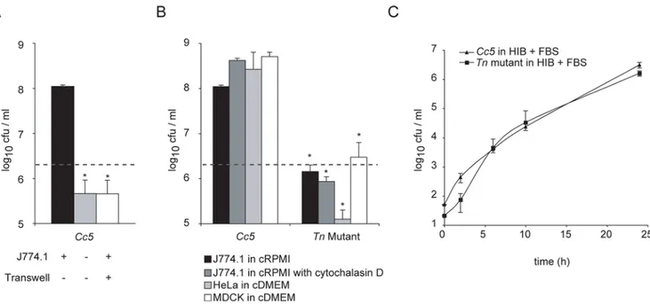
clone that was unable to grow in the presence of J774.1 cells, but grew normally on blood agar plates. Wild type (wt) and mutant bacteria grew equally well in serum enriched heart infusion medium (Fig. 1C). Impaired growth of this Tn mutant was not due to an increased phagocytic uptake by J774.1 since addition of cytochalasin D had little effect on bacterial growth (Fig. 1B).
Surface-localized sialidase is required for the growth of
Cc5in contact with cells
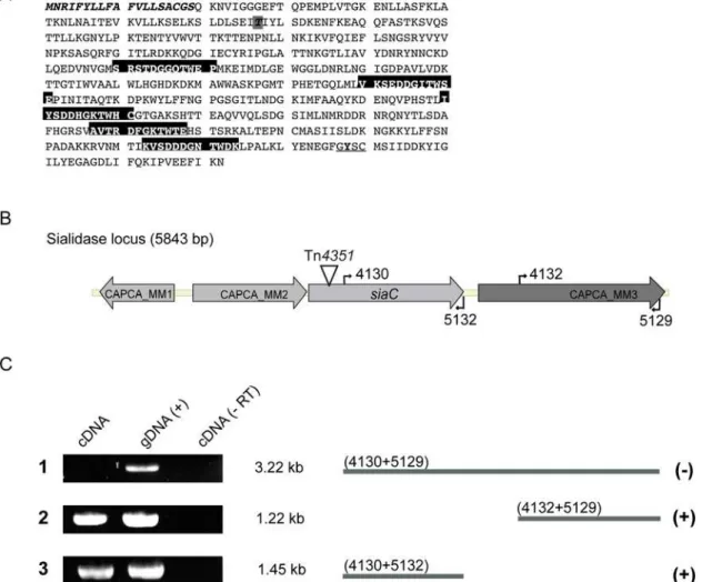
The transposon inserted at codon 77 within a gene encoding a protein with similarity to bacterial sialidase, glycosylhydrolase that cleaves terminal sialic acid from glycoconjugates (Fig. 2A). The mutated gene, designated siaC, was found to be located downstream of genes encoding a predicted transcriptional regulator and a putative N-acyl-glucosamine epimerase (accession number: EU329392). The first gene downstream was found to start 148 bp further from thesiaCstop codon (Fig. 2B). To exclude any polar effects of the Tn integration, we tested whether the downstream gene was transcriptionally linked tosiaC. Total RNA was isolated from wtCc5, reverse transcribed using two different primers annealing either at the end ofsiaC(5132) or at the end of the downstream gene (5129) and the cDNA was amplified by PCR. As shown in Fig 2C, even though transcripts were present for both genes separately, no transcript spanned siaC and the downstream gene. This result indicates thatsiaCis not transcrip-tionally linked to the downstream gene.
While intact Cc5 bacteria cleaved 29-(4-Methylumbelliferyl)-a -D-N-acetylneuraminic acid (MUAN), the Tn mutant could not, indicating that the mutated gene does indeed encode a sialidase (Fig. 3A). We engineered an expression shuttle vector by taking advantage of a cryptic plasmid isolated from another strain ofC. canimorsusand the promoter of an insertion sequence fromB. fragilis [16]. We constructed plasmids encoding full length (FL) SiaC, a variant deprived of the 21 N-terminal residues, predicted to be a signal peptide (D1–21), and a catalytic mutant (Y488C). Sialidase activity (Fig. 3A) and growth in the presence of J774.1 cells (Fig. 3B) was restored by introducingin trans siaCFL, but notsiaCD1–
21. Sialidase activity was not restored to wt levels bysiaCY488C, but
it was still significant (Fig. 3A), suggesting that this residual activity might account for elevated growth in comparison to the siaC
Figure 1. Growth ofC. canimorsus 5is dependent on cell contact.(A) Viable counts of 26106Cc5after 24 h in presence of J774.1 macrophages in RPMI supplemented with 10% FBS (moi = 20) (black) or in RPMI with FBS without cells (grey) and in a transwell system preventing physical contact between bacteria and macrophages in RPMI with FBS (white). (B) Viable counts ofCc5and Tn mutant after 24 h culture with macrophages in RPMI and FBS (black), with macrophages in RPMI and FBS in addition of cytochalasin D (grey), with HeLa cells (light grey) and MDCK cells in DMEM and FBS (white). The grey dotted line represents the bacterial number inoculated. The difference is statistically significant betweenCc5and Tn mutant (2-tailed unpaired Student’s t test p,0.05) in 3 or more experiments. (C) Growth curve of wtCc5(triangles) and Tn mutant (squares) in heart infusion broth (HIB) supplemented with 10% FBS, represented as the mean of 3 or more experiments with the error bars showing the s.d.
doi:10.1371/journal.ppat.1000164.g001 Author Summary
mutant (Fig. 3B). Using a sarcosyl extraction method, SiaCFLand
SiaCY488Cwere found to be associated with the outer membrane
(Fig. 3C), whereas SiaCD1–21 was only detected in total cells
(Fig. 3B). Indirect immunofluorescence using polyclonal anti-SiaC serum on paraformaldehyde fixed but unpermeabilized bacteria confirmed that SiaC is exposed on the bacterial surface unless the signal peptide is removed (Fig. 3D). Although it is surface exposed, no SiaC could be detected in the supernatant of infected J774.1 cultures, indicating that it is tightly associated with the outer membrane (Fig. 3C). Hence, surface-localized sialidase is required for growth ofCc5at the expense of mammalian cells.
Growth is sustained by N-acetyl glucosamine (GlcNAc) and N-acetyl galactosamine (GalNAc) but not by sialic acids
Since sialidases cleave terminal sialic acid from glycoconjugates, we first tested whether the addition of sialic acids could restore growth ofsiaC. Addition of neither sialic acid (Neu5Ac, N-Acetyl-2,3-dehydro-2-deoxyneuraminic acid) nor its activated form
(CMP-Neu5Ac, Cytidine-59-monophospho-N-acetylneuraminic acid) restored growth of siaCin presence of J774.1. In contrast, growth could be restored by the addition of purified recombinant SiaC or neuraminidase/sialidase NanH fromClostridium perfringens to the culture medium, but not by the addition of the catalytically inactive SiaCY488C (Fig. 4A). This suggested that removal of
terminal sialic acids from glycoconjugates is required to make other carbohydrates accessible. Indeed, N-acetyl glucosamine (GlcNAc) and N-acetyl galactosamine (GalNAc), common carbo-hydrate moieties of glycoconjugates, allowed growth ofsiaCin the presence of macrophages (Fig. 4B). Notably, addition of glucose (Glc), galactose (Gal), mannose (Man) or sialyl-lactose (N-acetylneuraminosyl-D-lactose) could not restore growth of siaC bacteria (Fig. 4C). As galactose (Gal) is a common sugar preceding GlcNAc in glycan molecules, we next tested addition of N-acetyl lactosamine (LacNAc), a disaccharide consisting of b-D-Gal
b(1R4) GlcNAc. LacNAc also restored the growth defect ofsiaC indicating the presence of an active b-galactosidase releasing monosaccharides Gal and GlcNAc in wt andsiaC Cc5(Fig. 4B).
Figure 2. Identification of the Tn integration site and analysis of mRNA present in wtC. canimorsus5.(A) Amino acid sequence of theC. canimorsussialidase showing the signal peptide (italics) and the BNR/asp repeats (Ser/Thr-X-Asp-X-Gly-X-Thr-Trp/Phe) of bacterial sialidases (boxed). Domain predictions were analyzed by InterProScan [42]. The residues conserved in sialidases at the C-terminus are underlined and the tyrosine 488 is bold [43]. The Tn4351integration site in SiaC at amino acid 77 is indicated, boxed in grey and bold. (B) Genetic locus of the sialidase gene (siaC) including its upstream genes, gntR-like gene (CAPCA_MM1) and putative N-acyl-glucosamine epimerase encoding gene (CAPCA_MM2); and downstream coding sequence (CAPCA_MM3). (C) Reverse transcription performed on total RNA with specific primers (5129 or 5132) followed by PCR to identify transcripts present in wtCc5(cDNA). PCR reactions were also performed using genomic DNA (gDNA) as template instead of cDNA as a positive control. As a control, reverse transcription was performed without reverse transcriptase in a parallel assay and used as template for the subsequent PCR reaction (-RT).
doi:10.1371/journal.ppat.1000164.g002
Sialidase desialylates macrophage and epithelial cell surfaces
J774.1 macrophages were incubated with either wt or siaC bacteria and thereafter analyzed for lectin binding to investigate desialyation process on the macrophage cell surface. We used Sambucus nigraagglutinin (SNA), which recognizes terminal sialic acids (2- 6 or 2- 3) linked to Gal or to GalNAc, and peanut agglutinin (PNA), a lectin specific for Gal (b 1–3) GalNAc, a disaccharide often forming the core unit of O-linked glycoconju-gates (Fig. 5A). As shown in Fig. 5B, wt bacteria greatly reduced the amount of sialic acids (SNA panel) and Gal (b 1–3) GalNAc (PNA panel) at the cell surface, whilesiaCbacteria had no effect on glycans masked by sialic acids. When cells were treated simultaneously with purified SiaC andsiaCbacteria, neither sialic acid nor Gal (b1–3) GalNAc were detected, indicating thatsiaC bacteria are still proficient in extensive deglycosylation of exposed glycans chains. The same deglycosylation of cell surfaces was observed when HeLa epithelial cells were used (Fig. 5C).
Sialidase inhibitor
N-Acetyl-2,3-dehydro-2-deoxyneuraminic acid (Neu5Ac2en) can inhibit growth of
Cc5
As bacterial and viral sialidases can share common ASP boxes that interact with sialic acid, we postulated that common sialidase
inhibitor might have sufficient specificity for the active site in SiaC to inhibit growth ofCc5wt bacteria in presence of cells. We tested the anhydro sialate derivative Neu5Ac2en, which is known to inhibit many viral and bacterial neuraminidases [17,18]. Approx-imately 150 cfu/ml of wt Cc5 were inoculated to a culture of J774.1 macrophages in the presence of 1mM Neu5Ac2en and growth was monitored after 2, 6, 10 and 24 h (Fig. 6A). Between 2 and 24 h post infection, counts of wt Cc5 were significantly reduced to values close to thesiaCmutant (Fig. 6B). These data indicate that Neu5Ac2en has affinity for the active site of SiaC and restricts the growth ofCc5in the presence of J774.1 cells.
Cc5but notsiaCis able to persist in murine tissue cages To test whether sialidase could play a role duringC. canimorsus systemic infection, we selected a murine tissue cage infection model [19]. Around 107 cfu of wt or siaC Cc5 bacteria were injected directly into Teflon cages, which had been subcutaneously implanted in C57BL/6 mice. Colony forming units (cfu) counts of wt decreased on day 2 and 5. However, on day 9 they increased by 1 to 3 logs in 4 out of 5 mice, and were able to persist in 3 of 5 mice after 27 days post infection with more than 107bacteria per ml fluid. ThesiaCbacteria were undetectable after the second day in 5 out of 5 infected mice (Fig. 7A). After infection, the total number of leukocytes in tissue cage fluid (1.86104+/21.36104 leukocytes/ml, mean +/2 standard deviation, s.d.) did not
Figure 3. Surface localized sialidase is required for growth.(A) Sialidase activity of intact bacteria, measured with the substrate MUAN as the mean of triplicate measurements and s.d. of a representative experiment. (B) Viable counts after challenge with 26106Cc5(black),siaC(light grey) or
siaCcomplemented with plasmids containingsiaCFL,siaCD1–21andsiaCY488Cafter 24 h in presence of J774.1 with the grey dotted line indicating the bacterial number inoculated. Sialidase was detected by immunoblotting with a polyclonal antibody against SiaC in total cells (TC). (C) Outer membrane protein fractions (OMP), cell free supernatants (SN) of the J774.1 cultures shown in (B) including as control TC ofCc5were analyzed by immunoblotting for the presence of SiaC. (D) Surface localization of SiaC was tested by immunofluorescence on paraformaldehyde fixed but not permeabilized bacteria using anti-SiaC followed by anti- rabbit IgG conjugated to FITC.
significantly increase and was not related to the bacterial load, suggesting that Cc5 infection did not lead to strong leukocyte recruitment. This was in agreement with the suppression of
inflammation, which we previously reported [13]. In mixed infections, the competitive index ofsiaCbacteria was 9.761024, 5.861027 and 4.761027 on day 5, 9 and 14, respectively. As
Figure 4. Aminosugars but not sialic acids sustain growth ofC. canimorsus.Viable counts after challenge with 26106wtCc5(black) orsiaC (grey) grown for 24 h with J774.1 in cRPMI (control) or in the same condition with the addition of Neu5Ac, Neu5Ac- CMP, 12.5 ng/ml enzyme SiaCFL, SiaCY488Cor NanH fromC. perfringens(A) or with the addition of GalNAc, GlcNAc or LacNAc (B) or with the addition of mannose, galactose, glucose or sialyl-lactose (C). Mean values from 3 or more experiments and s.d. are shown including statistical difference between wtCc5andsiaCwith * p,0.05, ** p,0.01 and *** p,0.001 for each pair of columns (2-tailed unpaired Student’s t test). The grey dotted line indicates the bacterial number inoculated.
doi:10.1371/journal.ppat.1000164.g004
observed during infection with wtCc5alone, 3 mice out of 5 that were infected by both strains developed a persistent infection (Fig. 7B).
The fluid from uninfected control cages was collected and the leukocytes and liquid were tested separately for their capacity to sustain growth ofCc5. Interestingly, wtCc5 did not grow in the presence of the cell-free liquid (data not shown) but they grew in presence of leukocytes whereas siaC bacteria did not (Fig. 7C). Both strains grew equally well in heart infusion broth
supple-mented with 10% FBS, indicating a similar fitnessin vitro(Fig. 1C). Mixed cultures in heart infusion broth supplemented with 10% FBS showed comparable growth of wt and siaC bacteria. Both strains reached 106cfu/ml after 24 h (Fig. 7D).
Our data from mixed infection in mice suggest that there is no cross-feeding of nutrients between wt andsiaC Cc5(Fig. 7B). We thus tested whether there would be cross-feeding between wt and siaCbacteria when inoculated to J774.1 cultures. When wt and siaCwere inoculated together at 1:1 ratio to J774.1 cells, wtCc5
Figure 5. C. canimorsus desialylates macrophage and epithelial cell surfaces. (A) The targets of the lectins used in this study are schematically represented (adapted from [44]). Surface carbohydrates of J774.1 macrophages (B) or HeLa epithelial cells (C) were analyzed by lectin binding after 2 h of infection with 46107wt (Cc5) orsiaCbacteria. Cells were fixed with paraformaldehyde and incubated for 1 h with lectin SNA, which recognizes terminal sialic acids (2- 6 or 2- 3) linked to Gal or to GalNAc or PNA that binds to the disaccharide Gal 1–3 GalNAc only after removal of terminal sialic acids. SiaC was added to cells alone or withsiaCbacteria at 100 ng/ml. Biotinylated lectins were visualized by FITC conjugated streptavidin.
reached 108cfu/ml whilesiaCbacteria only reached 36106cfu/ ml 24 h post infection (Fig. 7E).
Taken together, these results demonstrate that SiaC plays an essential role in allowing persistence of wtCc5in this tissue cage model and that clearance ofsiaCbacteria is not due to a growth defect per se but to an altered interaction of the mutant with the host. Since sialidase is surface-exposed, one could consider the possibility that it alters the susceptibility to complement. Hence, we checked the susceptibility of wt and siaC Cc5 to mouse complement and found no difference (data not shown). It is also very unlikely that siaC bacteria have an increased sensitivity to killing by mouse leukocytes. Indeed, we tested phagocytosis and killing by human polymorphonuclear leukocytes and found no difference between wt and siaCbacteria (Manuscript in prepara-tion). Hence, we conclude that the role of sialidase in infected mice is essentially nutritional.
Discussion
Sialic acids are a family of nine carbon acid sugars among which Neu5Ac is one of the most widespread variants. Sialic acids are predominantly found at the terminal position of cell-surface and secreted eukaryotic glycan structures and are involved in many physiological processes including binding to microbes and down-regulation of innate immunity [20–22]. Therefore it is not surprising that sialic acids play a role in a variety of host microbe interactions. Several pathogens have evolved ways to expose sialic acid on their surface and hence to escape complement killing and opsonization by mimicry. Sialic acids are incorporated into capsules byE. coliK1 [23], Group BStreptococci[24], Serogroups B, C, W135 and YNeisseria meningitidis[25]. The lipooligosacchar-ide of Neisseria gonorrhoeae, Neisseria meningitidis and Haemophilus influenzae are also sialylated [26]. In this case, a bacterial sialyltransferase uses CMP-Neu5Ac from the host as a substrate [26]. Sialic acids can also be synthesized from lactate byNeisseria itself, demonstrating a close link between metabolism and evasion of innate immune defenses [27].
Besides molecular mimicry, many microbes can utilize sialic acids as a source of carbon and nitrogen like E. coli K1, H. influenzae or C. perfringens [28]. Their metabolism comprises a permease for uptake and a neuraminiate lyase for conversion to N-acetyl mannosamine, which is either degraded or used in sialic acid biosynthesis. A number of commensal and pathogenic bacteria are also endowed with a sialidase, a glycosylhydrolase that cleaves sialic acid from sialo-glycoconjugates. Bacterial sialidases have been thought since a long time to contribute to virulence in bacteria that colonize mucosal surfaces such asVibrio cholerae, Streptococcus pneumoniae,group Bstreptococci,C. perfringensand B. fragilis but the exact role of sialidase on virulence remains controversial [29]. Recently, it was shown that a sialidase is involved in the formation of Pseudomonas aeruginosa biofilms and hence contributes to colonization of the lungs during the initial stages of infection in cystic fibrosis patients [30]. InS. pneumoniae,a sialidase initiates an extensive deglycosylation of different host proteins, including IgA1 and human secretory component [31]. Furthermore, the sequential action of exoglycosidases sustains growth ofS. pneumoniaeon human a-1 acid glycoprotein, though growth is not as robust as on sucrose and lactose. Although the genetic analysis suggests that sugars from the glycan chain would sustain growth, this has not been shown directly [32].
In the present study, which is among the very first on the pathogenesis mechanisms ofC. canimorsus,we demonstrate that a sialidase allows C. canimorsus to feed on glycan chains from glycoproteins. The role of sialidase is not to supply sialic acid since growth of the sialidase-deficient mutant could not be restored by adding sialic acid to the culture medium. Thus, we have a situation similar to that ofS. pneumoniae: the role of sialidase is to provide access to masked sugars of surface-exposed glycoproteins. Growth of the sialidase-deficient mutant could be restored by amino sugars like GalNAc, GlcNAc and LacNAc but not by glucose, galactose, mannose or sialyl-lactose, indicating that the nutritional require-ments ofC. canimorsusare very different from those ofS. pneumoniae. Our study thus confirms the importance of a sialidase to initiate a deglycosylation process for bacterial metabolism. Moreover, in
Figure 6. The sialidase inhibitor Neu5Ac2en decreases growth of wtC. canimorsus 5in presence of macrophages.(A) Viable counts of approximately 150 bacteria grown in cRPMI in the presence of J774.1 cells for 2, 6, 10 and 24 h:siaCbacteria (light grey); wtCc5bacteria with 1mM Neu5Ac2en (grey, dotted line); wtCc5in the absence of inhibitor (black). Mean values from 4 experiments and s.d. are shown including statistical difference betweenCc5andsiaCin grey or betweenCc5andCc5treated with Neu5Ac2en in black with * p,0.05, ** p,0.01 and *** p,0.001 (2-tailed unpaired Student’s t test). (B) Data from viable counts (mean) shown in (A) is represented as the fold difference compared to wtCc5.The statistical difference is depicted from (a) with * p,0.05, ** p,0.01 and *** p,0.001.
doi:10.1371/journal.ppat.1000164.g006
comparison withS. pneumoniae, C. canimorsususes sialidase to feed on glycoproteins exposed at the surface of epithelial cells or even of macrophages, in spite of the fact that they do not adhere to these cells. The observation of extracellular bacteria specifically feeding on the surface of epithelial cells is not unprecedented. It has been described for B. thetaiotaomicron, a major commensal from the intestine, which feeds on fucosylated intestinal cells. Colonization byB. thetaiotaomicroneven triggers the appearance of fucosyltrans-ferase and fucosylated glycan expression [33]. Recent studies showed that host acquired fucose is incorporated byB. fragilisinto capsular polysaccharide or glycoproteins, which in turn provides a survival advantage in the mammalian intestinal ecosystem [34]. As forC. canimorsus, it is likely that the capacity to feed on HeLa cells reflects the adaptation to feed on buccal epithelial cells.
Sialidase, which is pivotal in this feeding process, is surface localized and this surface localization is a prerequisite for unmasking glycan structures. It is not common to find enzymes anchored into the outer membrane, facing the outside of Gram-negative bacteria but there are examples like pullulanase a 116-kDa isoamylase ofKlebsiella oxytoca[35]. Not surprisingly, SiaC is endowed with an N-terminal signal sequence, which turned out to be critical for its targeting. Sialidase thus crosses the cytoplasmic membrane via the Sec pathway but we have at present no explanation on how it crosses the outer membrane and remains anchored. It is probably not by aC. canimorsusspecific mechanism since sialidase appeared to be also surface-exposed when expressed inE. coli(unpublished data). Sialidase could be a lipoprotein, like pullulanase. Alternatively, sialidase could be a surface-anchored
Figure 7. The sialidase mutant is hypo-virulent in a tissue cage mouse infection model.Tissue cages were implanted in C57BL/6 mice and infected with 107Cc5wt andsiaCbacteria (n = 5) singly (A) or in competition (B). Bacteria were counted in tissue cage fluid (TCF) during 27 days (Cc5 =black circles;siaC= open circles). Individual values are shown; horizontal lines indicate the median value of each group. The black dotted line is the detection limit of 20 bacteria per ml TCF. (A) Cfu numbers between groups were significantly different on days 2, 5 and 9 with p,0.01 and on days 14 and 27 with p,0.05 (Mann Whitney test). (B) 107cfuCc5and erythromycin resistantsiaCwere inoculated at a 1:1 ratio. Bacterial numbers were analyzed for 27 days (n = 5). Viable counts between wt andsiaCwere significantly different on day 2, 5 and 9 with p,0.01 and on day 14 with p,0.05. (C)Ex vivoisolated leukocytes were resuspended in serum free RPMI and inoculated at a moi of 20 (26106bacteria) or 0.2 (26104bacteria) indicated with grey dotted lines and bacterial viable count was monitored after 24 h. Values represent the mean using TCF cells from 3 uninfected mice. TCF leukocytes consist of 68%+/24.8% polymorphonuclear neutrophils (PMNs), 18%+/23.2% monocytes and 9.1%+/23.7% macrophages. Wt andsiaCnumbers were significantly different with p,0.05 (*) and p,0.001 (**) using 2-tailed unpaired student’s t test. (D)In vitro,Cc5andsiaC were tested in heart infusion broth with FBS inoculated at a 1:1 ratio with approximately 100 bacteria total and bacterial growth was monitored for 2, 6, 10 and 24 h. (E) Viable counts after challenge with 26106(grey dotted line)Cc5(black) orsiaC(grey) grown for 24 h with J774.1 in cRPMI singly (control) or at a 1:1 ratio (cross-feeding).
auto-transporter protein like the Y. enterocolitica YadA [36]. However, the fact that the C-terminus of sialidase is not involved in the surface localization (unpublished data) argues against this hypothesis. Work in progress tries to address the question of how sialidase is anchored in the outer membrane.
Unlike what is observed with pullulanase, our data indicate that extremely little sialidase is released from C. canimorsus. This observation is in perfect agreement with the fact thatC. canimorsus needs to be in direct contact with cells to feed on them. It also makes sense in the context of the mouth commensal microflora. Indeed, the oral cavity is occupied by some 500 different bacterial strains [37,38], creating a fierce competition for nutrition. The fact that C. canimorsus does not release this enzyme suggests thatC. canimorsus maximizes the benefit of sialidase by not sharing this fitness factor with competing bacteria. In agreement with this hypothesis, there is no cross-feeding when wt andsiaC Cc5bacteria are inoculated together in the presence of macrophages. This implies that wt C. canimorsus must be extremely efficient in capturing the aminosugars that it extracts from the surface of cells and we hypothesize thatC. canimorsushas dedicated high affinity transporters for these in its outer membrane.
Extracellular C. canimorsus replicated very efficiently not only when they were in direct contact with HeLa cells but also with J774.1 macrophages. Thus,C. canimorsus not only resists phago-cytosis by cultured macrophages [13,14], but they even take advantage of macrophages whose normal function is to engulf and kill microbes. To our knowledge, this is the very first report of a pathogen that can feed on phagocytic cells. This observation suggests that sialidase could contribute to virulence. We used a mouse tissue cage model in which the readout is bacterial persistence and we observed a dramatic difference in persistence between wt and sialidase-deficient C. canimorsus. Even more, we gained evidence thatin vivo, C. canimorsusalso feeds on phagocytes. These observations confirm our hypothesis that sialidase contrib-utes to virulence, at least in the mouse model. It seems reasonable to extrapolate that it also plays a critical role during human infections. We would however be reluctant to call sialidase a virulence factor since it most probably evolved as a fitness factor for commensalism in the dog’s mouth. Nevertheless, the mouse experiment shows that it may become a persistence factor ifC. canimorsusis introduced in the tissues from another host. Our study thus shows once again the link between metabolism and virulence, as already well documented in studies on Salmonella [39],Listeria [40] and Neisseria [27]. However, unlike what was seen with Salmonella, there seem to be no or very little redundancy in thein vivo metabolism of C. canimorsus since the loss of sialidase had dramatic consequences on growth. It is interesting to observe that nutrition in vivo may be quite specific in spite of a very rich nutritional environment. Indeed, only GlcNAc and GalNAc could rescue growth while glucose had no effect and galactose was even deleterious. This difference could result from the fact that unlike Salmonella,C. canimorsusis a commensal highly adapted to its niche and only exceptionally a pathogen. Specialization is probably the hallmark of a bacterium that is primarily a commensal and only rarely a pathogen. Finally, C. canimorsus represents one more example illustrating that the distinction between commensals and pathogens is illusive. Commensalism and pathogenesis are two faces of the same coin.
Influenza neuraminidases have been successfully targeted with chemotherapeutic inhibitors for prophylaxis and treatment [41]. Given the wide prevalence and important role of sialidases in microbial infections, inhibition of bacterial sialidases could also provide a mechanism to prevent bacterial spreading during infections. Here, we observed a significant inhibition of the growth
of C. canimorsusin the presence of macrophages by Neu5Ac2en. These preliminary data indicate that microbial sialidases could indeed serve as an attractive drug target to prevent bacterial dissemination.
Materials and Methods
Bacterial strains and growth conditions
C. canimorsus 5 was routinely grown on Heart Infusion Agar (HIA; Difco) supplemented with 5% sheep blood (Oxoid) for 2 days at 37uC in presence of 5% CO2. Bacteria were harvested
by gently scraping colonies off the agar surface, washed and resuspended in PBS.C. canimorsuswas also grown in Heart Infusion Broth (Difco) supplemented with 10% (v/v) fetal bovine serum (FBS; Invitrogen) for approximately 24 h without shaking in an 37uC incubator with 5% CO2. Selective agents were added at the
following concentrations: erythromycin, 10mg/ml; cefoxitin, 10mg/ml; gentamicin, 20mg/ml; ampicillin, 100mg/ml.
Cell Culture and Infection
Murine monocyte-macrophage J774A.1 cells (ATCC TIB-67) were cultured in RPMI 1640 (Invitrogen) supplemented with 10% (v/v) FBS (Invitrogen), 2 mM L-glutamine and 1 mM sodium pyruvate. Human epithelial HeLa cells (ATCC CCL-2) and canine epithelial MDCK kidney cells (ATCC CCL-34) were grown in DMEM (Invitrogen) with 10% (v/v) FBS. Cells were seeded in medium without antibiotics at a density of 105/cm2and cultured at 37uC in humidified atmosphere containing 5% CO2.
Unless otherwise indicated, infection was performed after 15 h at a moi of 20 representing 26106bacteria per ml in each well at 37uC. Monosaccharides and disaccharides (Sigma Aldrich) were added to 0.1% (w/v) final concentration. Neu5Ac and CMP-Neu5Ac were added to 0.01% final concentration.
Cc5was pretreated with 1mM Neu5Ac2en at 37uC for 30 min. Subsequently, infection of J774.1 was carried out in presence of 1 mM Neu5Ac2en during 24 h.
Arbitrarily Primed PCR
Primers specific to the ends of the transposon and primers of random sequence that may anneal to chromosomal DNA sequences in close proximity to the transposon insertions were used in two rounds of PCR before sequencing. The first round of amplification was carried out in 50ml containing 100 ng of genomic DNA, 1.5 mM MgCl2, 200mM of primers 59
CA-GAATTCTGTTGCATTTGCAAGTTG 39 complementary to Tn4351 and 59ggccacgcgtcgactagtacNNNNNNNNNNacgcc39, 2.5 U of DNA polymerase (DyNAzymeII, Finnzymes), 200mM of each dNTP, in 10 mM Tris HCl (pH 8.3) for 6 cycles (94uC for 1 min, 30uC for 1 min, 72uC for 2 min) and 30 cycles (94uC for 1 min, 45uC for 1 min, 72uC for 2 min) and final 10 min at 72uC. 10ml of PCR product containing random fragments was used as template in a second round of 30 cycles of amplification (94uC for 30 sec, 45uC for 30 sec, 72uC for 1 min) using primers 59
CAGAATTCTGTTGCATTTGCAAGTTG 39 and 59
GGCCACGCGTCGACTAGTAC 39, from the 59of the random primer. PCR products were purified using NucleoSpinH from Machery Nagel. 20- 50 ng of random sized products were sequenced using an ABI sequencer. The Tn integration site was further confirmed by using primers on chromosomal DNA by sequencing towards the Tn integration site. Primers used were 59
AATTGTTGTAACGATTGTCG 39 or 59
GCGAAGCGT-TATCCCAAAGC 39 complementary to the siaCsequence in a sequencing reaction containing 2mg genomic DNA of siaC, betaine 0.25 M and BigDye Terminator Ready Reaction (PE
Biosystems) with an initial denaturation step for 5 min and subsequent 99 cycles (95uC for 30 sec, 50uC for 20 sec, 60uC for 4 min).
RNA isolation and reverse transcription (RT) PCR
Cc5 were grown for 2 days on HIA blood plates. RNA was isolated from 56108 bacteria by a hot phenol/chloroform extraction method followed by DNase I (Amersham Pharmacia) treatment (0.5 U/mg RNA) for 2 h at 37uC. RNA was further cleaned by using a RNeasy kit (Quiagen) and stored at 280uC until use. An additional DNase I digest was introduced with 0.25 U/mg RNA for 15 min at 37uC and stopped by addition of final 2.5 mM EDTA and heat inactivation at 75uC for 10 min. Subsequent reverse transcription was performed with 50 U Superscript II/mg RNA in RT buffer (Invitrogen), 10 mM DTT and 50mM specific primer (5129: 59
GGGTAATCCG-CACTTGTCGGG39 or 5132: 59
GTTTAGTTCTTGA-TAAATTCC 39) for 60 min at 42uC and stopped at 70uC for 10 min. 10% of cDNA preparation or of a preparation made without addition of reverse transcriptase was subjected to PCR using following primer combinations: 4130 (59 GGGTAACAA-CAAAAACCACTG 39)+5129; 4132 (59 TATAAGAA-TAATTGGTGGGC 39)+5129; 4130+5132. 100 ng of genomic DNA from Cc5 was used as a positive control of the PCR reactions.
Construction of complementation and expression plasmids
Full length siaC was amplified with 59
CATACCATGG-GAAATCGAATTTTTTATCTT 39 and 59
GTTCTAGA-GAGTTCTTGATAAATTCCTCAACTG 39 primers and
cloned into the E. coli- C. canimorsus shuttle vector pMM47.A [16] withNcoI andXbaI, leading to the insertion of a glycine at position 2 and a C- terminal histidine 66tag in plasmid pMM52 (siaCFL). Forward primer 59
AAAGCCATGGGAAACG-TAATCGGCGGAGGCG 39 was used with the same reverse primer to construct pMM50 (siaCD1–21), deleting the first 63 bp of siaC, but still including methionine and glycine at position 1 and 2, respectively, and using a C-terminal His 66tag. The catalytic mutation in siaCof was introduced by site directed mutagenesis with an inverse PCR on pMM52, using primers 59 GAAG-GATTTGGGTGTTCGTGTATGTCG 39and 59 CGACATA-CACGAACACCCAAATCCTTC 39 leading to pMM59 ( sia-CY488C). Plasmids derived from pMM47.A contained thecfxAgene
originating fromBacteroides sp.and could be selected inC. canimorsus with 10mg/ml cefoxitin [16]. The beta-lactamase also present on pMM47.A was used as a selection marker inE. coli.
The cDNAs encoding SiaCD1–21 (pHS2) were subsequently
amplified using 59
GGAATTCCATATGAACGTAATCGGCG-GAGGC 39 plus 59
CGCGGATCCCTAGTTCTTGA-TAAATTCCTC 39 and cloned into the expression vector pET15b(+) (Novagen). Plasmid pHS3 encoding SiaCD1–21,Y488C
was constructed by site directed mutagenesis on template pHS2 using the same primers as described for pMM59. All constructs were sequenced with an ABI sequencer. The sequence of SiaC was deposited at GenBank (accession number: EU329392).
Purification of recombinant SiaC and immunoblotting Expression ofsiaCconstructs inE. coliBL21(DE3) was induced with 0.5 mM isopropyl-b-D-1-thiogalactopyranoside at A600= 0.5
for 3 h. Proteins were purified by affinity chromatography using chelating Sepharose (Pharmacia) charged with NiSO4 according
to the manufacturer’s instructions. Samples were analyzed by
SDS-PAGE by the system of Laemmli, and immunoblotted. Polyclonal serum from rabbit was generated against recombinant SiaCD1–21. The antigen was injected at days 0, 14, 28, and 56 with
a final bleeding at day 80 (Laboratoire d’Hormonologie, Marloie, Belgium).
MUAN hydrolysis
107bacteria were incubated with 0.006% 29 -(4-Methylumbelli-feryl)-a-D-N-acetylneuraminic acid (MUAN) in 0.25 M sodium acetate pH 7.5 at 37uC for 3 min. Reactions were stopped with 50 mM Na2CO3 pH 9.6 and fluorescence was determined at
445 nm with a Wallac Victor2 1420 Multilabel counter (Perkin Elmer).
Outer Membrane Preparation
Bacterial cells resuspended in PBS containing DNase and RNase (10mg/ml), were sonicated on ice. Unbroken cells were removed at 30006g for 15 min, and total membranes were collected at 20 0006g for 30 min at 4uC. The membranes were suspended in PBS and sarcosyl (N-Lauroylsarcosine sodium salt, Sigma) was added to a final concentration of 1% (v/v). After incubation on ice for 1 h, membranes were collected at 20 0006g for 30 min and resuspended in electrophoresis sample buffer and analyzed by SDS-PAGE by the system of Laemmli.
Immunofluorescence of bacteria
107bacteria were incubated on poly-D-lysine (BD) coated glass slides for 1 h at 37uC and subsequently fixed with 3% paraformaldehyde for 15 min. Anti- SiaC polyclonal serum (1:500) and a FITC conjugated secondary antibody (Goat Anti-Rabbit IgG, Southern Biotech) was used at 1mg/ml and fluorescence was measured with a Leica DMIRE2 microscope. Pictures were taken with a digital camera (Hamamatsu Photonics) and analyzed with OpenLab software (version 3.1.2) and Adobe Photoshop CS3 (version 10.0.1).
Lectin Staining
105J774.1 macrophages or HeLa epithelial cells were seeded on
poly-D-lysine coated slides. Infection was carried out with 46107 bacteria for 2 h. Uninfected cells were alternatively treated with purified recombinant SiaC at 100 ng/ml. Cells were fixed with 3% paraformaldehyde for 15 min. Biotinylated lectins SNA and PNA (Vector Laboratories) were incubated with cells at 2mg/ml and 2.5mg/ml, respectively, for 1 h. After washing with PBS, cells were treated with 1mg/ml fluorescein conjugated streptavidin (Vector Laboratories) and fluorescence was determined on mounted slides (Vectashield, Vector Laboratories).
Mice and tissue cage infection model
differentiated by Diff-Quick (Medion Diagnostics) Wright staining of cytospins and examined under light microscopy. The percentage of viable leukocytes was assessed by trypan blue exclusion.
The survival ofsiaCbacteria in the competition experiment was compared directly with wtCc5in individual animals giving a 1:1 ratio of wt to mutant bacteria. The number of mutant (erythromycin resistant) and wt bacteria recovered from the TCF of animals was established by plating to media with and without erythromycin. The competitive index was calculated as the (number of mutant/wild-type bacteria recovered from animals)/(number of mutant/wild-type bacteria in the inoculum).
Statistical analysis
For growth experiments, means and standard deviation (s.d.) were calculated and statistical significance was evaluated by using
a two- tailed, unpaired Student’s t test. Differences were determined to be significant when p,0.05. Forin vivoexperiments, individual mouse values are shown including the median value of each group. Mann Whitney test with the post hoc Bonferroni correction was used for comparison between Cc5 and siaC cfu numbers during infection.
Acknowledgments
We thank Z Rajacic, N Jann and M Schmaler for assistance during thein vivostudies.
Author Contributions
Conceived and designed the experiments: MM HS RL GRC. Performed the experiments: MM HS CP. Analyzed the data: MM HS RL GRC. Wrote the paper: MM HS RL GRC.
References
1. Bobo RA, Newton EJ (1976) A previously undescribed gram-negative bacillus causing septicemia and meningitis. Am J Clin Pathol 65: 564–569.
2. Brenner DJ, Hollis DG, Fanning GR, Weaver RE (1989) Capnocytophaga canimorsus sp. nov.(formerly CDC group DF-2), a cause of septicemia following dog bite, andC. cynodegmi sp. nov., a cause of localized wound infection following dog bite. J Clin Microbiol 27: 231–235.
3. Pers C, Gahrn-Hansen B, Frederiksen W (1996) Capnocytophaga canimorsus septicemia in Denmark, 1982–1995: review of 39 cases. Clin Infect Dis 23: 71–75.
4. Le Moal G, Landron C, Grollier G, Robert R, Burucoa C (2003) Meningitis due toCapnocytophaga canimorsusafter receipt of a dog bite: case report and review of the literature. Clin Infect Dis 36: e42–46.
5. Lion C, Escande F, Burdin JC (1996)Capnocytophaga canimorsusinfections in human: review of the literature and cases report. Eur J Epidemiol 12: 521–533. 6. Coyne MJ, Comstock LE (2008) Niche-specific features of the intestinal
bacteroidales. J Bacteriol 190: 736–742.
7. McBride MJ (2004) Cytophaga-flavobacteriumgliding motility. J Mol Microbiol Biotechnol 7: 63–71.
8. Duchaud E, Boussaha M, Loux V, Bernardet JF, Michel C, et al. (2007) Complete genome sequence of the fish pathogenFlavobacterium psychrophilum. Nat Biotechnol 25: 763–769.
9. Schuijffel DF, van Empel PC, Pennings AM, van Putten JP, Nuijten PJ (2005) Successful selection of cross-protective vaccine candidates forOrnithobacterium rhinotrachealeinfection. Infect Immun 73: 6812–6821.
10. Segers P, Mannheim W, Vancanneyt M, De Brandt K, Hinz KH, et al. (1993) Riemerella anatipestifer gen. nov., comb. nov., the causative agent of septicemia anserum exsudativa, and its phylogenetic affiliation within the Flavobacterium-CytophagarRNA homology group. Int J Syst Bacteriol 43: 768–776. 11. Subramaniam S, Huang B, Loh H, Kwang J, Tan HM, et al. (2000)
Characterization of a predominant immunogenic outer membrane protein of Riemerella anatipestifer.Clin Diagn Lab Immunol 7: 168–174.
12. Tierney DM, Strauss LP, Sanchez JL (2006)Capnocytophaga canimorsusmycotic abdominal aortic aneurysm: why the mailman is afraid of dogs. J Clin Microbiol 44: 649–651.
13. Shin H, Mally M, Kuhn M, Paroz C, Cornelis GR (2007) Escape from immune surveillance byCapnocytophaga canimorsus. J Infect Dis 195: 375–386.
14. Meyer S, Shin H, Cornelis GR (2008) Capnocytophaga canimorsus resists phagocytosis by macrophages and blocks the ability of macrophages to kill other bacteria. Immunobiology: in press.
15. Cooper AJ, Kalinowski AP, Shoemaker NB, Salyers AA (1997) Construction and characterization of aBacteroides thetaiotaomicron recAmutant: transfer ofBacteroides integrated conjugative elements is RecA independent. J Bacteriol 179: 6221–6227.
16. Mally M, Cornelis GR (2008) Genetic tools forCapnocytophaga canimorsus. Appl Environ Microbiol: in press.
17. Schreiner E, Zbiral E, Kleineidam RG, Schauer R (1991) 2,3-Didehydro-2-deoxysialic acids structurally varied at C-5 and their behaviour towards the sialidase fromVibrio cholerae. Carbohydr Res 216: 61–66.
18. Hoyer LL, Roggentin P, Schauer R, Vimr ER (1991) Purification and properties of clonedSalmonella typhimuriumLT2 sialidase with virus-typical kinetic preference for sialyl alpha 2----3 linkages. J Biochem 110: 462–467.
19. Kristian SA, Lauth X, Nizet V, Goetz F, Neumeister B, et al. (2003) Alanylation of teichoic acids protects Staphylococcus aureus against Toll-like receptor 2-dependent host defense in a mouse tissue cage infection model. J Infect Dis 188: 414–423.
20. Schauer R (2000) Achievements and challenges of sialic acid research. Glycoconj J 17: 485–499.
21. Traving C, Schauer R (1998) Structure, function and metabolism of sialic acids. Cell Mol Life Sci 54: 1330–1349.
22. Varki NM, Varki A (2007) Diversity in cell surface sialic acid presentations: implications for biology and disease. Lab Invest 87: 851–857.
23. Barry GT (1959) Detection of sialic acid in variousEscherichia colistrains and in other species of bacteria. Nature 183: 117–118.
24. Wessels MR, Rubens CE, Benedi VJ, Kasper DL (1989) Definition of a bacterial virulence factor: sialylation of the group Bstreptococcalcapsule. Proc Natl Acad Sci U S A 86: 8983–8987.
25. Bhattacharjee AK, Jennings HJ, Kenny CP, Martin A, Smith IC (1975) Structural determination of the sialic acid polysaccharide antigens ofNeisseria meningitidisserogroups B and C with carbon 13 nuclear magnetic resonance. J Biol Chem 250: 1926–1932.
26. Mandrell RE, Apicella MA (1993) Lipo-oligosaccharides (LOS) of mucosal pathogens: molecular mimicry and host-modification of LOS. Immunobiology 187: 382–402.
27. Exley RM, Shaw J, Mowe E, Sun YH, West NP, et al. (2005) Available carbon source influences the resistance ofNeisseria meningitidisagainst complement. J Exp Med 201: 1637–1645.
28. Vimr ER, Kalivoda KA, Deszo EL, Steenbergen SM (2004) Diversity of microbial sialic acid metabolism. Microbiol Mol Biol Rev 68: 132–153. 29. Corfield T (1992) Bacterial sialidases–roles in pathogenicity and nutrition.
Glycobiology 2: 509–521.
30. Soong G, Muir A, Gomez MI, Waks J, Reddy B, et al. (2006) Bacterial neuraminidase facilitates mucosal infection by participating in biofilm production. J Clin Invest 116: 2297–2305.
31. King SJ, Hippe KR, Weiser JN (2006) Deglycosylation of human glycoconju-gates by the sequential activities of exoglycosidases expressed byStreptococcus pneumoniae. Mol Microbiol 59: 961–974.
32. Burnaugh AM, Frantz LJ, King SJ (2007) Growth ofStreptococcus pneumoniaeon human glycoconjugates is dependent upon the sequential activity of bacterial exoglycosidases. J Bacteriol.
33. Bry L, Falk PG, Midtvedt T, Gordon JI (1996) A model of host-microbial interactions in an open mammalian ecosystem. Science 273: 1380–1383. 34. Coyne MJ, Reinap B, Lee MM, Comstock LE (2005) Human symbionts use a
host-like pathway for surface fucosylation. Science 307: 1778–1781. 35. Pugsley AP, Kornacker MG, Ryter A (1990) Analysis of the subcellular location
of pullulanase produced byEscherichia colicarrying thepulAgene fromKlebsiella pneumoniaestrain UNF5023. Mol Microbiol 4: 59–72.
36. Koretke KK, Szczesny P, Gruber M, Lupas AN (2006) Model structure of the prototypical non-fimbrial adhesin YadA ofYersinia enterocolitica. J Struct Biol 155: 154–161.
37. Kroes I, Lepp PW, Relman DA (1999) Bacterial diversity within the human subgingival crevice. Proc Natl Acad Sci U S A 96: 14547–14552.
38. Paster BJ, Boches SK, Galvin JL, Ericson RE, Lau CN, et al. (2001) Bacterial diversity in human subgingival plaque. J Bacteriol 183: 3770–3783. 39. Becker D, Selbach M, Rollenhagen C, Ballmaier M, Meyer TF, et al. (2006)
RobustSalmonellametabolism limits possibilities for new antimicrobials. Nature 440: 303–307.
40. Goetz M, Bubert A, Wang G, Chico-Calero I, Vazquez-Boland JA, et al. (2001) Microinjection and growth of bacteria in the cytosol of mammalian host cells. Proc Natl Acad Sci U S A 98: 12221–12226.
41. von Itzstein M (2007) The war against influenza: discovery and development of sialidase inhibitors. Nat Rev Drug Discov 6: 967–974.
42. Quevillon E, Silventoinen V, Pillai S, Harte N, Mulder N, et al. (2005) InterProScan: protein domains identifier. Nucleic Acids Res 33: W116–120. 43. Roggentin P, Rothe B, Kaper JB, Galen J, Lawrisuk L, et al. (1989) Conserved
sequences in bacterial and viral sialidases. Glycoconj J 6: 349–353.
44. Varki A (2007) Glycan-based interactions involving vertebrate sialic-acid-recognizing proteins. Nature 446: 1023–1029.