RESEARCH ARTICLE / ARTIGO
Fungi and bacteria associated with post-harvest rot of ginger
rhizomes in Espírito Santo, Brazil
Silvino Intra Moreira1, Deiziane da Consolação Dutra1, Augusto César Rodrigues2, José Rogério de Oliveira1, Onkar Dev Dhingra1 & Olinto Liparini Pereira1
1Departamento de Fitopatologia, Universidade Federal de Viçosa, 36570-000, Viçosa, MG, Brazil; 2Gaia Importação e Exportação Ltda, Rodovia BR-101, km 145, 29906-712, Linhares, ES, Brazil
Author for correspondence: Olinto Liparini Pereira, email: oliparini@ufv.br
ABSTRACT
The objective of this study was to identify fungi and bacteria associated with the post-harvest rot of ginger rhizomes (Zingiber officinale Roscoe) in the Serrana region of Espírito Santo, Brazil. Rhizomes with symptoms of rot were sampled in the packing-house and in the field soon after harvest. In the packing-house, we report positive pathogenicity tests for Acremonium murorum, Acrostalagmus luteo-albus, Fusarium sp., Fusarium oxysporum, Lasiodiplodia theobromae and Sclerotium rolfsii. For the rhizomes sampled during
harvest, the mean incidence of pathogens was as follows: F. oxysporum, 74%; Fusarium sp., 31%; Fusarium solani, 21%; Nigrospora oryzae, 5%; Fusarium semitectum and Nigrospora sphaerica, 6%; Alternaria tenuissima, 4%; Penicillium commune, Verticillium sp.(1)
and Verticillium sp.(2), 3%; A. luteo-albus, Aspergillusniger, Chaetomium sp. and Epicoccum sp., 2%; and Curvularia geniculata and Mucor hiemalis, 1%. The mean incidence of bacteria that cause soft rot was as follows: Enterobacter cloacae subsp. cloacae, 4%; and Pseudomonas fluorescens, 1%. The presence of Enterobacter cloacae subsp. cloacae indicated probable fecal contamination. This is the
first record of ginger rhizome rot caused by P. fluorescens in the world and the first from A. murorum,A. luteo-albus, L. theobromae and E. cloacae subsp. cloacae causing ginger rhizome rotin Brazil.
Key words: Zingiber officinale, etiology, horticulture, post-harvest losses, post-harvest pathology.
INTRODUCTION
The rhizome of ginger (Zingiber officinale Roscoe) is one of the most important spices in the world, and it is used as raw material in the food industry, pharmaceuticals and cosmetics. Although Brazil’s crop is much smaller than China’s, the world’s largest producer and exporter (ITC, 2010), ginger is one of the country’s most exported vegetables, reaching 9.1 t in 2005 (Brasil, 2008). Brazil’s ginger is recognized in the international market for its quality. The Giant variety has broad appeal on the international
market and is produced mainly by small holders in the state of Espírito Santo. An average annual yield is about 30 t per ha and can reach up to 60 t per ha, so ginger has significant socio-economic importance. The main region of production includes the cities of Santa Leopoldina and Santa Maria de Jetibá. The first seed-rhizomes originated in São Paulo state, and this propagation material came from Hawaii, USA.
The exported product is inspected by importing countries to verify the absence of pesticide residues, sprouting buds, enteric bacterial contamination, and plant health problems, especially rot. Loss from rot caused by pathogens begins in the field and continues in the packing-house, cold storage and refrigerated containers on ships, causing significant losses for the Brazilian ginger industry. Contaminated seed rhizomes used in planting as well as
infected plants discarded near the fields are the primary sources of inoculums for the new crop. The major causes of loss from decay of ginger rhizomes include Fusarium
oxysporum f.sp. zingiberi E.E. Trujilo, reported in Hawaii,
Australia and Korea (Trujilo, 1964; Stirling, 2004; Farr and Rossman, 2010), which also causes vascular wilt called “fusarium yellows”, and several species of Pythium,
causing “soft rot”, such as Pythium myriotylum Drechsler
(Wang et al., 2003) and Pythium aphanidermatum (Edson)
Fitzpatrick (Kavita and Thomas, 2008), reported in several countries including Taiwan, Malaysia, the USA, Japan (Farr and Rossman, 2010), India (Ravindran and Nirmal Babu, 2005) and Australia (Stirling, 2004). Another major cause of rot is Sclerotium rolfsii Saccardo, which causes a type of “cotton rot” in ginger (Stirling, 2004) and occurs in Australia, the USA, South Africa and Venezuela (Farr and Rossman, 2010). Dickeya chrysanthemi (Brenner et al.) Samson et al. is the main cause of “soft rot” in Australia (Stirling, 2002; 2004) and occurs in Brazil (Malavolta Jr. and Almeida, 1998). Lastly, Enterobacter cloacae (Jordan)
Hormaeche and Edwards causes “water-soaked” or “wet rot” decay (Nishijima et al., 2004), which has been reported in ginger in Hawaii.
etiological agents associated with rot. The goal of this work was to conduct a survey of etiologic bacteria and fungi causing post-harvest rot of ginger rhizomes in the main production region in the state of Espírito Santo, Brazil.
MATERIAL AND METHODS
Sampling of ginger rhizomes
Rhizomes with fungal rot were collected in the packing-house for direct isolation, identification and pathogenicity tests. The first samples were collected in November 2006, after 15 days in cold storage at 13±1ºC at a Gaia Importação e Exportação Ltda facility located in Santa Leopoldina, ES (20º06’04”S, 40º31’47”W). Another set of samples was collected in April 2009, after 90 days in cold storage at 13±1ºC at the Departamento de Fitopatologia, Universidade Federal de Viçosa (UFV). These rhizomes were from the fields of Santa Maria de Jetibá, ES (20º01’28”S, 40º44’08”W) and were provided by the Raízes Serranas Ltda company.
Rhizomes found with symptoms of rot during harvest were collected for direct and indirect isolation of fungi and bacteria as well as identification and pathogenicity tests of the isolated organisms. Samples were collected from 5 properties in the city of Santa Leopoldina in September 2008 - Califórnia (P1), Rio Bonito (P2), Holanda (P3), Caramurú (P4) and Rio das Farinhas (P5). From each property, 20 medium-sized (between 180 and 400 g) rhizomes with rot were collected, for a total of 100 rhizomes. They were stored in a cold chamber at 13±1ºC for a period of four months at UFV.
Fungi
All rhizomes were evaluated, and soon after, the direct and indirect isolations of fungi were performed following Alfenas et al. (2007), using Potato-Dextrose-Agar (PDA) medium plus rifamycin at 4 mg/L. The isolates were transferred to PDA and MEA (Malt Extract Agar) to induce mycelial growth and to VBA (Vegetable Broth Agar) (Pereira et al., 2003) to induce sporulation. The reproductive structures were mounted in lactophenol or lactofuccina and observed under a light microscope. The identification was based on the references Genera of Hyphomycetes (Carmichael et al., 1980), Compendium of Soil Fungi (Domsch et al., 2007), Combined Keys to Illustrated Genera of Ascomycetes Vols. I and II (Hanlin, 1998), Illustrated Genera of Ascomycetes Vol. I (Hanlin, 1990), Fusarium Species: An Illustrated
Manual for Identification (Nelson et al., 1983), The
Fusarium Laboratory Manual (Leslie and Summerell,
2006), Alternaria – An Identification Manual (Simmons,
2007).
The first pathogenicity test assessed the ability of the isolates to colonize the cut surface of rhizomes. Healthy rhizomes were washed in tap water and sanitized by immersion in sodium hypochlorite at 0.4 mg/L for 5
min and cut with a flame-sterilized knife into slices about 3 mm thick. A mycelial disc of 9 mm diameter from culture was grown in PDA for 14 days at 27ºC in a growth chamber and was then placed on each slice of ginger rhizome. These samples were incubated for 14 days at 27ºC in a humid chamber on a previously sterilized Petri dish with three layers of filter paper moistened with sterile distilled water and a glass bulkhead. For each species, we used three strains with three replicates for each isolate, for a total of nine replicates per fungal species. As a control, PDA discs were used. Only those fungi that were able to colonize the cut surface of ginger rhizomes were tested with whole rhizomes. Healthy rhizomes were sanitized as described above and were perforated with a 1 mm diameter metal rod that had been flame-sterilized. Each rhizome was punctured with three sets of five punctures each, upon which were placed three mycelia discs of 9 mm diameter colonies developed on PDA for 14 days at 27ºC in a growth chamber. The test was conducted using three rhizomes per fungal species. As a control, PDA discs were used. After positioning the mycelial discs over the punctures, they were covered with PVC film and the rhizomes were placed in a moist chamber made from plastic bags of low density polyethylene (LDPE) and cotton for 14 days at about 25ºC. The bags were opened daily for 1 min to release excess CO2. After the incubation period, the rhizomes were cut transversely at the injection site for the observation of symptoms. The test was considered positive for colonization when there was mycelial growth inside the punctures in the rhizomes.
Bacteria
Isolation was undertaken following Nishijima et al. (2004) for ginger rhizomes with modifications as follows: the immersion of the ginger pieces in absolute alcohol; flame-sterilization; the immersion of ginger in sterile distilled water for 2 min; the immersion of the platinum loop in the water; the creation of cross striations in the Petri dishes with a Nutrient Agar (NA) medium incubated at 27ºC for 48 h; and the subculturing of isolated colonies.
To determine the pathogenicity of bacterial isolates, inoculations were made on the cut surface of rhizomes according to Nishijima et al. (2004) with modifications. The process was as follows: immerse healthy rhizomes in a solution of sodium hypochlorite of 0.4 mg/L for 5 min; cut slices about 3 mm thick; and arrange four rhizome discs on a plate in a humid chamber formed by sterile filter paper moistened with sterile distilled water. The slices were inoculated with bacteria grown for 48 h in NA at 27ºC, and the results were evaluated after 6 days of incubation at 27ºC. The control was discs of non-inoculated rhizomes kept in the same conditions. Isolates with positive results were selected for identification.
described in the Laboratory Guide for Identification of Plant Pathogenic Bacteria (Schaad et al., 2001) and Bergey’s Manual of Systematic Bacteriology Vol.2 (Brenner et al., 2005). All tests were repeated at least twice. The fatty acid profile was also analyzed using the IR2A 1.0 and ITSA 1.0 libraries of the Sherlock Midi system (Midi, Newark, DE).
For identification at the genus level, the bacterial isolates were first seeded in NA and incubated for 48 h at 27ºC. The tests performed were Gram reaction (Ryu, 1940), anaerobic growth (Hugh and Leifson, 1953), fluorescence under UV light (King et al., 1954), and oxidase and urease (Schaad et al., 2001).
The tests for identification to the species level were performed using colonies previously grown for 48 h at 27ºC using liquid Kado and Heskett medium (Kado and Heskett, 1970). Identification tests were conducted using dehydrolase with arginine (Thornley, 1960); levan formation using sugars including sucrose, D-mannose, D-mannitol, D-maltose, D-trehalose, L-arabinose or erythritol as the sole carbon source; liquefaction of gelatin (Schaad et al., 2001); rot in potato tubers (Schaad et al., 2001); reduction of nitrate to N2 (Fahy and Parsley, 1983); lysine and ornithine decarboxylases (Moeller, 1955); and fatty acid profile analysis.
RESULTS
Associated fungi
The fungal species that occurred on rhizomes in cold storage in Santa Leopoldina and Santa Maria de Jetibá were Acrostalagmus luteo-albus (Link) Zare, W. Gams
and Schroers; Acremonium murorum (Corda) W. Gams;
Fusarium sp.; F. oxysporum; Lasiodiplodia theobromae
(Patouillard) Griffon and Maublanc; and S. rolfsii.
From the rhizomes collected in the field, about three hundred thirty fungal isolates were obtained, which were identified as the sixteen species listed in Table 1. The highest occurrence was F. oxysporum at about 74%, and the
occurrence on all properties was at least 60%. The highest incidence of F. oxysporum was observed on property
four (P4), where almost 86% of rhizomes tested positive. The second highest average incidence of a pathogen was approximately 31% for Fusarium sp., reaching 48% at P1.
Fusarium solani (Martius) Saccardo showed an average
occurrence of 21%, with a high occurrence at P2 and P3, but it was absent at P5. No other fungal species reached above a 6% occurrence rate.
A total of fourteen different species were found using the direct method of isolation, and ten species were found by indirect isolation (Table 1). Half of the species isolated
TABLE 1 - Occurrence of fungal species and number of strains from direct (D) and indirect (I) isolation from ginger harvested on five properties in Califórnia (P1), Rio Bonito (P2), Holanda (P3), Caramurú (P4), and Rio das Farinhas (P5) regions in Santa Leopoldina, ES
Fungi P1 P2 P3 P4 P5 Average
% D I % D I % D I % D I % D I % D I
Acrostalagmus luteo-albus
- - - 04.76 - 1 - - - 04.76 - 1 01.90 - 0.4
Alternaria tenuissima
14.28 2 1 04.76 1 - - - 03.81 0.6 0.2
Aspergillus niger - - - 04.76 1 - - - - 04.76 1 - - - - 01.90 0.4
-Chaetomiumsp. - - - 09.52 1 2 - - - 01.90 0.2 0.4
Curvularia geniculata
04.76 1 - - - 00.95 0.2
-Epicoccumsp. - - - 04.76 1 - 05.00 1 - - - 01.95 0.4
-Fusariumsp 47.62 7 4 23.81 5 1 20.00 3 - 28.57 4 4 33.33 5 2 30.67 4.8 3.8
Fusarium oxysporum
66.66 11 11 76.19 12 11 60.00 11 3 85.71 18 4 80.95 17 4 73.90 13.8 6.6
Fusarium semitectum
- - - 04.76 1 - 05.00 1 - 19.05 3 - - - - 05.76 1.0
-Fusarium solani 19.05 2 2 42.86 7 5 30.00 5 1 14.28 2 1 - - - 21.24 3.2 1.8
Mucor hiemalis - - - 04.76 1 - - - - 00.95 0.2
-Nigrospora oryzae
09.75 - 2 - - - 14.28 - 3 04.81 - 1.0
Nigrospora sphaerica
09.75 - 2 04.76 1 - 15.00 - 3 - - - 05.90 0.2 1.0
Penicillium commune
- - - 14.28 2 2 - - - 02.86 0.4 0.4
Verticilliumsp. (1)
- - - 10.00 2 - 09.52 2 - - - - 03.90 0.8
-Verti cilliumsp. (2)
were found through both methods equally. However, F.
oxysporum was found twice as often in direct isolation as
in indirect isolation.
Pathogenicity of associated fungi
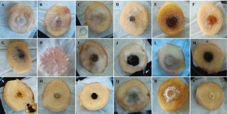
In the first inoculation test, sliced (cut surface) rhizomes tested positive for most fungi, and the only species that were not found were Mucor hiemalis Wehmer (Figure
1O), Nigrospora oryzae (Berkeley and Broome) Petch (Figure 1P) and Verticillium sp. (2) (Figure 1S).
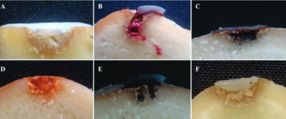
The perforated surface-colonization test was positive for all species evaluated; however, only six species were able to cause tissue rot (Figure 2).
Fusarium oxysporum caused the browning of the
epidermal tissue and cortical hypodermic tissue near the site of inoculation, surface depression, and white mycelia on the internal tissues and epidermal surface (Figure 2A). In rhizomes with natural infections, a range of symptoms were observed from darkening (tanning) to black rot of vascular tissue (Figure 3A). Fusarium sp. caused the browning of
the epidermis, hypodermis and cortex adjacent to the site of inoculation; the depression of the surface; the growth of red and white mycelia on the internal tissues and surface of the epidermis; and the abundant production of red pigment (Figure 2B). Rhizomes with natural infections were observed inside and outside and were found to have abundant red and white mycelia and abundant production of red pigment (Figure 3A).
Acremonium murorum produced black mycelia inside the
cavities of the rhizome and caused the darkening of epidermal and hypodermic tissues peripheral to the site of inoculation (Figure 2C). Colonization of black mycelia was observed in the vascular regions of rhizomes with natural infections of the fungus (Figure 3C). Acrostalagmus luteo-albus produced
abundant orange mycelia inside the cavities and caused the darkening of epidermal and hypodermic tissues near the inoculation site (Figure 2D). Natural infection resulted in orange mycelia in the inner tissues exposed by cuts (Figure 3D).
Lasiodiplodiatheobromae produced black mycelia in cavities
and on the surface of the rhizomes and caused the browning of epidermal and hypodermic tissues adjacent to the inoculation site (Figure 2E); Natural infections of the fungus resulted in black and white mycelia and pycnidia on the cut surfaces of rhizomes (Figure 3E). Sclerotium rolfsii caused white mycelia in the puncture, browning of epidermal, hypodermical and cortical tissues adjacent to the puncture, and depression of the surface site around the inoculation (Figure 2F). In natural infections, there was widespread colonization of white mycelia and numerous sclerotia (Figure 3F).
Bacteria
Pathogenicity of bacteria
Some rhizomes sampled from the field had softening, widespread tissue defragmentation, soaked appearance, skin browning, bacterial pus (Figures 4A, B) and fetid odor.
FIGURE 1 - Results of the pathogenicity tests for colonization of cut ginger. A.Fusarium oxysporum; B. Fusarium sp.; C.Fusarium solani (in sporodochia formation); D. Fusarium semitectum; E.Acremonium murorum; F.Acrostalagmus luteo-albus; G.Lasiodiplodia theobromae; H. Sclerotium rolfsii; I. Alternaria tenuissima; J. Aspergillus niger; L. Chaetomium sp.; M. Curvularia geniculata; N.
FIGURE 2 - A. Results of the pathogenicity tests for colonization of the punctured surface and rot of ginger rhizomes inoculated with mycelial discs with a cross-sectional view of the rhizomes inoculated with Fusarium oxysporum; B. Fusarium sp.;
C. Acremonium murorum; D. Acrostalagmus luteo-albus;
E. Lasiodiplodia theobromae
and F.Sclerotium rolfsii.
FIGURE 3 - A. Symptoms of natural decay caused by
Fusarium oxysporum; B.
Fusarium sp.; C.Acremonium murorum; D. Acrostalagmus luteo-albus; E. Lasiodiplodia theobromae; F. Sclerotium rolfsii.
FIGURE 4 - A. Symptoms of natural soft rot caused by Enterobacter cloacae
subsp. cloacae on surface
and B. internal tissues of ginger rhizome harvested in Holanda. C. Results of the pathogenicity test in ginger rhizome discs inoculated with
E. cloacae subsp. cloacae
Of the ninety-eight isolates from rhizomes sampled in the field, five were able to cause the symptoms of soft rot, including the characteristic fetid odor, which was found in about 5% of the isolates. All colonies took on a cream color when in the NA medium.
The results of pathogenicity tests can be seen in Figures 4C, D. Softening and maceration of tissue were observed during the test, followed by soaked appearance and fetid odor.
Identification of bacteria
The isolate BAC001 was identified as Pseudomonas
fluorescens (Trevisan) Migula, similar to the biovars II and IV (Schaad et al., 2001) (Table 2). The fatty acid profile showed two species, P. fluorescens, with similarity indices of 0.71 and 0.40 for the Sherlock® ITSA and
IR2A libraries, respectively, and Pseudomonas putida
(Trevisan) Migula, with similarity indices of 0.73 and 0.71 for the Sherlock® ITSA and IR2A libraries, respectively.
However, these data, together with the pathogenic history of these species, ruled out the possibility of the isolate being identified as P. putida (Schaad et al., 2001;
Brenner et al., 2005).
Isolates of BAC003, BAC005, BAC006 and BAC012 showed similar characteristics, except for the tests of urease, L-arabinose and lysine and ornithine decarboxylases (Table 3). The fatty acid profile showed high similarity to E. cloacae subsp. cloacae, from at least
one of the libraries consulted, ranging from 0.71 to 0.81. These tests allowed the identification of these isolates as E.
cloacae subsp. cloacae (Brenner et al., 2005; Nishijima et
al., 2004; Hoffmann et al., 2005).
DISCUSSION
There was a balanced distribution of the saprophytes and phytopathogenic species found through direct and indirect isolation. Using the two methods of isolation was complementary; for example, Acrostalagmus luteo-albus
was only isolated by indirect isolation of the field samples. When the samples were taken from the packing-house, we identified six fungal species, and from the field samples, we identified sixteen species. Despite the smaller number of samples and only one method of isolation, it was expected that samples taken from the packing-house would show a significantly lower number of fungal species because at the postharvest stage rhizomes have been selected, washed, cured, and healed and they are in cold storage.
The first test of fungal colonization was done on the cut surface of rhizomes. This surface consisted of cortical tissue, similar to what is usually exposed through cuts made during postharvest processing or, if there is a stage of healing, to the layer of suber (Lana, 1991). The fungi that were able to colonize these cuts were both pathogenic and opportunistic, a result of the fact that the inoculation was made in freshly-cut unhealed tissue. These results may indicate the importance of the healing process. In the second test, the fungi were able to grow some mycelia along the punctures, but few were able to cause rot, with the exception of F. oxysporum, F. sp., A. murorum, A. luteo-albus, L. theobromae and S. rolfsii. These results confirmed
that damage to the rhizomes contributes to infection by fungi that cause rot.
Fusarium oxysporum is considered a serious
problem in many ginger-producing countries such as the
TABLE 2 - Relationship and results of identification tests for Pseudomonas fluorescens isolated from ginger rhizomes with symptoms of rot harvested in Holanda
*Data from Brenner et al.(2005) and Schaad et al. (2001); ** A ITSA1.0 MIS library; BIR2A1.0 MIS library. S
Test Isolate
BAC001
*P. fluorescens *P.
marginalis
*P. viridiflava
*P. putida
bvI bvII bvIII bvIV bvV bvA bvB
Gram - - -
-Aerobic growth + + + + + + + + + +
Anaerobic growth - - -
-Fluorescence on King B + + + + + + + + + +
Oxidase + + + + + + + + +
-Urease - - -
-Levan + + + - + - + - -
-Dehydrolase arginine - + + + + + + - + +
NO3to N2 + - + + + - - - -
-Liquefaction of gelatina + + + + + - Nd + -
-Potato rot + + + + + + Nd + -
-Use of sucrose + + + - + D + - - D
Use of D-trehalose + + + D + D + - -
-**Similarity with the fatty acid prole ofP.fluorescens, in %
A 71 B
40 ** Similarity with the fatty acid
prole ofP.putida, in %
A 73 B
TABLE 3 - Relationship and results of identification tests for Enterobacter cloacae subsp. cloacae isolated from ginger rhizomes with
symptoms of rot harvested in Holanda (isolates BAC003 and BAC005), Caramurú (BAC006), and Rio das Farinhas (BAC012)
*Data from Brenner et al. (2005) and Schaad et al. (2001); ** A ITSA1.0 MIS library; BIR2A1.0 MIS library. S
Test Isolate *E. cloacae
subsp. cloacae
*Pectobacterium
spp.
BAC003 BAC005 BAC006 BAC012
Gram - - -
-Aerobic growth + + + + + +
Anaerobic growth - - -
-Fluorescence on King B - - -
-Oxidase - - - Nd
Urease - - - + V
-Liquefaction of gelatina - - - Nd
Ornithine decarboxylase + - + + +
-Lysine decarboxylase - - + - -
-Use of the following sugars
D-Mannose + + + + +
D-Mannitol + + + + +
D-Maltose + + + + +
D-Trehalose + + + + +
L-Arabinose - - - + +
Erythritol - - - -
-** Similarity with the fatty acid prole of
E. cloacaesubsp.cloacae, in %
A
00
B81 A
71
B78 A
77
B00 A
00
B79
USA (Trujilo, 1963), India (Dake and Edison, 1989) and Australia (Stirling, 2004). Because twice as many isolates of this species were found with direct isolation as with indirect, it is likely that infections occur from inoculums in the soil, but also in large part from fungi entering wounds caused by postharvest processing. Acremonium murorum was reported
in ginger in the USA and in Chinese rhizomes in 1946. The species A. stromaticum was found in ginger in 1975 in India
(Farr and Rossman, 2010). Acrostalagmus luteo-albus was
reported in ginger in 1941 and was identified at the time as
A. cinnabarinus in rhizomes from China imported to the
USA (Farr and Rossman, 2010). Lasiodiplodia theobromae
was reported as being associated with ginger in India in 1979 (Farr and Rossman, 2010). Sclerotium rolfsii can
infect many tubers and rhizomes and was reported on ginger in South Africa in 2000 (Farr and Rossman, 2010). This study is the first report of ginger rhizome rot caused by
A. murorum, A. luteo-albus and L. theobromae in Brazil.
Several species of Fusarium were found to be
associated with ginger rhizomes, but not all were capable of causing decay. As discussed earlier, F. oxysporum and
Fusarium sp. had the highest occurrence and were able to
cause rot. However, it is important to note that F. solani,
which had a high incidence in the field, did not cause rot in rhizomes. There was also sporulation with the development of sporodochia characteristic of the species, which produced a green color in the rhizome discs (Figure 1C). Finally, there were a few occurrences of F. semitectum, but it was not able
to cause rot during our tests.
Decay-causing bacteria were detected in only 5% of rhizomes, with an incidence of E. cloacae subsp.
cloacae in 4% and P. fluorescens in 1%. For these bacteria,
occurrence rates of 5% and 3%, respectively, were found in the ‘Holanda’ region, indicating that this region has more problems with ginger rot caused by these bacteria (Tables 2 and 3). However, it is possible that a larger number of isolates of E. cloacae subsp. cloacae was associated with
rhizomes. According to Nishijima et al. (2004), this bacteria can be found in healthy tissues and may cause rot when conditions are favorable. In studies of other plant species, McInroy and Kloepper (1995) and Magnani (2005) found that P. fluorescens may behave similarly.
In Brazil, P. fluorescens has been reported on lettuce, garlic, potatoes, onions, tomatoes and philodendron (Malavolta Jr. et al., 2008). In the UK, it has been associated with post-harvest rot of broccoli (Cui and Harling, 2006). It is noteworthy that the species known to cause rot in the reserve plant organs are P. fluorescens, Pseudomonas
marginalis (Brown) Stevens and Pseudomonas viridiflava
(Burkholder) Dowson (Hunter and Cigna, 1981; Krejzar et al., 2008; Malavolta Jr. et al., 2008).
Some species of Enterobacter can cause diseases
in humans, and some can also cause plant diseases. Ginger rhizome rot caused by E. cloacae subsp. cloacae has been
reported in the USA, and that caused by Enterobacter sp.
has been reported in Australia (Stirling, 2004). Netoet al. (2003) evaluated plant and clinical case isolates and observed that clinical strains showed a high similarity with plant pathogenic isolates in standard serological total protein, RAPD and with phytopathogenic capacity in onions. The presence of
E. cloacae subsp. cloacae may indicate a contamination of
This study is the first reported occurrence of P.
fluorescens associated with and causing the decay of ginger rhizomes in the world and the first reported occurrence of E.
cloacae subsp. cloacae in association with and causing the
decay of ginger rhizomes in Brazil.
The identification of fungi and bacteria causing the decay of ginger rhizomes is the first step towards further studies to develop an integrated crop management program to prevent and control ginger rot in fields and post-harvest in the highlands of the Serrana region of Espírito Santo and other regions of Brazil.
ACKNOWLEDGEMENTS
We thank Conselho Nacional de Desenvolvimento Científico e Tecnológico - CNPq, Fundação de Amparo à Pesquisa do Estado de Minas Gerais - FAPEMIG, Coordenação de Aperfeiçoamento de Pessoal de Nível Superior - CAPES, Gaia Importação and Exportação Ltda and Raízes Serranas Ltda for financial support. We also thank the technical assistance of Maria Sueli de Oliveira Cardoso.
REFERENCES
Alfenas AC, Ferreira FA, Mafia RG, Gonçalves RC (2007) Isolamento de fungos fitopatogênicos. In: Alfenas AC, Mafia RG (Eds.) Métodos em Fitopatologia. Viçosa MG. Editora UFV. Brasil. MDIC, Ministério do Desenvolvimento, Indústria e Comércio Exterior. SECEX, Secretaria do Comércio Exterior. Available at: http://www.mdic.gov.br/sitio . Accessed on January 26, 2008.
Brenner DJ, Krieg NR, Staley JT (2005) The Proteobacteria. Part B - The Gammaproteobacteria. In: Garryt GM (Ed.) Bergey’s Manual of Systematic Bacteriology. Vol. 2. 2nd Ed. Ann Arbor MI,
USA. Springer.
Carmichael JW, Kendrick WB, Conners LI, Singler L (1980) Genera of Hyphomycetes. Edmonton Canada. The University of Alberta Press.
Cui X, Harling R (2006) Evaluation of bacterial antagonists for biological control of broccoli head rot caused by Pseudomonas fluorescens. Phytopathology 96:408-416.
Dake GN, Edison S (1989) Association of pathogens with rhizome rot of ginger in Kerala. Indian Phytopathology 42:116-119. Domsch KH, Gams W Anderson T (2007) Compendium of Soil Fungi. 2nd Ed. Eching Germany. IHW-Verlag.
Fahy PC, Parsley GL (1983) Plant Bacterial Diseases: A Diagnostic Guide. Sidney Australia. Academic Press.
Farr DF, Rossman AY (2010) Fungal Databases, Systematic Mycology and Microbiology Laboratory, USDA-ARS. Available at: http://nt.ars-grin.gov/fungaldatabases. Accessed on February 13, 2008.
Hanlin RT (1998) Combined Keys to Illustrated Genera of Ascomycetes. Vols. I and II. St. Paul MN, USA. APS Press. Hanlin RT (1990) Illustrated Genera of Ascomycetes. Vol. I. 2nd
Ed. St. Paul MN, USA. APS Press.
Hoffmann H, Stindl S, Ludwig W, Stumpf A, Mehlen A, Heesemann J, Monget D, Schleifer KH Roggenkamp A (2005) Reassignment of Enterobacter dissolvens to Enterobacter cloacae as E. cloacae
subspecies dissolvens comb. nov. and emended description of
Enterobacter asburiae and Enterobacter kobei. Systematic and
Applied Microbiology28:196-205.
Hugh R, Leifson E (1953) The taxonomic significance of fermentative versus oxidative metabolism of carbohydrates by various Gram-negative bacteria. Journal of Bacteriology 66:24-26.
Hunter JE, Cigna JA (1981) Bacterial blight incited in parsnip by Pseudomonas marginalis and Pseudomonas viridiflava.
Phytopathology 71:1238-1241.
ITC. International Trade Centre UNCTAD/WTO. Global Spice Market: Imports – 1996-2000. Available at: http://www. fao.org/fileadmin/user_upload/inpho/docs/Post_Harvest_ Compendium_-_Ginger.pdf. Accessed on July 15, 2008.
Kado CI, Heskett MG (1970) Selective media for isolation of
Agrobacterium, Coryneobacterium, Erwinia, Pseudomonas and Xanthomonas. Phytopathology 60:969-976.
Kavita PG, Thomas G (2008) Population genetic structure of the clonal plant Zingiberzerumbet (L.) Smith (Zingiberaceae),
a wild relative of cultivated ginger, and its response to Pythium aphanidermatum. Euphytica160:89-100.
King EO, Ward MK, Raney DE (1954) Two simple media for the demonstration of pyocyanin and fluorescin. Journal of Laboratory and Clinical Medicine 44:301-307.
Krejzar V, Mertelík J, Pánková I, Kloudová K, Václav K (2008)
Pseudomonas marginalis associated with soft rot of Zantedeschia
spp. Plant Protection Science 44:85-90.
Lana, MM (1991) Estudo da conservação pós-colheita de rizomas de gengibre (Zingiber officinale Roscoe). M.Sc Dissertation,
Universidade Federal de Viçosa. Viçosa MG.
Leslie JF, Summerell BA (2006) The Fusarium Laboratory
Manual. Ames IA, USA. Blackwell Publishing.
Magnani GS (2005) Diversidade de bactérias endofíticas em cana-de-açúcar. M.Sc Dissertation, Universidade Federal do Paraná. Curitiba PR.
Malavolta Jr VA, Almeida IMG (1998) Podridão em gengibre causada por bactérias pectinolíticas do gênero Erwinia. Summa
Phytopathologica24:67.
Malavolta Jr VA, Beriam LOS, Almeida IMG, Rodrigues Neto J, Robbs CF (2008) Bactérias fitopatogênicas assinaladas no Brasil: uma atualização. Summa Phytopatologica 34:1-188.
McInroy JA, Kloepper JW (1995) Survey of indigenous bacterial endophytes from cotton and sweet corn. Plant and Soil 173:337-342.
Moeller V (1955) Simplified tests for some amino acid descarboxylases and for arginine dihydrolase system. Acta Pathologica et Microbiologia Scandinavica 36:158-172.
Nelson PE, Toussoun TA, Marasas WF (1983) Fusarium Species:
An Illustrated Manual for Identification. State College PA, USA. Pennsylvania State University Press.
Rosato YB (2003) Comparative RFLP-ITS Analysis between
Enterobacter cloacae strains isolated from plants and clinical
origin. Arquivos do Instituto Biológico 70:367-372.
Nishijima KA, Alvarez AM, Hepperly PR, Shintaku MH, Keith LM, Sato DM, Bushe, Armstrong JW, Zee FT (2004) Association of Enterobater cloacae with rhizome rot of edible ginger in
Hawaii. Plant Disease 88:1318-1327.
Nishijima KA, Couey HM, Alvarez, AM (1987) Internal yellowing, a bacterial disease of papaya fruits caused by Enterobacter cloacae.
Plant Disease 71:1029-1034.
Nishijima KA, Wall MM, Siderhurst MS (2007) Demonstrating pathogenicity of Enterobacter cloacae on macadamia and
indentifying associated volatiles of gray kernel of macadamia in Hawaii. Plant Disease 91:1221-1228.
Pereira JM, Barreto RW, Ellison CA, Maffia LA (2003)
Corynespora cassiicola f.sp. lantanae: A potencial biocontrol
agente from Brazil for Lantanacamara. Biological Control
26:21-31.
Ryu EA (1940) A simple method for differentiation between Gram-positive and Gram-negative organisms without staining. Kitazato Archives of Experimental Medicine 17:58-63.
Schaad NW, Jones JB, Chun W (2001) Laboratory Guide for
TPP 2012-0023 - Received 26 May 2012 - Accepted 28 January 2013 Section Editor: Silvaldo Felipe da Silveira Identification of Plant Pathgenic Bacteria. 3rd Ed. St. Paul MN,
USA. APS Press.
Simmons EG (2007) Alternaria – An Identification Manual.
Utrecht The Netherlands. CBS Fungal Biodiversity Centre. Souza JL, Resende PL (2001) Cultivo orgânico de gengibre, taro e inhame. Viçosa MG. CPT.
Stirling AM (2002) Erwinia chrysanthemi, the cause of soft rot
in ginger (Zingiber officinale) in Australia. Australasian Plant Pathology 31:419-420.
Stirling AM (2004) The causes of poor establishment of ginger (Zingiber officinale) in Queensland, Australia. Australasian Plant Pathology 33:203-210.
Thornley MJ (1960) The differentiation of Pseudomonas from
other gram-negative bacteria on the basis of arginine metabolism. Journal of Applied Bacteriology 23:37-52.
Trujilo EE (1964) Fusarium yellows and rhizome rot of common
ginger. Phytopathology53:1370-1371.
Wang PH, Chung CY, Lin YS, Yeh Y (2003) Use of polymerase chain reaction to detect the soft rot pathogen, Pythium myriotylum,