To wards unde rstanding the
kallikre in-kinin syste m : insights
fro m m e asure m e nt o f kinin pe ptide s
St. Vincent’s Institute of Medical Research, Fitzroy, Victoria, Australia D.J. Campbell
Abstract
The kallikrein-kinin system is complex, with several bioactive pep-tides that are formed in many different compartments. Kinin peppep-tides are implicated in many physiological and pathological processes including the regulation of blood pressure and sodium homeostasis, inflammatory processes, and the cardioprotective effects of precondi-tioning. We established a methodology for the measurement of indi-vidual kinin peptides in order to study the function of the kallikrein-kinin system. The levels of kallikrein-kinin peptides in tissues were higher than in blood, confirming the primary tissue localization of the kallikrein-kinin system. Moreover, the separate measurement of bradykallikrein-kinin and kallidin peptides in man demonstrated the differential regulation of the plasma and tissue kallikrein-kinin systems, respectively. Kinin peptide levels were increased in the heart of rats with myocardial infarction, in tissues of diabetic and spontaneously hypertensive rats, and in urine of patients with interstitial cystitis, suggesting a role for kinin peptides in the pathogenesis of these conditions. By contrast, blood levels of kallidin, but not bradykinin, peptides were suppressed in patients with severe cardiac failure, suggesting that the activity of the tissue kallikrein-kinin system may be suppressed in this condition. Both angiotensin converting enzyme (ACE) and neutral endopepti-dase (NEP) inhibitors increased bradykinin peptide levels. ACE and NEP inhibitors had different effects on kinin peptide levels in blood, urine, and tissues, which may be accounted for by the differential contributions of ACE and NEP to kinin peptide metabolism in the multiple compartments in which kinin peptide generation occurs. Measurement of the levels of individual kinin peptides has given important information about the operation of the kallikrein-kinin system and its role in physiology and disease states.
Co rre spo nde nce
D.J. Campbell St. Vincent’s Institute of Medical Research 41 Victoria Parade Fitzroy Victoria 3065 Australia
Fax: + 61-3-9416-2676 E-mail:
J.Campbell@ medicine.unimelb.edu.au
Presented at the III International Symposium on Vasoactive Peptides, Belo Horizonte, MG, Brasil, O ctober 8-10, 1999.
Received November 26, 1999 Accepted February 3, 2000
Ke y wo rds ·Bradykinin
·Kallidin
·Kallikrein
·Kininogen
Intro ductio n
Kinins are potent vasodilator oligopep-tides that contain the nonapeptide bradyki-nin [BK-(1-9)] as part of their sequence. Kinins have effects on many different as-pects of physiology, including the regulation of blood pressure and of renal and cardiac
function, and have been implicated in the pathogenesis of hypertension and inflamma-tion (for a review, see 1). Kinins may medi-ate in part the effects of angiotensin convert-ing enzyme (ACE) and neutral endopepti-dase 24.11 (NEP) inhibitors.
The B2 receptor normally predominates, whereas B1 receptors are induced by tissue injury, such as that which occurs following myocardial ischaemia (2) and inflammation (3). A complex variety of kinin peptides acts through these receptors. In man, plasma kal-likrein forms bradykinin [BK-(1-9)] from high molecular weight (HMW) kininogen, whereas tissue kallikrein forms kallidin [Lys0
-BK-(1-9), KBK-(1-9)] from low molecular weight (LMW) and HMW kininogens (1). Moreover, bradykinin peptides may be gen-erated by aminopeptidase-mediated cleav-age of kallidin peptides (Figure 1). These peptides are more potent at the B2 receptor (4) (Figure 2). A proportion of kinins is hydroxylated on proline3 of the BK-(1-9)
sequence (5), and hydroxylated kinins have similar biological activity to non-hydroxy-lated kinins (4). Enzymes collectively called kininases metabolize these kinins (Figure 3). The carboxypeptidase (kininase I) metabo-lites of BK-(1-9) and KBK-(1-9) are brady-kinin-(1-8) [BK-(1-8)] and Lys0
-bradykinin-(1-8) [KBK--bradykinin-(1-8)], respectively, which are also bioactive, but more potent on B1 recep-tors (4), whereas the ACE and NEP metabo-lites bradykinin-(1-7) [BK-(1-7)] and Lys0
-bradykinin-(1-7) [KBK-(1-7)] are inactive (Figure 2). Kinin peptides are less complex in the rat than in man. In the rat both plasma and tissue kallikrein generate BK-(1-9), and kinin peptides are not hydroxylated in ro-dents.
Despite its long history, many aspects of the physiology of bradykinin peptides and their role in disease states are yet to be defined. The sole sources of kinin peptides are the kininogens. Kininogen deficiency in man is reported to be relatively asymptomat-ic, suggesting that kinin peptides may have little role in normal physiology. Although the detection of these patients is due to their severe abnormality in surface-activated in-trinsic coagulation, fibrinolytic and kinin-generating pathways, these patients have little or no bleeding abnormality (6,7). However, studies in experimental animals provide ev-idence for an important role for kinin pep-tides in the regulation of blood pressure and sodium homeostasis, and in contributing to inflammatory processes (8-11). The B2 re-ceptor gene knockout mouse is hypertensive with cardiac hypertrophy (8). Both the B2 receptor gene knockout mouse and the ki-ninogen-deficient Brown-Norway Katholiek
Figure 1 - Generation of kinin peptides by tissue and plasma kallikrein. In man tissue kallikrein generates kallidin w hereas plasma kallikrein generates bradykinin (BK). How ever, in the rat both tissue and plasma kallikrein generate bradykinin. LM WK, HM WK, Low and high molecular w eight kininogens, respectively; KBK, kallidin (Lys0-bradykinin); Hyp3,
hy-droxylated proline.
non-hydroxylated
Plasma kallikrein
Kallidin LM WK
Kininogen gene
HM WK
Tissue kallikrein
KBK-(1-9) Hyp3-KBK-(1-9)
BK-(1-9) Hyp3-BK-(1-9)
Bradykinin Aminopeptidase
hydroxylated
Figure 2 - Kinin peptides and kinin receptors. Whereas bradykinin [BK-(1-9)] and kallidin [KBK-(1-9)] are more potent agonists of the B2 receptor, BK-(1-8) and KBK-(1-8) are more potent
agonists of the B1 receptor. ACE, Angiotensin converting enzyme; NEP, neutral
endopepti-dase; BK, bradykinin; KBK, Lys0-bradykinin.
BK-(1-5) KBK-(1-5)
KBK-(1-7) BK-(1-7)
BK-(1-9)
BK-(1-8) B1 receptor KBK-(1-8) Carboxypeptidase
ACE, NEP ACE
rat strain show increased sensitivity to the pressor effects of increased dietary salt, min-eralocorticoid administration, and angio-tensin II (Ang II) infusion, and an impair-ment of the cardioprotective effects of pre-conditioning (8-11). In addition, the Brown-Norway Katholiek rat strain shows a re-duced response to inflammatory stimuli (10). There have been many different ap-proaches to the study of the kallikrein-kinin system. One approach has been the measure-ment of kallikrein and kininogen levels in blood, urine and tissues. However, there may be difficulties in the interpretation of such studies. For example, alternative pathways of kinin formation involving enzymes other than kallikreins may operate in disease states. Although LMW kininogen is a poor sub-strate for plasma kallikrein, it will form BK-(1-9) in the presence of neutrophil elastase, which, by cleaving a fragment from LMW kininogen, renders LMW kininogen much more susceptible to cleavage by plasma kal-likrein (12). Moreover, the combination of mast cell tryptase and neutrophil elastase releases BK-(1-9) from oxidized kininogens that are resistant to cleavage by kallikreins (13). Furthermore, endogenous inhibitors of kallikrein activity may modulate the activity of kallikrein in vivo, and kallikrein and
pre-kallikrein may be activated during sample processing. In addition, kininase activity is a
critical determinant of the activity of the kallikrein-kinin system through the modula-tion of kinin peptide levels in vivo.
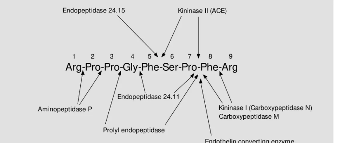
My laboratory focussed on the measure-ment of kinin peptides to better understand the functioning of the kallikrein-kinin sys-tem. Initially, we developed amino-termi-nal-directed radioimmunoassays (RIA) for BK-(1-9), its kininase I metabolite BK-(1-8), and the biologically inactive ACE and NEP metabolite (1-7) (14,15). (1-7), BK-(1-8), and BK-(1-9) were separated by high performance liquid chromatography (HPLC) before RIA. Calculation of the BK-(1-7)/ BK-(1-9) and BK-(1-8)/BK-(1-9) ratios pro-vided indices of the rate of BK-(1-9) me-tabolism to BK-(1-7) and BK-(1-8), respec-tively. We applied these assays to the meas-urement of kinin peptides in blood, urine, and tissues of rats and dogs (14-27). Subse-quently, we developed amino-terminal-di-rected RIA for the corresponding kallidin peptides (28-30). These bradykinin and kal-lidin peptide RIA were also able to measure hydroxylated bradykinin and kallidin pep-tides. We were able to separate non-hydroxy-lated and hydroxynon-hydroxy-lated bradykinin and kalli-din peptides by HPLC, thus enabling meas-urement of 6 bradykinin peptides [non-hy-droxylated and hy[non-hy-droxylated (1-7), BK-(1-8), BK-(1-9)] and 6 kallidin peptides [non-hydroxylated and [non-hydroxylated KBK-(1-7),
Arg-Pro-Pro-Gly-Phe-Ser-Pro-Phe-Arg
Aminopeptidase P Kininase I (Carboxypeptidase N)
Prolyl endopeptidase Endopeptidase 24.11
Carboxypeptidase M
Endothelin converting enzyme
1 2 3 4 5 6 7 8 9
Endopeptidase 24.15 Kininase II (ACE)
KBK-(1-8), KBK-(1-9)] in blood, urine, and tissue of man.
The purpose of this brief review is to summarize what we have learnt about the kallikrein-kinin system from measurement of kinin peptide levels, and to discuss these findings in relation to data obtained from alternative approaches to the study of the kallikrein-kinin system.
Kinin pe ptide le ve ls in blo o d and tissue s
Circulating levels of kinin peptides are very low, usually less than 3 fmol/ml. We attribute the higher levels reported in previ-ous studies to a failure to adequately prevent artefactual generation of kinin peptides dur-ing sample collection and processdur-ing. Much higher levels of kinin peptides are measured during acute episodes of angio-oedema (31). The maintenance of low levels of kinin pep-tides is relevant to the success of ACE and NEP inhibitor therapy. There is considerable evidence for a role for kinin peptides in mediating some of the effects of ACE and NEP inhibition. However, these inhibitors are generally free of the side effects one might expect from increased kinin peptide levels. For example, increased endothelial
permeability, such as that manifest in angio-oedema, is an infrequent complication of ACE inhibitor therapy. The low incidence of side effects with ACE and NEP inhibitor therapy indicates that kinin peptide levels are low, even in the presence of ACE and/or NEP inhibition, and thus any effect of ACE and NEP inhibition on kinin peptide levels must be subtle. This is to be expected, given the many different kininases that metabolize kinin peptides (Figure 3). The relative pro-portions of these different kininases may vary between different tissue compartments. Any effect of ACE or NEP inhibition on kinin peptide levels in a particular compart-ment will depend on the relative contribu-tion of ACE and NEP to kinin peptide me-tabolism in that compartment.
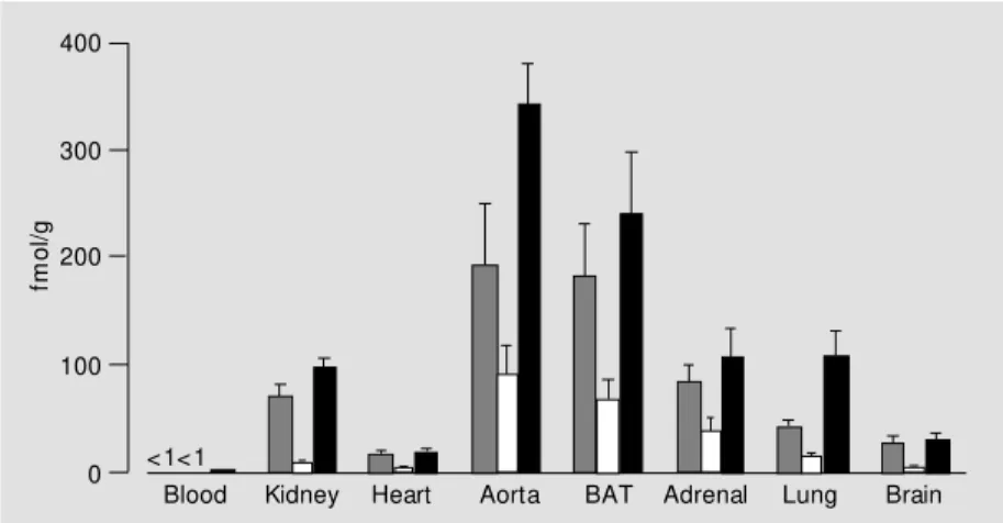
In studies in normal rats, tissue levels of kinin peptides were higher than circulating levels, consistent with tissues being the main site of formation of kinin peptides (Figure 4). We found increased levels of kinin pep-tides in tissues of rats with myocardial in-farction, in diabetic rats, and in spontane-ously hypertensive rats (SHR) (21,26,32). In rats with myocardial infarction, BK-(1-9) levels were increased in heart on day 2 and 3 post-infarction, associated with a decrease in the BK-(1-7)/BK-(1-9) ratio, suggesting reduced metabolism of BK-(1-9) to BK-(1-7) in this tissue. The increased cardiac BK-(1-9) levels in the acute phase of myocardial infarction were consistent with a role for this peptide in cardioprotection and limitation of infarct size (33,34). In diabetic rats, the lev-els of BK-(1-9) and its metabolite BK-(1-7) were increased in kidney, aorta, and heart. The increased BK-(1-9) levels were consist-ent with the participation of this peptide in the vascular and metabolic abnormalities of diabetes, in particular the glomerular hyper-filtration, increased glomerular plasma flow, and elevated glomerular capillary hydraulic pressure of early insulin-dependent diabetes (35). The increased levels of BK-(1-9) and its metabolites in kidney, adrenal, lung, and
fm
o
l/g
400
300
200
100
0
Blood Kidney Heart Aorta BAT Adrenal Lung Brain <1<1
heart of young SHR suggested increased kallikrein activity in these tissues. More-over, the reduced BK-(1-7)/BK-(1-9) and BK-(1-8)/BK-(1-9) ratios in kidney of SHR indicated reduced endopeptidase and car-boxypeptidase-mediated metabolism of BK-(1-9), that may have contributed to the in-creased BK-(1-9) levels in this tissue. When taken together with the previously reported hypotensive effect of B2 receptor antago-nism in young SHR (36), and the cosegrega-tion of the SHR kallikrein gene with blood pressure (37), the increased BK-(1-9) levels in tissues of young SHR were consistent with a role for this peptide in the pathogen-esis of hypertension in these rats.
In man, the generation of bradykinin and kallidin peptides by plasma and tissue kal-likrein, respectively, enabled differentiation between the role of plasma and tissue kal-likrein in kinin peptide formation. In blood and atrial tissue we found bradykinin tides to be more abundant than kallidin pep-tides, whereas kallidin peptides were more abundant in urine (28-30). Bradykinin and kallidin peptide levels were higher in venous than arterial blood, consistent with the for-mation of these peptides in tissues. Further evidence for the formation of kinin peptides in tissue was the higher level of bradykinin and kallidin peptides in atrial tissue than in blood.
We investigated whether the kallikrein-kinin system is activated in interstitial cysti-tis, a chronic inflammatory condition of the bladder wall, by measuring urinary excre-tion rates of kinin peptides. Catheter urine was collected from subjects during a water diuresis (~10 ml/min) before and after dis-tension of the bladder with 100 ml water, and the contribution of the bladder wall to urinary kinin peptides was assessed by meas-uring the change in kinin peptide levels after 2 min of bladder stasis before and after distension. We found increased urinary ex-cretion rates of bradykinin, but not kallidin, peptides after 2-min bladder stasis after
blad-der distension (28). These findings provid-ed evidence for increasprovid-ed bradykinin pep-tide levels in the bladder wall of subjects with interstitial cystitis, where these pep-tides may participate in the pathogenesis and symptomatology of this condition.
In comparison with normal subjects, pa-tients with severe cardiac failure, who were receiving ACE inhibitor therapy, had in-creased blood levels of bradykinin peptides but suppressed blood levels of kallidin pep-tides (30). This suppression of blood kalli-din peptide levels despite ACE inhibitor therapy suggested that the activity of the tissue kallikrein-kinin system may be sup-pressed in severe cardiac failure.
Fe e dback re gulatio n o f the kallikre in-kinin syste m
The role of endogenous kinin peptides has been determined mainly by study of the effects of kinin antagonists, most often the B2 receptor antagonist icatibant (D-Arg-[Hyp3, Thi5, D-Tic7, Oic8]-bradykinin).
In contrast to these findings, Siragy et al. (38) reported that icatibant increased immu-noreactive kinin peptide levels in renal inter-stitial fluid collected by microdialysis probe from sodium-deficient dogs. The difference between our findings and those of Siragy et al. (38) may be due to differences between species, or to differences in sodium status. Moreover, the identity of the immunoreac-tive material measured by Siragy et al. (38) is uncertain, as is the effect of the chronically indwelling microdialysis probe on kinin pep-tide formation.
Effe cts o f ACE inhibitio n o n kinin pe ptide le ve ls
Many studies implicate kinin peptides in the effects of ACE inhibitors, including their effects on blood pressure and cardiac hyper-trophy (39-41). We studied the effects of ACE inhibition on kinin peptide levels in both experimental animals and in man. The dose-related effects of ACE inhibitors on circulating and tissue levels of angiotensin
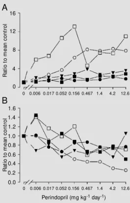
and bradykinin peptides were examined by administration of the ACE inhibitor perindo-pril or lisinoperindo-pril to rats in the drinking water for 7 days (17). Perindopril increased circu-lating BK-(1-9) levels ~8-fold with a thresh-old dose of 0.052 mg kg-1 day-1, and
in-creased BK-(1-9) levels in kidney, heart and lung in parallel with the changes observed for blood (Figure 5). By contrast, aortic and brown adipose tissue BK-(1-9) and BK-(1-7) levels increased several-fold for perindopril doses as low as 0.006 mg kg-1 day-1. Lisinopril
also increased aortic BK-(1-9) and BK-(1-7) levels at doses below the threshold for de-crease in Ang II/Ang I ratio. These data indicated that vascular BK-(1-9) levels re-sponded to low doses of ACE inhibitor, and may be important mediators of the effects of these compounds. Moreover, the increase in vascular kinin peptide levels at doses of ACE inhibitor below the apparent threshold for ACE inhibition led us to propose that these compounds increase kinin peptide lev-els by inhibition of a non-ACE kininase. This concept is discussed later in this sec-tion.
We investigated the effects of ACE inhi-bition on kinin peptide levels in rats with myocardial infarction. Perindopril increased BK-(1-9) levels in heart by 45%, which did not achieve statistical significance, although there was a statistically significant reduction in the BK-(1-7)/BK-(1-9) ratio in heart, indi-cating inhibition of cardiac metabolism of BK-(1-9) by perindopril. We also showed that ACE inhibition with either perindoprilat or enalaprilat increased blood levels of kinin peptides in dogs with pacing-induced heart failure (27).
In both rats and dogs we found that ACE inhibition increased the levels of both BK-(1-9) and its metabolite BK-(1-7), although there was suppression of the BK-(1-7)/BK-(1-9) ratio (17,27). As shown in Figure 2, BK-(1-7) is both the product of BK-(1-9) metabolism by ACE and the substrate for its subsequent metabolism to BK-(1-5). The
in-Figure 5 - Dose-response effects of perindopril on BK-(1-9) levels (A) and BK-(1-7)/BK-(1-9) ratios (B) in plasma (open circles), kid-ney (filled circles), lung (tri-angles), heart (filled squares), and aorta (open squares) of nor-mal nor-male Sprague Daw ley rats administered perindopril in the drinking w ater for 7 days. Data are reported as the ratio to the mean of the respective control group. Error bars and signifi-cance levels are not show n. (Re-draw n from Ref. 17).
R
at
io
t
o
m
e
an
c
o
n
tr
o
l
16
12
8
4
0
R
at
io
t
o
m
e
an
c
o
n
tr
o
l
1.6 1.4
0.8 0.6
0.0 1.2 1.0
0.4 0.2
A
B
0 0.006 0.017 0.052 0.156 0.467 1.4 4.2 12.60 0.006 0.017 0.052 0.156 0.467 1.4 4.2 12.6
crease in BK-(1-7) levels during ACE inhi-bition suggests that ACE inhiinhi-bition has greater impact on BK-(1-7) metabolism than BK-(1-7) formation. Thus, in comparison with other kininases, ACE has a more domi-nant role in (1-7) metabolism than BK-(1-9) metabolism.
Our studies in man demonstrated a dif-ferential regulation of bradykinin and kalli-din peptides in blood, and differential regu-lation of kinin peptide levels in blood and atrial tissue. ACE inhibition increased blood levels of bradykinin but not kallidin peptides in man (30). Moreover, ACE inhibition did not modify bradykinin or kallidin peptide levels or the BK-(1-7)/BK-(1-9) ratio in atrial tissue of patients undergoing cardiac sur-gery, although Ang II levels and Ang II/Ang I ratio in atrial tissue were reduced (29). We consider it unlikely that the increase in blood levels of bradykinin, but not kallidin, pep-tides was due to artefactual generation of bradykinin peptides by plasma kallikrein during sample collection because ACE inhi-bition would be expected to protect bradyki-nin and kallidin peptides to a similar degree. A more likely explanation for the differen-tial effects of ACE inhibition on blood levels of bradykinin and kallidin peptides is the formation of bradykinin and kallidin pep-tides in different compartments, where ACE may make a greater or lesser contribution to kinin metabolism. Thus, if ACE were a ma-jor kininase in the compartment where brady-kinin peptides were formed, one would ex-pect ACE inhibition to increase bradykinin peptide levels. By contrast, if non-ACE kininases were predominant in the compart-ment where kallidin peptides were formed, ACE inhibition may not affect kallidin pep-tide levels.
Our finding that ACE inhibition did not modify bradykinin or kallidin peptide levels in atrial tissue is evidence against a role for changes in cardiac kinin peptide levels in mediating the therapeutic effects of ACE inhibition. It is possible that other enzymes
such as NEP (42), neutral endopeptidase 24.15 (43), and aminopeptidase P (44) play a greater role than ACE in kinin metabolism in the interstitium of atrial tissue.
BK-(1-9) disappears in one passage through the pulmonary vascular bed (48). If lung kininase activity were reduced by 2% such that 96% of circulating BK-(1-9) disappeared in one passage through the pulmonary vas-cular bed, then the biological effects of intra-venously administered BK-(1-9) would be increased 2-fold.
Recent studies support the proposal that ACE inhibitors may interact with the kinin receptor, although this interaction appears to be indirect, and mediated by cross-talk be-tween ACE and the B2 receptor (49,50). There is evidence that this cross-talk may involve more than one mechanism. Marcic et al. (49) found that a variety of ACE inhibi-tors and ACE substrates, including angio-tensin-(1-7), were able to resensitise the B2
receptor. By contrast, Benzing et al. (50) found that the ACE substrate hippuryl-L-histidyl-L-leucine did not reproduce the ef-fects of ramiprilat on B2 receptor transloca-tion and reactivatransloca-tion of signaling events ini-tiated by the B2 receptor. Thus, it would appear that different aspects of B2 receptor function show differential modulation by ACE substrates.
Effe cts o f co mbine d ACE and NEP inhibitio n
The combination of NEP and ACE inhi-bition is a candidate therapy for hyperten-sion and cardiac failure. Given that NEP and ACE metabolize angiotensin and bradykinin peptides, we investigated the effects of NEP inhibition and combined NEP and ACE inhi-bition on the levels of these peptides in both normal Sprague Dawley rats and in rats with myocardial infarction (23,24). For normal rats we administered the NEP inhibitor ecadotril (0, 0.1, 1, 10, 100 mg kg-1 day-1),
either alone or together with a submaximal dose of perindopril (0.2 mg kg-1 day-1), to
rats by 12 hourly gavage for 7 days (23). Ecadotril produced diuresis, natriuresis, and increased urine cyclic GMP and BK-(1-9) levels. Perindopril increased BK-(1-9) lev-els in blood, kidney, and aorta. Combined NEP/ACE inhibition produced the summa-tion of these effects of separate NEP and ACE inhibition. In addition, perindopril po-tentiated the ecadotril-mediated diuresis, natriuresis and decrease in urine BK-(1-7)/ BK-(1-9) ratio. This study demonstrated that combined NEP/ACE inhibition produced greater inhibition of kinin peptide metabo-lism than either inhibitor alone, leading to enhanced effects on diuresis and natriuresis. In rats with myocardial infarction we administered ecadotril (0, 0.1, 1, 10, 100 mg kg-1 day-1), either alone or together with a
submaximal dose of perindopril (0.2 mg kg-1
day-1) by 12 hourly gavage from day 2 to 28
post-infarction (24). Neither perindopril nor
m
g
/g
3.6
3.4
3.2
3.0
2.8
0 0.1 1.0 10 100
%
o
f
co
n
tr
o
l
300
200
100
0
0 0.1 1.0 10 100 Heart BK-(1-9)
Heart w eight/body w eight ratio
*
* * * *
Ecadotril dose (mg kg-1 day-1)
Figure 6 - Effects of ecadotril ad-ministration alone (open col-umns) and together w ith 0.2 mg kg-1 day-1 perindopril (closed
ecadotril reduced cardiac hypertrophy when administered separately, whereas the combi-nation of perindopril and 10 or 100 mg kg-1
day-1 ecadotril reduced heart weight/body
weight ratio by 10%. Moreover, administra-tion of ecadotril to perindopril-treated rats increased cardiac BK-(1-9) levels by more than 2-fold (Figure 6). BK-(1-9) has cardio-protective actions and many studies demon-strate a role for kinin peptides in mediating the cardiac effects of ACE inhibitors (39,41), including the prevention of cardiac hyper-trophy by these compounds (51,52). Thus, the increased cardiac BK-(1-9) levels we observed in rats with myocardial infarction receiving combined NEP/ACE inhibition may have contributed to the reduction of cardiac hypertrophy in these rats.
Inte ractio n be twe e n the re nin-angio te nsin syste m and the kallikre in-kinin syste m
Antagonists of the type 1 Ang II (AT1) receptor increase plasma renin levels (19), and the resultant increase in Ang II levels would be expected to stimulate type 2 Ang II (AT2) receptors. A number of studies sug-gest that the AT2 receptor may interact with the kallikrein-kinin system (53). Wiemer et al. (54,55) reported that the Ang II-mediated increase in cyclic GMP production by endo-thelial cells was blocked by icatibant, by a nitric oxide synthase inhibitor, and by an AT2 receptor antagonist. Furthermore, these authors found that the combined administra-tion of Ang II and an AT1 receptor antagonist reduced reperfusion arrhythmias of ischaemic rat heart, effects that were blocked by icatibant or nitric oxide synthase inhibition; moreover, arrhythmias were aggravated by AT2 receptor antagonism. These findings led Wiemer et al. (54,55) to suggest that the effect of AT1 receptor antagonists may in part be mediated by increased endogenous bradykinin peptide levels consequent to stim-ulation of AT2 receptors by the increased
endogenous Ang II levels that accompany AT1 receptor antagonism.
More recently, Liu et al. (41) reported that the reduction of left ventricular volumes by AT1 receptor antagonism in rats with myocardial infarction-induced heart failure was partially prevented by either AT2 recep-tor or B2 receptor antagonism. Furthermore, Tsutsumi et al. (56) reported that AT2 recep-tor over-expression in vascular smooth mus-cle cells of transgenic mice prevented the pressor effects of Ang II, and the pressor effect of Ang II was restored by either AT2 or B2 receptor antagonism. Moreover, these transgenic mice had increased kinin-form-ing activity in their vasculature. Further evi-dence for a role for the AT2 receptor in regulation of kinin production was obtained in studies by Siragy et al. (57,58). Siragy et al. (57) reported that renin inhibition, but not AT1 receptor antagonism, decreased immu-noreactive kinin peptide levels in renal inter-stitial fluid collected by microdialysis probe from sodium-deficient dogs, suggesting that Ang II tonically stimulates renal kinin pep-tide production by a non-AT1 receptor mech-anism. Moreover, both a low sodium diet and Ang II infusion increased immunoreac-tive kinin peptide levels in renal interstitial fluid of control mice but not in AT2 receptor gene knockout mice (58).
Dawley rat with widespread tissue expres-sion of the mouse Ren-2 gene (18). The TGR(mRen-2)27 rat has increased Ang II levels in blood and tissues. In comparison with normal Sprague Dawley rats, TGR(mRen-2)27 rats had increased BK-(1-9) levels in brown adipose tissue (1.9-fold) and lung (1.6-fold). Together, these studies sug-gest that Ang II may tonically stimulate kinin peptide formation by an AT1 receptor mech-anism.
In addressing the discrepancies between our own studies and those suggesting inter-action between AT2 receptor activation and the kallikrein-kinin system, it should be noted that, apart from the studies of Siragy et al. (57,58), kinin peptide levels were not meas-ured. Moreover, Siragy et al. measured im-munoreactive kinin peptides in fluid col-lected from an indwelling microdialysis probe. As noted above, the identity of the immunoreactive material is uncertain, as is the effect of the chronically indwelling mi-crodialysis probe on kinin peptide forma-tion. Given that our studies do not support the proposed stimulation of kinin peptide formation by AT2 receptor activation, it is appropriate to consider other mechanisms of interaction between the AT2 receptor and the B2 receptor. As discussed above, there is evidence for cross-talk between the ACE enzyme and the B2 receptor and a similar cross-talk may exist between the AT2 recep-tor and the B2 receptor.
Another potential interaction between the renin-angiotensin system and the kallikrein-kinin system may be the modulation of the renin-angiotensin system by kinin peptide levels. Kinin peptide administration increases renin secretion (59,60), possibly mediated by increased nitric oxide formation (61), and icatibant is reported to decrease plasma re-nin levels in anaesthetized rabbits (62), sug-gesting that endogenous kinin peptides may tonically stimulate renin secretion. More-over, the location of B2 receptors in the kidney is predominantly in the renal tubules,
vascular endothelium, and renomedullary in-terstitial cells of the renal medulla (63), loca-tions appropriate for the modification of re-nin secretion, possibly by modifying sodium delivery to the macula densa. We examined whether endogenous kinin peptides modu-late angiotensin peptide levels by adminis-tering icatibant (1 mg/kg) to rats by intraper-itoneal injection and measuring circulating and tissue levels of angiotensin peptides af-ter 4 h (25). Icatibant did not influence plasma levels of renin, angiotensinogen, ACE, NEP, or circulating or tissue levels of angiotensin peptides. This study demonstrated that en-dogenous kinin peptide levels acting through the B2 receptor did not modulate the renin-angiotensin system. Our findings are sup-ported by the failure of icatibant administra-tion for 7 days to modify renin mRNA levels in kidney of adult rats (64), and the normal plasma renin levels and normal renin and AT1 receptor mRNA levels in kidney of the B2 receptor gene knockout mouse (8).
Summary and co nclusio ns
myocar-dial infarction, and increased tissue BK-(1-9) levels in diabetic rats and in SHR are con-sistent with a role for BK-(1-9) in the patho-genesis of these conditions. Our studies in man suggest a role for bradykinin peptides in the pathogenesis of interstitial cystitis, whereas we found evidence for suppression of the activity of the tissue kallikrein-kinin system in patients with severe cardiac fail-ure. The differential effects of ACE and NEP
inhibition on kinin peptide levels in different tissues probably reflect the varying contri-bution of ACE and NEP to kinin metabolism in different tissues and tissue compartments. New methodologies are required to further elucidate the regulation of kinin peptide lev-els in individual tissue compartments, and the role of local tissue kinin peptides in physiology and disease states.
Re fe re nce s
1. Bhoola KD, Figueroa CD & Worthy K (1992). Bioregulation of kinins: Kallikreins, kininogens, and kininases. Pharmacologi-cal Review s, 44: 1-80.
2. Foucart S, Grondin L, Couture R & Nadeau R (1997). M odulation of noradrenaline re-lease by B1 and B2 kinin receptors during
metabolic anoxia in the rat isolated atria. Canadian Journal of Physiology and Phar-macology, 75: 639-645.
3. Schrem m er-Danninger E, Öf f ner A, Siebeck M & Roscher AA (1998). B1 bradykinin receptors and carboxypepti-dase M are both upregulated in the aorta of pigs after LPS infusion. Biochemical and Biophysical Research Communica-tions, 243: 246-252.
4. Regoli D, Rhaleb N-E, Drapeau G, Dion S, Tousignant C, D’ Orléans-Just e P & Devillier P (1989). Basic pharmacology of kinins: pharmacologic receptors and other mechanisms. Advances in Experimental M edicine and Biology, 247A: 399-407. 5. Kato H & Enjyoji K (1992). Hydroxylated
kininogens and kinins. In: Fritz H, M üller-Esterl W , Jochum M , Roscher A & Luppertz K (Editors), Recent Progress on Kinins. Biochemistry and M olecular Biol-ogy of the Kallikrein-Kinin System. Birk-häuser Verlag, Basel, 217-224.
6. Stormorken H, Briseid K, Hellum B, Hoem NO, Johansen HT & Ly B (1990). A new case of total kininogen deficiency. Throm-bosis Research, 60: 457-467.
7. Colman RW, Bagdasarian A, Talamo RC, Scott CF, Seavey M , Guimaraes JA, Pierce JV & Kaplan AP (1975). Williams trait. Hu-man kininogen deficiency w ith diminished levels of plasminogen proactivator and prekallikrein associated w ith abnormali-ties of the Hageman factor-dependent pathw ays. Journal of Clinical Investiga-tion, 56: 1650-1662.
8. M adeddu P, Varoni M V, Palomba D, Emanueli C, Demontis M P, Glorioso N, Dessì-Fulgheri P, Sarzani R & Anania V (1997). Cardiovascular phenotype of a mouse strain w ith disruption of bradyki-nin B2-receptor gene. Circulation, 96: 3570-3578.
9. Yang XP, Liu YH, Scicli GM , Webb CR & Carretero OA (1997). Role of kinins in the cardioprotective effect of preconditioning - Study of myocardial ischemia/reperfu-sion injury in B2 kinin receptor knockout
mice and kininogen-deficient rats. Hyper-tension, 30: 735-740.
10. Damas J (1996). The Brow n Norw ay rats and the kinin system. Peptides, 17: 859-872.
11. Katori M & M ajima M (1996). Pivotal role of renal kallikrein-kinin system in the de-velopm ent of hypert ension and ap-proaches to new drugs based on this rela-tionship. Japanese Journal of Pharmacol-ogy, 70: 95-128.
12. Sato F & Nagasaw a S (1988). M echanism of kinin release from human low -molecu-lar-mass-kininogen by the synergistic ac-tion of human plasma kallikrein and leuko-cyte elastase. Biological Chemistry Hop-pe-Seyler, 369: 1009-1017.
13. Kozik A, M oore RB, Potempa J, Imamura T, Rapala-Kozik M & Travis J (1998). A novel mechanism for bradykinin produc-tion at inflammatory sites. Diverse effects of a mixture of neutrophil elastase and mast cell tryptase versus tissue and plasma kallikreins on native and oxidized kininogens. Journal of Biological Chemis-try, 273: 33224-33229.
14. Campbell DJ, Kladis A & Duncan A-M (1993). Bradykinin peptides in kidney, blood, and other tissues of the rat. Hyper-tension, 21: 155-165.
15. Campbell DJ, Law rence AC, Kladis A &
Duncan A-M (1995). Strategies for mea-surement of angiotensin and bradykinin peptides and their metabolites in central nervous system and other tissues. In: Smith AI (Editor), M ethods in Neuroscien-ces. Vol. 23. Peptidases and Neuropep-tide Processing. Academic Press, Or-lando, 328-343.
16. Duncan A-M & Campbell DJ (1993). M eas-urement of bradykinin peptides in plasma and tissues. Current Opinion in Investiga-tional Drugs, 2: 1181-1190.
17. Campbell DJ, Kladis A & Duncan A-M (1994). Effects of converting enzyme in-hibitors on angiotensin and bradykinin peptides. Hypertension, 23: 439-449. 18. Campbell DJ, Rong P, Kladis A, Rees B &
Skinner SL (1995). Angiotensin and brady-kinin peptides in the TGR(mRen-2)27 rat. Hypertension, 25: 1014-1020.
19. Campbell DJ, Kladis A & Valentijn AJ (1995). Effects of losartan on angiotensin and bradykinin peptides, and angiotensin converting enzyme. Journal of Cardiovas-cular Pharmacology, 26: 233-240. 20. Duncan A-M , Burrell LM , Kladis A &
Campbell DJ (1996). Effects of angio-tensin converting enzyme inhibition on an-giotensin and bradykinin peptides in rats w ith myocardial infarction. Journal of Car-diovascular Pharmacology, 28: 746-754. 21. Duncan AM , Burrell LM , Kladis A &
Campbell DJ (1997). Angiotensin and bradykinin peptides in rats w ith myocar-dial infarction. Journal of Cardiac Failure, 3: 41-52.
Experimen-tal Therapeutics, 284: 799-805.
23. Campbell DJ, Anastasopoulos F, Duncan A-M , James GM , Kladis A & Briscoe TA (1998). Effects of neutral endopeptidase inhibition and combined angiotensin con-verting enzyme and neutral endopepti-dase inhibition on angiotensin and brady-kinin peptides in rats. Journal of Pharma-cology and Experimental Therapeutics, 287: 567-577.
24. Duncan AM , James GM , Anastasopoulos F, Kladis A, Briscoe TA & Campbell DJ (1999). Interaction betw een neutral en-dopeptidase and angiotensin converting enzyme inhibition in rats w ith myocardial infarction: effects on cardiac hypertrophy and angiotensin and bradykinin peptide levels. Journal of Pharmacology and Ex-perimental Therapeutics, 289: 295-303. 25. Campbell DJ, Kladis A, Briscoe TA & Zhuo
J (1999). Type 2 bradykinin-receptor an-tagonism does not modify kinin or angio-tensin peptide levels. Hypertension, 33: 1233-1236.
26. Campbell DJ, Kelly DJ, Wilkinson-Berka JL, Cooper M E & Skinner SL (1999). In-creased bradykinin and “ normal” angio-tensin peptide levels in diabetic Sprague-Daw ley and transgenic (mRen-2)27 rats. Kidney International, 56: 211-221. 27. Su JB, Barbe F, Crozatier B, Campbell DJ
& Hittinger L (1999). Increased bradykinin levels accompany the hemodynamic re-sponse to acute inhibition of angiotensin converting enzyme in dogs w ith pacing-induced heart failure. Journal of Cardio-vascular Pharmacology, 34: 700-710. 28. Rosamilia A, Clements JA, Dw yer PL,
Kende M & Campbell DJ (1999). Activa-tion of the kallikrein-kinin system in inter-stitial cystitis. Journal of Urology, 162: 129-134.
29. Campbell DJ, Duncan A-M & Kladis A (1999). Angiotensin converting enzyme inhibition modifies angiotensin, but not kinin peptide levels in human atrial tissue. Hypertension, 34: 171-175.
30. Duncan A-M , Kladis A, Jennings GL, Dart AM , Esler M & Campbell DJ (2000). Ki-nins in humans. American Journal of Phys-iology, 278: R897-R904.
31. Nussberger J, Cugno M , Amstutz C, Cicardi M , Pellacani A & Agostoni A (1998). Plasm a bradykinin in angio-oedema. Lancet, 351: 1693-1697. 32. Campbell DJ, Duncan AM , Kladis A &
Harrap SB (1995). Increased levels of bradykinin and its metabolites in tissues of young spontaneously hypertensive rats. Journal of Hypertension, 13: 739-746.
33. Schölkens BA, Linz W & Konig W (1988). Effects of the angiotensin converting en-zyme inhibitor, ramipril, in isolated is-chemic rat heart are abolished by a brady-kinin antagonist. Journal of Hypertension, 6 (Suppl 4): S25-S28.
34. Linz W, M artorana PA & Schölkens BA (1990). Local inhibition of bradykinin deg-radation in ischemic hearts. Journal of Car-diovascular Pharmacology, 15 (Suppl 6): S99-S109.
35. Zatz R, M eyer TW, Rennke HG & Brenner BM (1985). Predominance of hemody-namic rather than metabolic factors in the pathogenesis of diabetic glomerulopathy. Proceedings of the National Academy of Sciences, USA, 82: 5963-5967.
36. O’Sullivan JB & Harrap SB (1995). Reset-ting blood pressure in spontaneously hy-pertensive rats: the role of bradykinin. Hypertension, 25: 162-165.
37. Pravenec M , Kren V, Kunes J, Scicli AG, Carretero OA, Simonet L & Kurtz TW (1991). Cosegregation of blood pressure w ith a kallikrein gene family polymor-phism. Hypertension, 17: 242-246. 38. Siragy HM , Jaffa AA & M argolius HS
(1997). Bradykinin B2 receptor modulates
renal prostaglandin E2 and nitric oxide.
Hypertension, 29: 757-762.
39. Linz W, Wiemer G, Gohlke P, Unger T & Schölkens BA (1995). Contribution of ki-nins to the cardiovascular actions of an-giotensin-converting enzyme inhibitors. Pharmacological Review s, 47: 25-49. 40. Gainer JV, M orrow JD, Lovelend A, King
DJ & Brow n NJ (1998). Effect of bradyki-nin-receptor blockade on the response to angiotensin-converting-enzyme inhibitor in normotensive and hypertensive sub-jects. New England Journal of M edicine, 339: 1285-1292.
41. Liu YH, Yang XP, Sharov VG, Nass O, Sabbah HN, Peterson E & Carretero OA (1997). Effects of angiotensin-converting enzyme inhibitors and angiotensin II type 1 receptor antagonists in rats w ith heart failure - Role of kinins and angiotensin II type 2 receptors. Journal of Clinical Inves-tigation, 99: 1926-1935.
42. Graf K, Koehne P, Gräfe M , Zhang M , Auch-Schw elk W & Fleck E (1995). Regu-lation and differential expression of neu-tral endopeptidase 24.11 in human endo-thelial cells. Hypertension, 26: 230-235. 43. Rosenbaum C, Cardozo C & Lesser M
(1995). Degradation of lysylbradykinin by endopeptidase 24.11 and endopeptidase 24.15. Peptides, 16: 523-525.
44. Ersahin Ç & Simmons WH (1997). Inhibi-tion of both aminopeptidase P and
angio-tensin-converting enzyme prevents brady-kinin degradation in the rat coronary circu-lation. Journal of Cardiovascular Pharma-cology, 30: 96-101.
45. Greene LJ, Camargo ACM , Krieger EM , Stew art JM & Ferreira SH (1972). Inhibi-tion of the conversion of angiotensin I to II and potentiation of bradykinin by small peptides present in Bothrops jararaca ven-om. Circulation Research, 30 & 31 (Suppl II): II-62-II-71.
46. Campbell DJ (1995). Angiotensin convert-ing enzyme (ACE) inhibitors and kinin me-tabolism: Evidence that ACE inhibitors may inhibit a kininase other than ACE. Clinical and Experimental Pharmacology and Physiology, 22: 903-911.
47. Hooper NM , Hryszko J, Oppong SY & Turner AJ (1992). Inhibition by converting enzyme inhibitors of pig kidney aminopep-tidase P. Hypertension, 19: 281-285. 48. Stew art JM , Ferreira SH & Greene LJ
(1971). Bradykinin potentiating peptide PCA-Lys-Trp-Ala-Pro. An inhibitor of the pulmonary inactivation of bradykinin and conversion of angiotensin I to II. Bio-chemical Pharmacology, 20: 1557-1567. 49. M arcic B, Deddish PA, Jackman HL &
Erdos EG (1999). Enhancement of brady-kinin and resensitization of its B2 recep-tor. Hypertension, 33: 835-843.
50. Benzing T, Fleming I, Blaukat A, M üller-Esterl W & Busse R (1999). Angiotensconverting enzyme inhibitor ramiprilat in-terferes w ith the sequestration of the B2 kinin receptor w ithin the plasma mem-brane of native endothelial cells. Circula-tion, 99: 2034-2040.
51. Linz W & Schölkens BA (1992). A specific B2-bradykinin receptor antagonist HOE
140 abolishes the antihypertrophic effect of ramipril. British Journal of Pharmacolo-gy, 105: 771-772.
52. M cDonald KM , M ock J, D’Aloia A, Parrish T, Hauer K, Francis G, Stillman A & Cohn JN (1995). Bradykinin antagonism inhibits the antigrow th effect of converting en-zyme inhibition in the dog myocardium after discrete transmural myocardial ne-crosis. Circulation, 91: 2043-2048. 53. Searles CD & Harrison DG (1999). The
interaction of nitric oxide, bradykinin, and the angiotensin II type 2 receptor: les-sons learned from transgenic mice. Jour-nal of Clinical Investigation, 104: 1013-1014.
54. Wiemer G, Schölkens BA, Wagner A, Heitsch H & Linz W (1993). The possible role of angiotensin II subtype AT2
Hyperten-sion, 11 (Suppl 5): S234-S235.
55. Wiemer G, Schölkens BA, Busse R, Wagner A, Heitsch H & Linz W (1993). The functional role of angiotensin II-sub-type AT2-receptors in endothelial cells and
isolated ischemic rat hearts. Pharmaceuti-cal and PharmacologiPharmaceuti-cal Letters, 3: 24-27. 56. Tsutsumi Y, M atsubara H, M asaki H, Kurihara H, M urasaw a S, Takai S, M iyazaki M , Nozaw a Y, Ozono R, Nakagaw a K, M iw a T, Kaw ada N, M ori Y, Shibasaki Y, Tanaka Y, Fujiyama S, Koyama Y, Fujiyama A, Takahashi H & Iw asaka T (1999). Angio-tensin II type 2 receptor overexpression activates the vascular kinin system and causes vasodilation. Journal of Clinical In-vestigation, 104: 925-935.
57. Siragy HM , Jaffa AA, M argolius HS & Carey RM (1996). Renin-angiotensin sys-tem modulates renal bradykinin
produc-tion. American Journal of Physiology, 271: R1090-R1095.
58. Siragy HM , Inagami T, Ichiki T & Carey RM (1999). Sustained hypersensitivity to angiotensin II and its mechanism in mice lacking the subtype-2 (AT2) angiotensin receptor. Proceedings of the National A-cademy of Sciences, USA, 96: 6506-6510. 59. Yamamoto A, Keil LC & Reid IA (1992). Effect of intrarenal bradykinin infusion on vasopressin release in rabbits. Hyperten-sion, 19: 799-803.
60. Beierw altes WH, Prada J & Carretero OA (1985). Kinin stimulation of renin release in isolated rat glomeruli. American Jour-nal of Physiology, 248: F757-F761. 61. Schricker K, Hegyi I, Hamann M , Kaissling
B & Kurtz A (1995). Tonic stimulation of renin gene expression by nitric oxide is counteracted by tonic inhibition through
angiotensin II. Proceedings of the National Academy of Sciences, USA, 92: 8006-8010.
62. Chiu N & Reid IA (1997). Role of kinins in basal and furosemide-stimulated renin se-cretion. Journal of Hypertension, 15: 517-521.
63. Dean R, M urone C, Lew RA, Zhuo JL, Casley D, M üller-Esterl W, Alcorn D & M endelsohn FAO (1997). Localization of bradykinin B2 binding sites in rat kidney
follow ing chronic ACE inhibitor treatment. Kidney International, 52: 1261-1270. 64. Yosipiv IV & El-Dahr SS (1995).
INTRODUCTION
The Novartis Foundation bursary scheme is linked to the Novartis Foundation symposia. This scheme enables one young scientist to attend a Novartis Foundation symposium (in London except where otherwise specified) and immediately
following the meeting to spend a period in the laboratory of one of the participants.
DURATION AND FORMAT
The bursary is granted for a period of up to three months which includes travel, attendance at a Novartis symposium and from four to twelve weeks visiting a
participants laboratory. The actual period will be agreed between the host and bursar.
BURSARS
Bursars must be aged 23-35 and actively engaged in research in the field in question. They should not already have accepted an invitation to participate in that symposium. Successful candidates are expected to submit a short report following their return home.
PROCEDURE
Making the bursaries known: The availability of the bursaries is advertised by circular to overseas members of the Foundations Scientific Advisory Panel, to invited symposiasts and by an advertisement in Nature, or other journal if more
appropriate. Bursaries are advertised every 3-6 months, and at least six months before the date of the relevant meeting.
Applying: We do not issue application forms. Written applications should be made to the Bursary Scheme Administrator at the Novartis Foundation, including the following information:
· Full name, address, birthdate · Career history, including full list
· Title of symposium for which of publications
applying · Full details of current research
· Qualifications and short · Aims of future career
resumé of further education · Name & address of two referees
Selection of the bursar: This is made by the Foundations senior staff, usually around four months before the symposium.
Which laboratory/department? Offers to host a bursar are sought from symposiasts at the time of invitation to the symposium. The successful bursar is asked to select three names from the membership list of the symposium and every effort is made by the Novartis Foundation to accommodate the bursars choice.
VALUE OF THE BURSARY
The bursary covers the following:
· All the bursars travel expenses using the cheapest, most practicable and direct means of travel (i.e. economy class
fares using APEX fares whenever possible).
· Bed and breakfast accommodation during the symposium week, with an allowance for meals.
Board and lodging during the candidates visit to the host laboratory. It is expected that the bursar will be able to stay in university
accommodation or, if that is not available, in a modestly priced hotel.
CONTACT
![Figure 2 - Kinin peptides and kinin receptors. Whereas bradykinin [BK-(1-9)] and kallidin [KBK- [KBK-(1-9)] are more potent agonists of the B 2 receptor, BK-(1-8) and KBK-(1-8) are more potent agonists of the B 1 receptor](https://thumb-eu.123doks.com/thumbv2/123dok_br/15807755.650373/2.918.83.540.691.1012/peptides-receptors-bradykinin-kallidin-agonists-receptor-agonists-receptor.webp)


