Circulating Cells of Young Type 1 Diabetic Patients
Nicolle Kra¨nkel1*, Stephen Paul Armstrong2, Craig Alexander McArdle2, Colin Dayan2, Paolo Madeddu1
1Experimental Cardiovascular Medicine, University of Bristol, Bristol, United Kingdom,2Laboratories for Integrated Neuroscience and Endocrinology, University of Bristol, Bristol, United Kingdom
Abstract
Aims/Hypothesis:We aimed to understand early alterations in kinin-mediated migration of circulating angio-supportive cells and dysfunction of kinin-sensitive cells in type-1 diabetic (T1D) patients before the onset of cardiovascular disease.
Methods:Total mononuclear cells (MNC) were isolated from peripheral blood of 28 T1D patients free from cardiovascular complications except mild background retinopathy (age: 34.861.6 years, HbA1C: 7.960.2%) and 28 age- and sex-matched
non-diabetic controls (H). We tested expression of kinin receptors by flow cytometry and migratory capacity of circulating monocytes and progenitor cells towards bradykinin (BK) in transwell migration assays. MNC migrating towards BK (BKmig) were assessed for capacity to support endothelial cell function in a matrigel assay, as well as generation of nitric oxide (NO) and superoxide (O22*) by using the fluorescent probes diaminofluorescein and dihydroethidium.
Results:CD14hiCD16neg, CD14hiCD16pos and CD14loCD16posmonocytes and circulating CD34pos progenitor cells did not
differ between T1D and H subjects in their kinin receptor expression and migration towards BK. T1D BKmigfailed to generate
NO upon BK stimulation and supported endothelial cell network formation less efficiently than H BKmig. In contrast, O 22*
production was similar between groups. High glucose disturbed BK-induced NO generation by MNC-derived cultured angiogenic cells.
Conclusions/Interpretation: Our data point out alterations in kinin-mediated functions of circulating MNC from T1D patients, occurring before manifest macrovascular damage or progressed microvascular disease. Functional defects of MNC recruited to the vessel wall might compromise endothelial maintenance, initially without actively promoting endothelial damage, but rather by lacking supportive contribution to endothelial regeneration and healing.
Citation:Kra¨nkel N, Armstrong SP, McArdle CA, Dayan C, Madeddu P (2010) Distinct Kinin-Induced Functions Are Altered in Circulating Cells of Young Type 1 Diabetic Patients. PLoS ONE 5(6): e11146. doi:10.1371/journal.pone.0011146
Editor:Alma Zernecke, Universita¨t Wu¨rzburg, Germany
ReceivedJanuary 26, 2010;AcceptedMay 19, 2010;PublishedJune 17, 2010
Copyright:ß2010 Kra¨nkel et al. This is an open-access article distributed under the terms of the Creative Commons Attribution License, which permits unrestricted use, distribution, and reproduction in any medium, provided the original author and source are credited.
Funding:The study was supported by a grant from the European Foundation for the Study of Diabetes (www.europeandiabetesfoundation.org), co-sponsored by the Juvenile Diabetes Research Foundation and NovoNordisk. N.K. is supported by a Wellcome Trust project grant (#083018, www.wellcome.ac.uk). S.A. is supported by a Doctoral Training Award of the U.K. Biotechnology and Biological Sciences Research Council (www.bbsrc.ac.uk). The funders had no role in study design, data collection and analysis, decision to publish, or preparation of the manuscript, and do not participate in intellectual property arising from this research. None of the investigators are employed by the funders. Adherence to the PLoS ONE policies on sharing data and materials is not affected.
Competing Interests:The authors have declared that no competing interests exist.
* E-mail: [email protected]
Introduction
In patients with diabetes mellitus, vascular function deteriorates faster and cardiovascular complications occur more frequently than in the non-diabetic population. Enhanced and continuous recruitment of circulating inflammatory cells characterizes devel-oping atherosclerotic lesions. At the same time, circulating progenitor cells (CPC) and distinct monocyte subtypes - which are able to support endothelial homeostasis, modulate inflamma-tion and mediate repair - become dysfuncinflamma-tional and their recruitment is disturbed [1–3].
Although diabetes-associated alterations, like enhanced glycox-idative stress and insulin deficiency, directly affect endothelial cell (EC) survival and function, recruited cells have a critical role in further modulating vascular function by secretion of cytokines, proteases and radicals, like nitric oxide (NO) or superoxide (O22*).
Reduced availability of NO, important for angiogenesis and maintenance of endothelial integrity, together with increased
generation of O22*, a marker of inflammation and mediator of
atherosclerosis, are implicated in decline of vascular function in diabetes [4–7]. Distinct types of recruited cells can generate differential amounts of NO and O22*, depending on their specific
processing of stimuli, as well as pathology-induced dysfunction. In the vessel wall, one of the mechanisms generating NO and O22* is the kallikrein-kinin-system (KKS). The KKS regulates a
of both receptors in cardiovascular pathologies, which still need to be further elucidated [10].
We have recently demonstrated the importance of the B2R in the recruitment of circulating pro-angiogenic cell types as well as in the subsequent mounting of revascularization and recovery of blood flow in ischemic tissue [11]. Furthermore, the B2R ligand bradykinin (BK) is able to induce NO generation in resident EC, as well as O22*, depending on the (patho-)physiological context
[9,12–14]. Deregulation of kinin signaling in diabetes might therefore underlie the observed alterations in recruitment of circulating cells, as well as paracrine effects of recruited cells upon the endothelium, e.g. via generation of O22* rather than NO.
In the present study, we investigate alterations in kinin receptor expression on angio-supportive circulating cell types, namely CD34pos CPC and monocytes, and kinin-induced cellular functions, such as migration and generation of O22* and NO,
in type 1 diabetic patients (T1D) prior to the onset of cardiovascular disease. Results indicate the presence of functional alterations in circulating MNC which does not affect their homing in response to kinins, but may render them less efficient in supporting endothelial homeostasis by paracrine ways well before clinical manifestation of cardiovascular complications.
Results
Patients’ characteristics
T1D and H subjects did not differ with regard to factors influencing cardiovascular risk(Table 1). All T1D patients were free from cardiovascular complications as ascertained by clinical evaluation, except for the presence of mild background retinop-athy according to the scale of the UK national screening committee for diabetic retinopathy. Furthermore, HbA1C was
significantly higher in T1D as compared to H (Table 1). As the amount of blood available was limited due to restrictions imposed by the local ethics committee, assays were performed on subgroups. No difference regarding cardiovascular risk factors was found between T1D and H subjects when analyzing for subgroups, while HbA1C was still significantly elevated in T1D
(data not shown).
Availability and characterization of circulating progenitor cells (CPC) and monocytes
Monocyte and lymphocyte counts did not differ between H and T1D in the whole study groups (Table 2) or in subgroups (data not shown). Numbers of CD14hiCD16neg‘‘classical’’ monocytes, as well as CD14hiCD16posand CD14loCD16posinflammatory/regulatory monocytes in blood did not differ between H and T1D donors (Fig. 1A). The fractalkine receptor CX3CR1, previously associ-ated with recruitment of pro-atherosclerotic cells and their retention in the vessel wall [15], was co-expressed on the majority of CD16posmonocyte subtypes and less frequent on CD14hiCD 16neg monocytes, with no significant differences between H and T1D (Fig. 1B). In contrast, percentage of CD34posCPC was lower in the T1D as compared to the H group(Fig. 1C). Co-expression of KDR or CXCR4, previously associated with homing capacity of circulating endothelial progenitor cells, was slightly, but not significantly higher in T1D as compared to CD34posCPC from H subjects (Fig. 1D&E).
Kinin receptor expression on circulating cells
Kinin receptor expression was low on lymphocytes and CD14hiCD16negmonocytes(Fig. 2). In contrast, both kinin receptors were expressed more frequently on CD16posmonocyte subtypes and CD34posCPC co-expressing KDR or CXCR4, with a prevalence of the B2R over the B1R(Fig. 2). No differences in the expression of kinin receptors were detected between patient groups. Likewise, the ratio of kinin receptor subtypes (B1R vs. B2R) was comparable between H and T1D (data not shown). Similarly, mRNA levels of B1R were low in CD14posmonocytes and CD34posCPC isolated from H and T1D PBMC by magnetic sorting. B2R mRNA levels were higher in CD34pos CPC than in CD14+
monocytes, with no difference between H and T1D groups (Fig. S1).
Kinin-induced migration of circulating cells
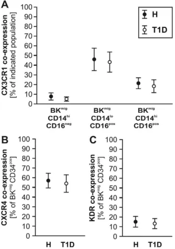
We previously reported the involvement of the B2R in CPC recruitment to ischemic tissue and subsequent promotion of reparative neovascularization [11]. Moreover, we demonstrated that by ex vivo migration towards BK, a cell population with enhanced pro-angiogenic characteristics (BKmig) can be enriched from total PB-MNC of healthy human subjects and patients with acute myocardial infarction, but not from patients with stable angina [11]. Now, comparing BKmigfrom T1D to H, we detected an enrichment of CPC and monocyte subtypes in BKmig and depletion of lymphocytes, with no significant differences between patient groups (Fig. 3). No significant differences in co-expression of CX3CR1, CXCR4, or KDR were detected by comparing H vs. T1D BKmigmonocytes or CPC (Fig. 4).
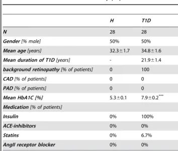
Table 1.Characteristics of the study populations.
H T1D
N 28 28
Gender[% male] 50% 50%
Mean age[years] 32.361.7 34.861.6
Mean duration of T1D[years] - 21.961.4
background retinopathy[% of patients] 0 100
CAD[% of patients] 0 0
PAD[% of patients] 0 0
Mean HbA1C [%] 5.360.1 7.960.2***
Medication[% of patients]
Insulin 0% 100%
ACE-inhibitors 0% 0%
Statins 0% 6.7%
AngII receptor blocker 0% 0%
Values are mean6SEM (where applicable). ***P,0.001 vs. H.
doi:10.1371/journal.pone.0011146.t001
Table 2.Blood cell count of patient groups.
H T1D
erythrocytes[n per mL PB] 4.8610968.76107 4.8610968.06107
lymphocytes[n per mL PB] 1.86106
61.16105 1.9
6106
61.16105
monocytes[n per mL PB] 4.4610563.26104 4.2610562.86105
neutrophils[n per mL PB] 3.4610662.46105 3.2610661.86105
eosinophils[n per mL PB] 1.06105
61.36104 1.9
6105
62.26104***
basophils[n per mL PB] 3.1610463.26103 3.1610463.26103
Values are mean6SEM. ***P
,0.001 vs. H.
Support of endothelial cell function by BKmig
Various mechanisms might contribute to the overall pro-angiogenic and endothelial-supportive effect seen before for healthy BKmig cells and some of those could be altered in T1D patients. Both, trans-differentiation of CPC and monocytes into vascular cells - thereby replacing defect resident endothelial cells (EC) - as well as paracrine effects might play a role, with recent reports accrediting more relevance to the later [16,17].
No differences between T1D- and H-derived BKmigwere found in their potential to give rise to acLDL+
UEAI+ , CD31+
and vWF+
EC or CD68+
macrophages in respective specific culture conditions(Fig. 5).
We have previously shown that BKmig, but not BKnon, support the formation of network structures by mature EC in an extracellular matrix gel [11]. Now, we observed more extensive network formation by HUVEC when they were co-cultured with H BKmigas compared to T1D BKmig(Fig. 6A). BKmigMNC from H preferentially integrated to or associated with EC network structures, while a larger percentage of T1D BKmigwithin the gels were visible as single cells, not in contact with other cells (Fig. 6B). Furthermore, T1D BKmig were preferentially located around branching points, while H BKmig also covered the branches of HUVEC networks, indicating altered inter-cellular communica-tion of T1D cells with EC (Fig. 6C&D).
Generation of NO and O22*
We next analyzed BK-induced NO and O22* synthesis, whose
balance is considered critical for EC survival and function. Kinins stimulate NO generation but might also induce O22* generation
by NADPH oxidase [9,12–14]. BKmig derived from H PBMC generated NO in response to BK stimulation through a mechanism involving both kinin receptors (being similarly blunted by B1R antagonist LdA-BK and B2R antagonist icatibant) and eNOS (being totally blocked by L-NIO), while T1D BKmig did not (Fig. 7A). In contrast, O22* generation
evoked by BK stimulation was not different between H and T1D groups (Fig. 7B). Intriguingly, both kinin receptor antagonists increased the O22* generation evoked by BK, suggesting the
existence of a cross talk between the two receptors in the control of oxidative stress following exposure to kinin. In this respect, we could not detect any difference between H and T1D. Further-more, BK stimulation led to elevated O22* levels when
endothelial NO synthase (eNOS) was inhibited (Fig. 7B), agreeing with previous reports on the anti-oxidative effects of eNOS/NO signaling [18,19].
Influence of differential glucose levels on CAC function
To test the hypothesis that moderately increased glucose levels (MG) influence kinin-related angiogenic cell functions differently
Figure 1. Characterization of monocyte and CPC in peripheral blood.No significant alterations were detected in the percentage of monocyte subtypes between non-diabetic (H, full circles) and T1D (empty circles) donors (A). CX3CR1 was mainly expressed by CD16posmonocyte subtypes, with no difference between study groups (B). CD34posCPC percentage among PB-MNC was lower in T1D patients (C, * P,0.05 vs. H), while co-expression of CXCR4 (D) and KDR (E) was slightly, but not significantly increased on CD34posCPC of T1D patients as compared to H subjects. Values are mean6SEM, n = 14 (monocytes inA & B) and n = 20 (CPC inC–E) per group.
than high glucose levels (HG), we studied the migratory activity as well as NO and O22* generation of H CAC cultured under
increasing glucose concentrations. After one week of culture in endothelial-specific medium, the majority of CAC expressed CD31 and CD11b and took up acetylated low density lipoproteins with no significant difference between glucose concentration-groups (Fig. S2). B1R and B2R expressions were not significantly
altered in CAC under different glucose conditions (Fig. 8A). CAC migration towards BK was unaltered when cells were grown in low or moderately increased glucose concentrations, while CAC grown in high glucose medium migrated less (Fig. 8B). Addition of insulin to CAC during culture under increasing glucose conditions did not improve BK-induced migration of HG-cultured CAC (Fig. S3).
Figure 2. Kinin receptor expression on distinct cell types in peripheral blood.Both kinin receptors were mainly expressed on CD16pos monocytes and KDR or CXCR4 co-expressing CPC, but no significant changes in the co-expression of the B1R (A) or the B2R (B) were detected on monocytes or CPC of T1D vs. H donors. Values are mean6SEM, n = 14 (monocytes), n = 20 (CPC) and n = 17 (lymphocytes) per group.
doi:10.1371/journal.pone.0011146.g002
Figure 3. Enrichment of distinct blood cell populations by migration towards bradykinin.Monocytes (A, B) and CPC (C) were enriched in BKmig, as compared to BKnonin both donor groups, while lymphocytes were depleted (A). Values are mean
6SEM of n = 20 (A, C) or n = 12 (B) donors per group. Dotted line indicates BKmig/BKnonratio = 1 (i.e. no enrichment/depletion).
Culture of CAC in increasing glucose concentrations progres-sively reduced the cells’ capacity to generate NO upon BK-stimulation and increased BK-induced O22* generation
(Fig. 8C&D). Different from what was observed in BKmigPBMC,
kinin receptor blockade only slightly increased O22* levels in LG
cultured CAC and even seemed to reduce oxidative stress in HG-cultured CAC.
Discussion
Several types of circulating angio-supportive cells are reduced and/or dysfunctional in diabetes mellitus, including hematopoietic stem cells, endothelial progenitor cells and pro-angiogenic monocytes [1,2]. Recent studies, addressing the question whether function and liberation of angio-supportive cells are disturbed prior to, along with, or as a result of microangiopathy, attribute high impact to the bone marrow and report dysfunctional cell liberation to underlie and precede later vascular complications [20,21]. Besides liberation, recruitment is the other crucial step a circulating cell has to perform before exerting its effect on the target tissue. Theoretically, cell recruitment could work as another selection step, preferring still-functional cells over dysfunctional ones. In this scenario, the regenerative potential of the vessel wall would be preserved even when CPC start to get dysfunctional, as long as enough CPC are still available, able to home, and do not yet themselves contribute to endothelial dysfunction, e.g. through the exaggerated generation of oxygen radicals. Unfortunately, information about initial alterations in cell homing early in the onset of diabetes-induced vascular dysfunction is scarce, as previous studies investigated mainly the late stages of vascular disease, when CPC are dysfunctional and macrovascular damage is already evident.
So far, mechanistic research has focused mainly on SDF-1/ CXCR4 signaling during the recruitment of angio-supportive cells [22–24]. Nevertheless, kinins play a key role in governing processes of the vessel wall and we recently demonstrated their necessity for recruiting circulating pro-angiogenic cells [8,9,11]. Given the relevance of BK-induced cellular functions, both during migration/cell recruitment and after migration, we aimed in the current study to analyze alterations in migratory and post-migratory activity of diabetic cells attracted by BK.
Unlike previous studies, where patients with higher HbA1C
and/or progressed vascular disease were tested, we compared young T1D patients with minimal microvascular disease and no clinically symptomatic macrovascular damage to age- and sex-matched non-diabetic controls. Results newly document that alterations in kinin-related post-migratory functions - but not migration itself - precede the development of diabetes-induced vasculopathy.
Figure 4. Co-expression of chemokine and growth factor receptors on migrating cells.CX3CR1 co-expression of migrating monocyte subtypes was comparable between T1D and H donors (A, n = 14 per group). Likewise, CXCR4 (B) and KDR (C) did not differ in their co-expression by migrating CD34pos CPC between T1D and healthy subjects (n = 20 per group).
doi:10.1371/journal.pone.0011146.g004
Figure 5. Outgrowth potential of migrating cells.BKmigof both study groups gave rise to equal numbers of UEAIposacLDLpos(A), CD31posor vWFposEC (B and C) or CD68posmacrophages (D). Values are mean
Two cell populations were studied in particular: CD34posCPCs
and monocytes, both reportedly involved in regulation of angiogenesis and endothelial repair [25–27]. In contrast to our earlier study, where patients with acute myocardial infarction or stable angina showed diminished abundance of B2R on CPCs, we could not detect any significant difference in kinin-receptor expression between cells from T1D and H donors [11]. Likewise, migratory response of the studied cell types to BK was preserved in T1D patients and the similar composition of migrating cell populations was confirmed by an outgrowth assay which did not reveal any differences in the capacity of H or T1D migrating cells
to give rise to EC or macrophages in culture. This result was surprising, given previous reports of migratory deficits of CD34pos CPC or cultured endothelial progenitor cells, associated with impaired NO generation, and is likely to be explained by more advanced stages of vascular disease in those studies [28,29].
Despite phenotypic similarities, BKmig from H and T1D differed in their paracrine activity and support of endothelial function: T1D BKmigwere less potent in supporting EC network formationin vitroand they generated less NO than H BKmig.
Enhanced production of reactive oxygen species and reduced NO generation are a hallmark of diabetic EC/CPC dysfunction and considered causal for their reduced angiogenic capacity, as well as in the progression of atherosclerosis [6,11,30]. Surprisingly, O22* generation by migrating T1D PB-MNC was not different
from the respective H-derived cells. In our in vitro model, no difference in the enrichment of inflammatory monocytes or granulocytes (data not shown), nor in the outgrowth of monocyte-derived CD68posmacrophages was detected between H and T1D PB cells, agreeing with absent changes in O22* generation in T1D
BKmig. Taken together, our data might shed some light on early vascular events prior to the establishment of macrovascular endothelial dysfunction: Under homeostatic conditions, EC-derived kinins support endothelial function and vascular integrity autocrinally and via the recruitment of angio-supportive cells, mainly mediated by the B2R [9]. BK-induced NO generation represents a major mechanism supporting endothelial cell function in this context, serving as a signaling molecule as well as an antioxidant [9,18,19]. Although BK has also been described to induce the formation of O22* via NADPH oxidase [14], the
anti-oxidant effect seems to prevail at this stage, indicated by increased levels of O22* under kinin receptor blockade. In early stages of
T1D, kinin receptor expression and migration towards BK remain unaltered in various types of circulating MNC. While BK-responsive cells still do not produce higher levels of O22*, they
lost their ability to generate sufficient levels of NO in response to BK stimulation and to support EC function. Presumably, additional deregulations in downstream molecular pathways (breakdown of oxidative defense, eNOS uncoupling) occur with additional aggravation of conditions, diverting BK-induced signaling and allowing oxygen radical production to prevail. In those conditions – like our CAC cultured under high glucose for one week – blockade of kinin receptors leads to a reduction of BK-generated oxidative stress. While future studies need to further investigate how diabetes affects intracellular processing of BK signals, our data indicate the involvement of both kinin receptors in antioxidative defense.
Patients participating in this study showed mildly elevated HbA1clevels, compatible with incomplete control of
hyperglyce-mia. Testing differentially elevated glucose concentrations in cell culture experiments, we were able to partly reproduce thein vivo data: Similar to the T1D patients’ cells, CAC grown in moderately increased glucose (10mM) retained their migratory capacity towards BK, which was only impaired at high concentrations of glucose (25mM) during the one week culture period. Upon BK administration, CAC generated NO, but not O22*, when cultured
at normal/low glucose concentrations (5mM). Under moderately increased or high glucose concentrations, BK-induced NO generation was abrogated, and O22* generation was induced. In
addition to the phenotypic differences between CAC and BKmig, culture conditions might be harsher than the environment within the T1D patients, explaining that BK apparently induced pro-oxidative signaling in HG-cultured cells – which is reduced by receptor blockade – while in the T1D patients’ BKmigcells, kinin receptor blockade led to an increase in oxidative stress, indicating
Figure 6. Support of endothelial network formation by migrating cells.The extension of networks (A) was lower if HUVEC were co-cultured with BKmig from T1D donors as compared to H-derived BKmig. BKmig MNC from H preferentially integrated to or associated with EC network structures, while a larger percentage of T1D BKmigwithin in the gels were visible as single cells, not in contact with other cells (B). While T1D cells (C) mainly surrounded branching points, H cells (D) also covered branches. Values are mean6SEM. n = 10 per group, size bar equals 100mm (C&D) * P,0.05 vs. H.
rather anti-oxidant effects of BK. Additional studies are needed to better elucidate differential signaling events initiated by moder-ately increased or high glucose levels as well as in early and longer persistent diabetes.
In summary, our data indicate a preservation of certain cellular functions, such as migration and low O22* generation in well
controlled young T1D patients, while NO generation and overall paracrine support of EC is already affected early on in disease onset. We therefore conclude that initially, the lack of paracrine support provided to EC by recruited cells, rather than actively inflicted damage, contributes to the loss of endothelial integrity in T1D. The increased state of inflammation, perpetuated by recruited inflammatory cells and generation of oxygen radicals, might only occur at a later stage.
Methods
Patient Recruitment
T1D patients (n = 28) with a background retinopathy according to the UK national screening committee for diabetic retinopathy were recruited at the Joint Clinical Research Unit of the Bristol Royal Infirmary. T1D patients with pre-proliferative and proliferative retinopathy, HbA1C above 10%, or macrovascular
complications (PAOD, CAD) were excluded. Respective healthy control subjects (H, n = 28) were recruited in parallel with diabetic patients to match age and gender of T1D patients in order to reduce variability. T1D and H donors with conditions reported to affect function of angiogenic cells (pregnancy, tumors, peripheral or coronary artery disease, hypertension, habitual smoking, extensive exercise training (e.g. marathon runners), over 55 years of age or taking medication affecting kinin signaling (ACE inhibitors, angiotensin receptor blockers) were excluded from both study groups. All procedures were performed in accordance with the Declaration of Helsinki and after obtaining approval of
the local ethics committee (Bath Research Ethics Committee, Bath, U.K.) and written informed consent from all donors.
Cell preparation and culture conditions
Blood was collected from matched pairs of T1D and H donors, anonymized by the study nurse and processed in parallel by a researcher blinded for group affiliation. Peripheral blood mono-nuclear cells (PB-MNC) were prepared from fresh EDTA-anticoagulated blood within 1 hour after withdrawal using density gradient centrifugation, as before [11]. Cells migrating towards BK (BKmig) and the respective non-migrating cells (BKnon) were derived from PB-MNC as described previously [11]. Cultured angiogenic cells (CAC) were obtained from PB-MNC by culture in endothelial-specific medium (EGM-2 MV, 10% bovine serum) for 5 days as previously described [11]. For some experiments, CAC of H donors were cultured in normal/low (LG, 5mM), medium (MG, 10mM), and high (HG, 25mM) glucose medium. Recom-binant human insulin (1U/mL) was added to duplicate LG, MG and HG groups during the culture period in some experiments.
Antigenic characterization
Individual cell populations in PB, BKmig and BKnon were identified according to their surface expression levels of CD34 (Miltenyi), KDR (R&D systems), CXCR4 (BD), CD14 (Miltenyi), CD16 (BD), CX3CR1 (Caltag) by flow cytometry (Fig. S4). Unlabelled primary antibodies against kinin B1 (B1R) and B2 receptor (B2R) were revealed by FITC-labeled anti-rabbit antibody (all Sigma). Isotype and secondary antibody controls were performed for each staining to verify specificity. 26105 to 36105 total events were acquired on a FACSCanto II flow cytometer and analyzed with FACSDiva 6.1.2 software (both BD). Enrichment or depletion of distinct cell types within BKmigwas calculated versus BKnonof the same experiment as before [11]. Expression of endothelial cell (von Willebrand Factor, CD31) and
Figure 7. Generation of nitric oxide and superoxide by migrating cells in response to bradykinin.H BKmigcells generated NO in response to BK in an eNOS dependent fashion, as indicated by inhibition by L-NIO. NO generation from T1D BKmigwas lower than from H cells and not inhibitable by L-NIO (A). Neither H nor T1D BKmiggenerated surplus O22* in response to BK addition (B). Inhibition of B1R (by LdA-BK) or B2R (by icatibant) partially inhibited NO generation (A) and increased O22* generation (B). eNOS blockade by L-NIO enhanced O22* levels per cell, underlining the antioxidant role of eNOS (B). Values are mean6SEM of n = 9 (A) or n = 10 (B) per group. * P,0.05 vs. H.
macrophage antigens (CD68), uptake of acetylated low-density lipoproteins (acLDL) and binding of ulex europaeus agglutinin I (UEAI) were studied by immunofluorescence microscopy in adherent BKmig-derived cells cultured under cell-type specific conditions.
Quantitative RT-PCR
CD14posmonocytes were isolated by magnetic sorting following the manufacturers recommendations (Miltenyi). Cells were enriched using 2 successive columns to increase purity. CD14neg cells were labeled with anti-CD34 magnetic beads (Miltenyi) and CD34posCD14negCPC isolated likewise. 105isolated cells of each population were lysed and reverse transcribed using the Power
SYBRH Green Cells-to-CtTMkit (Ambion). cDNA levels of B1R and B2R, as well as 18S rRNA were quantified by qPCR using the same kit and a DNA Engine Opticon 2 (BioRad).
Functional cell characterization
The ability of BKmig to support network formation of human umbilical vein EC (HUVEC) was studied in a matrigel assay as reported previously [11]. Generation of nitric oxide (NO) and superoxide (O22*) was assessed using the fluorescent probes
diaminofluorescein (DAF-2DA) and dihydroethidium (DHE, both invitrogen), respectively, by fluorescence microscopy or analysis by the IN Cell Analyzer (GE Healthcare). Briefly, nuclei were labeled with 5nM Hoechst 33342 (Sigma), and cells pre-incubated with either
Figure 8. Cellular function in differential glucose conditions.Expression of B1R and B2R was not significantly different in CAC cultured under moderately increased (MG, 10mM)) or high glucose (HG, 25mM) concentrations (A). Migration of CAC, derived from H donors, towards BK was not affected by MG, but reduced after culture in HG (B). CAC cultured in low/normal glucose (LG) generated NO upon BK addition, while CAC cultured in MG or HG did not generate NO (C). In contrast, LG-cultured CAC did not generate O22* upon BK stimulation, while CAC cultured in MG or HG did (D). Blockade of kinin receptors reduced NO generation even more in MG and HG (C), but also reduced O22* production (D). Values are mean6SEM of n = 5 (A), n = 11 (B), or n = 9 (C&D) per group.
antagonists of the B1R (Lys-(des-Arg9, Leu8)-Bradykinin, LdA-BK, Sigma, final concentration 0.5mM), the B2R (icatibant, Sigma, final concentration 0.5mM), or eNOS (L-NIO, 1mM). DAF or DHE (final concentration 5mM) and BK (final concentration 0.1mM) were added. Controls contained no stimulus, but the respective fluorescent probe. Fluorescence intensity was measured using an IN Cell Analyzer 1000 (GE Healthcare, Amersham, UK). Images were acquired using a 106objective, q505LP dichroic mirror (Chroma Technology Corp, Rockingham, VT) and 360-40nm (Hoechst) or 475-20nm (DHE / DAF) excitation filters with 460-40nm, 570-30nm (DHE) or 535-50nm (DAF) emission filters. Three independent experiments were performed in triplicate wells each with two fields of view (each 0.6mm2) per well. Fluorescence intensity (FI) was assessed, initially at baseline (0 min) then after 15min for O22* measurement or after 1h
and 3h for NO measurement. Image analysis was performed using IN Cell Analyzer Workstation 3.5 software (IN Cell Investigator, GE Healthcare) and a Dual Area Object Analysis algorithm as previously described [31]. Only cells with a cytoplasmic FI greater than 10% of background intensity were considered. Imaging data is reported as the mean cytoplasmic FI per cell.
Similarly, BK-induced NO and O22* generation by BKmigor
BKnon of H and T1D was studied by using fluorescence microscopy. Vehicle controls contained DAF or DHE, but no inhibitor or BK. Experiments were performed in duplicate for each patient. Fluorescence microscopic images were taken after 15min (DHE, red filter) and 1h (DAF, green filter), together with Hoechst-stained nuclei (blue filter) of the same viewfield in a Zeiss microscope/camera system with fixed exposure settings. Fluores-cence intensity per cell was then assessed using ImageJ v. 1.41 (NIH). Mean FI per cell is reported.
Statistical analysis
Normally distributed values were compared by paired t-test (two groups) or repeated measures ANOVA (multiple groups, followed by Holm-Sidakpost hoctest). Not normally distributed values were compared by Mann-Whitney U test (two groups) or ANOVA on Ranks (multiple groups, followed by Dunn’spost hoctest). A p-value below 0.05 was considered significant.
Supporting Information
Figure S1 mRNA levels of B1R and B2R in magnetically isolated CD14posmonocytes and CD34posCD14neg CPC. Values are mean6S.E.M. of n = 4 values.
Found at: doi:10.1371/journal.pone.0011146.s001 (0.07 MB JPG)
Figure S2 Expression of CD11b (B&E) and CD31 (C&F), as well as uptake of DiI-labelled acetylated low density lipoproteins (D&G) by CAC was not altered by glucose concentration in the medium. A: typical FSC/SSC plot of CAC at day 5 of culture. Values are mean6S.E.M. of n = 4 values.
Found at: doi:10.1371/journal.pone.0011146.s002 (0.35 MB JPG)
Figure S3 Impaired migration of CAC cultured under high glucose was not rescued by additional presence of insulin. Values are mean6S.E.M. of n = 4 values.
Found at: doi:10.1371/journal.pone.0011146.s003 (0.16 MB JPG)
Figure S4 Lymphocytes (ly), monocytes (mo) and granulocytes (gra) were identified based on their light scatter characteristics (A). Gates were set up for each fluorophore and antibody separately, using secondary antibody (B) and isotype controls (E, G, I & L). Representative examples show analysis of kinin B1 and B2 receptor expression (C&D), CD14hiCD16neg(M1), CD14hiCD16pos(M2) and CD14loCD16pos (M3) monocyte subpopulations (F), CD34pos (H), CXCR4 (K) and KDR (M). Co-expression was analyzed by logically combining gates for fluorophores with lympho- and monocytes the analysis, e.g. CD34pos CPC = (mo OR ly) AND CD34pos, CD14hiCD16negmonocytes = mo AND M1 during.
Found at: doi:10.1371/journal.pone.0011146.s004 (0.71 MB JPG)
Acknowledgments
The authors thank Mrs. N. McLintock and Dr. J. Rowlinson (University of Bristol, U.K.) for excellent technical assistance, PD Dr. V. Adams (University of Leipzig, Germany) and Dr. Paola Campagnolo (University of Bristol, U.K.) for critical review of the manuscript, Dr. W. Woltersdorf (Bristol Royal Infirmary, U.K.) for advice on hematological and pathophysiological issues, and Dr. M. Alhadj Ali (University of Bristol, U.K.) for help with recruitment of participants.
Author Contributions
Conceived and designed the experiments: NK CAM CD PM. Performed the experiments: NK SPA. Analyzed the data: NK SPA. Contributed reagents/materials/analysis tools: CAM CD PM. Wrote the paper: NK PM.
References
1. Waltenberger J (2001) Impaired collateral vessel development in diabetes: potential cellular mechanisms and therapeutic implications. Cardiovasc Res 49: 554–560. 2. Fadini GP, Sartore S, Agostini C, Avogaro A (2007) Significance of Endothelial
Progenitor Cells in Subjects with Diabetes. Diabetes Care 30: 1305–1313. 3. Sambuceti G, Morbelli S, Vanella L, Kusmic C, Marini C, et al. (2009) Diabetes
impairs the vascular recruitment of normal stem cells by oxidant damage, reversed by increases in pAMPK, heme oxygenase-1, and adiponectin. Stem Cells 27: 399–407.
4. Sessa WC (2009) Molecular control of blood flow and angiogenesis: role of nitric oxide. J Thromb Haemost 7: 35–37.
5. Brandes RP, Schro¨der K (2008) Differential vascular functions of Nox family NADPH oxidases. Curr Opin Lipidol 19: 513–518.
6. Thum T, Fraccarollo D, Schultheiss M, Froese S, Galuppo P, et al. (2007) Endothelial nitric oxide synthase uncoupling impairs endothelial progenitor cell mobilization and function in diabetes. Diabetes 56: 666–674.
7. Sorrentino SA, Bahlmann FH, Besler C, Mu¨ller M, Schulz S, et al. (2007) Oxidant stress impairs in vivo reendothelialization capacity of endothelial progenitor cells from patients with type 2 diabetes mellitus: restoration by the peroxisome proliferator-activated receptor-gamma agonist rosiglitazone. Circu-lation 116: 163–173.
8. Costa-Neto CM, Dillenburg-Pilla P, Heinrich TA, Parreiras-e-Silva LT, Pereira MG, et al. (2008) Participation of kallikrein-kinin system in different pathologies. Int. Immunopharmacol 8: 135–142.
9. Leeb-Lundberg LM, Marceau F, Mu¨ller-Esterl W, Pettibone DJ, Zuraw BL (2005) International union of pharmacology. XLV. Classification of the kinin receptor family: from molecular mechanisms to pathophysiological consequenc-es. Pharmacol Rev 57: 27–77.
10. Emanueli C, Bonaria Salis M, Stacca T, Pintus G, Kirchmair R, et al. (2002) Targeting kinin B(1) receptor for therapeutic neovascularization. Circulation 105: 360–366.
11. Kra¨nkel N, Katare RG, Siragusa M, Barcelos LS, Campagnolo P, et al. (2008) Role of kinin B2 receptor signaling in the recruitment of circulating progenitor cells with neovascularization potential. Circ Res 103: 1335–1343.
12. Danielisova´ V, Gottlieb M, Ne´methova´ M, Burda J (2008) Effects of bradykinin postconditioning on endogenous antioxidant enzyme activity after transient forebrain ischemia in rat. Neurochem Res 33: 1057–1064.
13. Tscho¨pe C, Walther T, Escher F, Spillmann F, Du J, et al. (2005) Transgenic activation of the kallikrein-kinin system inhibits intramyocardial inflammation, endothelial dysfunction and oxidative stress in experimental diabetic cardiomy-opathy. FASEB J 19: 2057–2059.
14. Larsen BT, Bubolz AH, Mendoza SA, Pritchard KA, Jr., Gutterman DD (2009) Bradykinin-induced dilation of human coronary arterioles requires NADPH oxidase-derived reactive oxygen species. Arterioscler Thromb Vasc Biol 29: 739–745.
CCR22/2Mice: Evidence for Independent Chemokine Functions in Athero-genesis. Circulation 117: 1642–1648.
16. Yoder MC, Ingram DA (2009) The definition of EPCs and other bone marrow cells contributing to neoangiogenesis and tumor growth: Is there common ground for understanding the roles of numerous marrow-derived cells in the neoangiogenic process? Biochim Biophys Acta 1796: 50–54.
17. Purhonen S, Palm J, Rossi D, Kaskenpa¨a¨ N, Rajantie I, et al. (2008) Bone marrow-derived circulating endothelial precursors do not contribute to vascular endothelium and are not needed for tumor growth. Proc Natl Acad Sci U S A 105: 6620–6625.
18. Fujii H, Ichimori K, Hoshiai K, Nakazawa H (1997) Nitric oxide inactivates NADPH oxidase in pig neutrophils by inhibiting its assembling process. J Biol Chem 272: 32773–32778.
19. Selemidis S, Dusting GJ, Peshavariya H, Kemp-Harper BK, Drummond GR (2007) Nitric oxide suppresses NADPH oxidase-dependent superoxide produc-tion by S-nitrosylaproduc-tion in human endothelial cells. Cardiovasc Res.75: 349–358. 20. Busik JV, Tikhonenko M, Bhatwadekar A, Opreanu M, Yakubova N, et al. (2009) Diabetic retinopathy is associated with bone marrow neuropathy and a depressed peripheral clock. J Exp Med 206: 2897–2906.
21. Oikawa A, Siragusa M, Quaini F, Mangialardi G, Katare RG, et al. (2010) Diabetes Mellitus Induces Bone Marrow Microangiopathy. Arterioscler Thromb Vasc Biol 30: 498–508.
22. Kubo M, Li TS, Kamota T, Ohshima M, Qin SL, et al. (2009) Increased expression of CXCR4 and integrin alphaM in hypoxia-preconditioned cells contributes to improved cell retention and angiogenic potency. J Cell Physiol 220: 508–514.
23. Seeger FH, Rasper T, Koyanagi M, Fox H, Zeiher AM, et al. (2009) CXCR4 expression determines functional activity of bone marrow-derived mononuclear
cells for therapeutic neovascularization in acute ischemia. Arterioscler Thromb Vasc Biol 29: 1802–1809.
24. Zemani F, Silvestre JS, Fauvel-Lafeve F, Bruel A, Vilar J, et al. (2008) Ex vivo priming of endothelial progenitor cells with SDF-1 before transplantation could increase their proangiogenic potential. Arterioscler Thromb Vasc Biol 28: 644–650.
25. Matsumoto T, Kuroda R, Mifune Y, Kawamoto A, Shoji T, et al. (2008) Circulating endothelial/skeletal progenitor cells for bone regeneration and healing. Bone 43: 434–439.
26. Wojakowski W, Tendera M, Michałowska A, Majka M, Kucia M, et al. (2004) Mobilization of CD34/CXCR4+, CD34/CD117+, c-met+ stem cells, and mononuclear cells expressing early cardiac, muscle, and endothelial markers into peripheral blood in patients with acute myocardial infarction. Circulation 110: 3213–3220.
27. Vo¨o¨ S, Eggermann J, Dunaeva M, Ramakers-van Oosterhoud C, Waltenberger J (2008) Enhanced functional response of CD133+circulating progenitor cells in patients early after acute myocardial infarction. Eur Heart J 29: 241–250. 28. Fadini GP, Sartore S, Albiero M, Baesso I, Murphy E, et al. (2006) Number and
function of endothelial progenitor cells as a marker of severity for diabetic vasculopathy. Arterioscler Thromb Vasc Biol 26: 2140–2146.
29. Segal MS, Shah R, Afzal A, Perrault CM, Chang K, et al. (2006) Nitric oxide cytoskeletal-induced alterations reverse the endothelial progenitor cell migratory defect associated with diabetes. Diabetes 55: 102–109.
30. Csa´nyi G, Taylor WR, Pagano PJ (2009) NOX and inflammation in the vascular adventitia. Free Radic Biol Med 47: 1254–1266.