Extracts o f
Ascaris suum
e gg and adult
wo rm share sim ilar im m uno suppre ssive
pro pe rtie s
Departamento de Imunologia, Instituto de Ciências Biomédicas, Universidade de São Paulo, São Paulo, SP, Brasil
V.M.O . Souza, E.L. Faquim-Mauro and M.S. Macedo
Abstract
Adult Ascaris suum body extract (Asc)prepared from male and female worms (with stored eggs) down-regulates the specific immune re-sponse of DBA/2 mice to ovalbumin (OA) and preferentially stimu-lates a Th2 response to its own components, which is responsible for the suppression of the OA-specific Th1 response. Here, we investi-gated the participation of soluble extracts prepared from male or female worms or from eggs (E-Asc) in these immunological events. Extracts from either sex (1 mg/animal) or E-Asc (0.35 or 1 mg protein/ animal) suppressed the delayed-type hypersensitivity (DTH) reaction (60-85%), proliferative response (50-70%), IL-2 and IFN-g secretion (below detection threshold) and IgG1 antibody production (70-90%) of DBA/2 mice to OA. A dose of 0.1 mg E-Asc/animal did not change DTH or proliferation, but was as effective as 0.35 mg in suppressing IL-2 and IFN-g, and OA-specific IgG1 antibodies. Lymph node cells from DBA/2 mice injected with Asc (1 mg/animal) or a high dose of E-Asc (1 mg protein/animal) secreted IL-4 upon in vitro stimulation
with concanavalin A. As previously demonstrated for Asc, the cy-tokine profile obtained with the E-Asc was dose dependent and changed towards Th1 when a low dose (0.1 mg protein/animal) was used. Taken together, these results suggest that adult worms of either sex and eggs induce the same type of T cell response and share similar immunosuppressive properties.
Co rre spo nde nce
M.S. Macedo
Departamento de Imunologia ICB/USP
Av. Prof. Lineu Prestes, 1730 05508-900 São Paulo, SP Brasil
Fax: + 55-11-3818-7224 E-mail: mamacedo@ usp.br
Research supported by FAPESP. V.M.O . Souza was the recipient of a fellowship from FAPESP (No. 96/8101-3).
Received May 7, 2001 Accepted O ctober 10, 2001
Ke y wo rds
·Ascaris suum
·Immunosuppression ·Adult worms ·Eggs ·Cytokines
Intro ductio n
Helminth parasites induce a predominant Th2-type response in the host, which is asso-ciated with both protective immunity and chronic states of disease (1-3). It was ob-served in some infections that the develop-ment of Th2 cells is specific for certain stages of the parasite life cycle and can down-regulate the Th1 response. In murine schisto-somiasis only egg antigens stimulate IL-4, IL-5 and IL-10 secretion (4,5), while mice
later on (10,11). A Th2 response to Brugia malayi, previously induced by continuous stimulation, alters the typical Th1 response to mycobacterial antigen purified protein de-rivative, inducing IL-4 and IL-5 production, besides IFN-g (12). In addition, in concur-rent infections the expansion of the Th2 response changes the course of disease me-diated by Th1 cytokines. For example, AKR mice become resistant to Trichuris muris
when infected with Schistosoma mansoni
(13) and attenuated L3 of B.pahangi down-regulates the murine susceptibility to cere-bral malaria induced by Plasmodium berghei
in BALB/c mice (14).
We have demonstrated that adult Ascaris suum body extract, prepared from male and female worms (with stored eggs), impairs the humoral and cell-mediated immune re-sponses to the unrelated antigen ovalbumin (OA). High doses of adult worm extract preferentially induce a Th2 response to its own antigens, with high levels of IL-4 and IL-10 (15). The extract was previously frac-tionated by gel filtration and three peaks, named PI, PII and PIII, were obtained. PI components were responsible for the sup-pression of the OA-specific response, whereas PIII components were not suppres-sive and stimulated anti-adult worm extract IgE antibodies (16,17). In vivo treatment with anti-IL-4 and anti-IL-10 monoclonal antibodies in OA plus adult worm extract-immunized mice abolishes the effects of adult
A. suum body extract on the Th1-related parameters of the immune response to OA. These results indicate the important role of these cytokines in the down-regulation of this response (18).
Although the suppressive effects of adult
A. suum body extract have been quite well established, it is not clear which of its com-ponents is responsible for this immunosup-pression. Therefore, in the present study we worked with soluble extracts prepared from
A. suum male or female worms or eggs ob-tained from female uteri (M-Asc, F-Asc and
E-Asc, respectively) and assessed their ef-fects on the OA-specific response and the pattern of cytokines and antibody isotypes elicited by them. Our results indicate that adult worms and eggs possess similar immu-nosuppressive properties.
Mate rial and Me tho ds
Anim als
Seven- to eight-week-old male DBA/2 mice were used for immunization. The ani-mals were bred in the animal house facilities of the Department of Immunology, ICB/Uni-versity of São Paulo, São Paulo, SP, Brazil.
Antige ns and antibo die s
OA (grade II and V) was obtained from Sigma (St. Louis, MO, USA). Adult worm A. suum extracts: live male and female worms were separated by sex and the extracts pre-pared as previously described (19). The ex-tracts contained 50% protein. Egg extract: briefly, uteri obtained from female worms were homogenized in an Ultra-Turrax appa-ratus (Janke and Kunkel, Staufen, Germany). After centrifugation at 13,000 g for 1 h, the pellet was frozen in liquid nitrogen and mac-erated in the presence of glass powder. This mixture was diluted in borate-buffered sa-line, pH 8.0 (equal volumes), stirred over-night at 4ºC, and then centrifuged at 13,000
Im m unizatio n
Groups of 5-7 mice were injected subcu-taneously (sc) with 100 µg OA plus 1 mg F-Asc or M-F-Asc emulsified in complete Freunds adjuvant (CFA) (Sigma) on both sides of the base of the tail (0.2 ml/animal). Another group received only OA in CFA. For E-Asc, 100 µg OA plus 1, 0.35 or 0.1 mg protein/animal emulsified in CFA were used. The experiments were repeated three times.
Hype rse nsitivity re actio ns
Hypersensitivity reactions were elicited 8 days after immunization by injection of 30 µl of 2% aggregated OA (1 h, 70ºC) into one of the hind footpads and the same volume of saline in the other. Nonimmunized mice were equally injected and used as test control for nonspecific swelling. Footpad swelling was monitored periodically from 3 to 24 h using a pocket thickness gauge (Mitutoyo Mfg. Co. Ltd., Tokyo, Japan) and is reported as the increase in thickness relative to the sa-line-injected paw. The results are reported as the arithmetic mean ± SEM for each group. Two-way analysis of variance followed by the multiple comparisons Tukey test (20) was used to compare the differently immu-nized groups.
Pro life ratio n assay
Nine days after immunization, cell sus-pensions from inguinal and periaortic lymph nodes (LN) of 5 mice were prepared in RPMI 1640 (Sigma) supplemented with 10 mM HEPES, 50 µM M2-mercaptoethanol, 216 mg L-glutamine/l, and 5% FCS. Triplicate preparations of cells (5 x 10⁵/well) were distributed into 96-well flat-bottomed mi-croplates (Costar, Cambridge, MA, USA) and incubated with OA, F-Asc, M-Asc (100 µg/ml each), E-Asc (50 µg protein/ml) or 2.5 µg/ml concanavalin A (Con A; Sigma) in a humidified CO2 incubator for 62 or 96 h.
Cell proliferation was measured by [3 H]-thymidine incorporation (2 Ci/ml; DuPont, Boston, MA, USA). One µCi/well was added 18 h before cell harvesting and incorporated [3H]-thymidine was determined by liquid scintillation spectrometry. The SD of mean count/min incorporated by triplicate cultures was less than 10%. The results are reported as stimulation index (mean count/min of [3H]-thymidine incorporated by stimulated LN cells divided by mean count/min incor-porated by unstimulated cells).
Cyto kine assays
The cell suspensions prepared for the proliferation assay were added to 24-well tissue culture plates (Costar) at a final con-centration of 10 or 6 x 10⁶ cells/ml. The cells were stimulated with OA, F-Asc, M-Asc (500 µg/ml each), E-Asc (250 µg protein/ml) or Con A (5 µg/ml). Supernatants were har-vested after 24 h (cultures of 10 x 106 cells) or 72 h (cultures of 6 x 10⁶ cells) and assayed for cytokine content. All cytokines were measured by specific two-site sandwich ELISA using the following monoclonal anti-bodies: XMG 1.2 and biotinylated AN 18 for IFN-g; IA12 and biotinylated JES6-5H4 for IL-2, and BVD-1D11 and biotinyl-ated BVD6-24G2 for IL-4. Binding of the biotinylated antibodies was determined us-ing the streptavidin-peroxidase conjugate (Sigma) and ABTS (2,2-azinobis(3-ethyl-benzthiazoline-6-sulfonic acid; Sigma) so-lution in 0.1 M citrate buffer plus H2O2. The plates were read (410 nm) in an automated ELISA reader (Dynatech MR 5000). Samples were quantified by comparison with stan-dard curves of purified recombinant cyto-kines.
D e te ctio n o f antibo dy iso type s
pare the antibody response among groups (20).
Re sults
Effe ct o f wo rm and e gg e xtracts o n
O A-spe cific hype rse nsitivity re actio ns
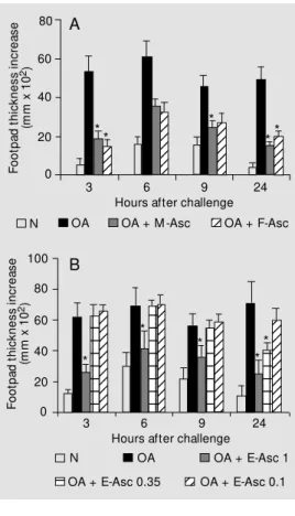
The development of hypersensitivity re-actions in mice immunized with OA alone, OA plus adult worm extracts or different doses of E-Asc after challenge with aggre-gated OA in the footpad were compared on day 8. As shown in Figure 1A, the immediate (3 h) and delayed-type hypersensitivity (DTH) (24 h) reactions to OA were reduced in animals that received OA + F-Asc or M-Asc compared with OA-immunized mice. Regarding E-Asc (Figure 1B), the group im-munized with OA + 1 mg E-Asc showed a reduced immediate reaction and a DTH re-sponse that did not differ significantly from that of the nonimmunized group. Mice im-munized with OA + 0.35 mg E-Asc showed only a reduced DTH reaction to OA. The hypersensitivity reactions of mice immunized with OA + 0.1 mg E-Asc were similar to those obtained in mice injected with OA alone.
Effe ct o f wo rm and e gg e xtracts o n
lym pho pro life rative re spo nse to O A
The proliferation of LN cells from mice immunized with OA and the different A. suum extracts was then measured after in vitro stimulation with the antigen or mito-gen. The results presented in Table 1 show that simultaneous immunization with OA + F-Asc or M-Asc prevented LN cells from responding to OA in vitro when compared with the response of cells from mice immu-nized with OA alone. The proliferation of cells from the former groups when stimulat-ed with Con A for 96 h was low. The same suppressive effect was caused by E-Asc, irrespective of the protein concentration of µg/ml OA, 10 µg/ml F-Asc or M-Asc, and 1
µg/ml E-Asc) and biotinylated goat anti-mouse monospecific antisera. The reactions were developed with the streptavidin-per-oxidase conjugate (Sigma), o -phenylenedia-mine and H2O2. The results are reported as the mean absorbance of samples/group (± SEM) at various plasma dilutions. Analysis of variance followed by the multiple parisons Tukey test was employed to
com-F o o tp a d t h ic k n e s s i n c re a s e (m m x 1 0 2) 80 12 12 12 12 12 12 12 12 12 12 12 12 12 12 12 12 12 12 12 12 12 12 12 12 12 60 40 20 0
3 6 9 24
Hours after challenge
12 12 12 12 12 12 12 12 12 12 12 12 12 12 12 12 12 12 12 12 12 12 12 12 12 12 12 12 12 12 12 12 12 12 12 12 12 12 12 12 12 12 12 12 12 12 12 12 12 12 12 12 12 12 12 12 12 12 12 12 12 12 12 12 12 12 12 12 12 12 12 12 12 12 12 12 12 12 12 12 12 12 100 80 40 20 0 60
3 6 9 24
Hours after challenge * * * ** * * * * *
N OA OA + M -Asc
12
12OA + F-Asc
F o o tp a d t h ic k n e s s i n c re a s e (m m x 1 0 2) A B Figure 1. Hypersensitivity
reac-tions in mice immunized w ith ovalbumin (OA), OA + Ascaris suum female (FAsc) or male (M -Asc) extract (A), or egg (E--Asc) extract (B) at different protein doses (1, 0.35 and 0.1 mg). The mice w ere challenged 8 days later w ith aggregated OA in the footpad. Nonim m unized m ice (N) w ere also challenged in the same w ay for nonspecific sw ell-ing. The results represent the mean ± SEM for 5-7 animals/ group. * P<0.05 compared w ith the OA-immunized group (Tukey test).
Table 1. Proliferative response of lymph node cells from mice immunized w ith ovalbu-min (OA) or OA plus female or male adult w orm extract.
Cells OA F-Asc M -Asc Con A
OA 7.9* 1.9 2.5 3.3
OA + F-Asc 1.0 13.2 14.4 5.2
OA + M -Asc 1.1 10.3 11.8 3.6
Inguinal and periaortic lymph node cells from mice immunized 9 days before w ith OA, OA + F-Asc or M -Asc in complete Freund’s adjuvant w ere stimulated w ith 100 µg/ml OA, 100 µg/ml F-Asc or M -Asc, or 2.5 µg/ml Con A for 96 h. F-Asc, M -Asc = female and male w orm extract, respectively; Con A = concanavalin A.
* Stimulation index calculated as described in M aterial and M ethods.
N OA OA + E-Asc 1
12 12
OA + E-Asc 0.1
12
the extract used for immunization (Table 2). In this experiment, the cells were stimulated for 62 h and, therefore, the proliferation of OA-primed cells was lower in response to OA and higher in response to Con A. Conse-quently, a dose-dependent suppression could also be observed after Con A stimulation. The stimulation index of LN cells from mice immunized with OA + F-Asc or OA + M-Asc was similar when cell cultures were restimulated in vitro with either F-Asc or M-Asc. In comparison, the nonspecific prolif-eration of cells from OA-immunized mice when in vitro restimulated with these ex-tracts was negligible (Table 1). The same was true for E-Asc that stimulated just the proliferation of OA + E-Asc-primed cells (Table 2).
Effe ct o f wo rm and e gg e xtracts o n cyto kine
and antibo dy pro ductio n in re spo nse to O A
LN cells from the above groups were also cultured with OA or Con A for 24 or 72 h and the supernatants collected for quantification of secreted cytokines. Cytokine assays for adult worm-injected groups and for
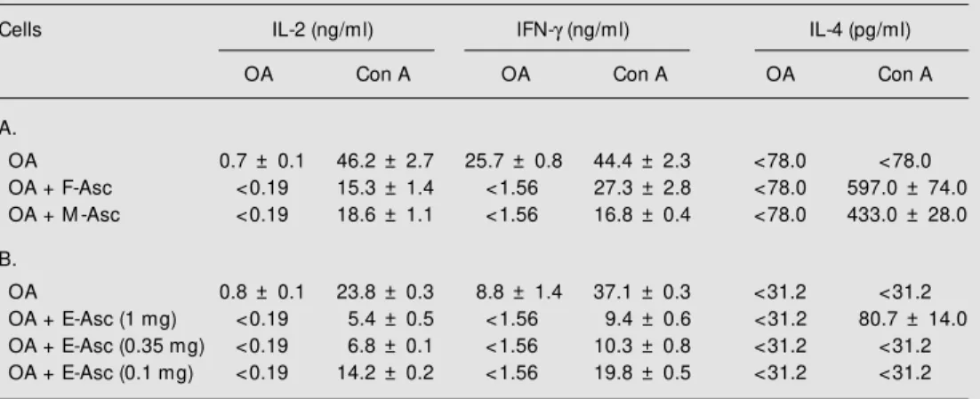
egg-in-jected groups were not performed at the same time. Cultures from mice immunized with OA + F-Asc, M-Asc or different doses of E-Asc did not have any detectable IL-2 or IFN-g when restimulated in vitro with OA compared to those from OA-immunized mice (Table 3). The levels of these cytokines were also reduced upon stimulation with Con A, but the lowest dose (0.1 mg) of E-Asc was less suppressive. Regarding IL-4, high levels of this cytokine were only detected in
cul-Table 2. Proliferative response of lymph node cells from mice immunized w ith ovalbumin (OA) or OA plus Ascaris suum egg extract.
Cells OA E-Asc Con A
OA 3.0* 0.0 37.0
OA + E-Asc (0.1 mg) 1.9 13.0 30.0 OA + E-Asc (0.35 mg) 1.5 11.0 21.0 OA + E-Asc (1 mg) 1.6 11.0 19.0
Inguinal and periaortic lymph node cells from mice immunized 9 days before w ith OA or OA + E-Asc at different protein doses w ere stimulated w ith 100 µg/ml OA, 50 µg protein/ml E-Asc or 2.5 µg/ ml Con A for 62 h. E-Asc = Ascaris suum egg extract; Con A = concanavalin A.
* Stimulation index calculated as described in M a-terial and M ethods.
Table 3. Synthesis of IL-2, IFN-g and IL-4 by lymph node cells from mice immunized w ith ovalbumin (OA) or OA plus female, male or egg extract.
Cells IL-2 (ng/ml) IFN-g (ng/ml) IL-4 (pg/ml)
OA Con A OA Con A OA Con A
A.
OA 0.7 ± 0.1 46.2 ± 2.7 25.7 ± 0.8 44.4 ± 2.3 <78.0 <78.0 OA + F-Asc <0.19 15.3 ± 1.4 <1.56 27.3 ± 2.8 <78.0 597.0 ± 74.0 OA + M -Asc <0.19 18.6 ± 1.1 <1.56 16.8 ± 0.4 <78.0 433.0 ± 28.0
B.
OA 0.8 ± 0.1 23.8 ± 0.3 8.8 ± 1.4 37.1 ± 0.3 <31.2 <31.2 OA + E-Asc (1 mg) <0.19 5.4 ± 0.5 <1.56 9.4 ± 0.6 <31.2 80.7 ± 14.0 OA + E-Asc (0.35 mg) <0.19 6.8 ± 0.1 <1.56 10.3 ± 0.8 <31.2 <31.2 OA + E-Asc (0.1 mg) <0.19 14.2 ± 0.2 <1.56 19.8 ± 0.5 <31.2 <31.2
the highest dose (1 mg) of E-Asc after Con A stimulation (Table 3B).
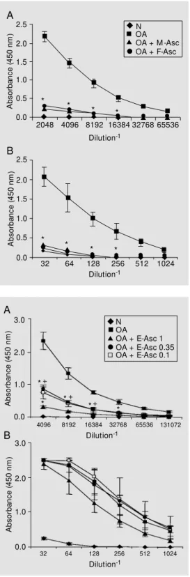
Antibody levels were also assessed in plasma obtained from all groups on day 9. IgG1 and IgG2a antibody production was strongly inhibited in mice immunized with OA plus adult worm extracts (Figure 2). The egg extract also had a suppressive effect on anti-OA IgG1 antibody production, with the highest dose being more effective (Figure 3A). IgG2a levels were not significantly dif-ferent among the immunized groups, although the group injected with 1 mg of E-Asc re-peatedly responded less than the other two (Figure 3B).
Pro file o f cyto kine s and antibo dy iso type s in
re spo nse to adult wo rm and e gg e xtracts
Cytokines were also measured in super-natants of LN cell cultures from mice in-jected with the different adult worm extract preparations after in vitro restimulation with the respective extract. Table 4 shows that F-Asc induced more IFN-g and IL-4 than M-Asc. In addition, more IL-2 and IFN-g, and less IL-4 were produced as the immuniz-ing dose of E-Asc was reduced.
To analyze the isotypes elicited by each extract the ELISA plates were coated with the respective antigens. F-Asc and M-Asc induced the same amounts of antigen-specif-ic IgG1 and IgG2a antibodies (Figure 4). A dose-dependent production of IgG1 antibod-ies was observed in mice immunized with E-Asc (Figure 5A). IgG2a antibodies were mostly induced by the highest doses of E-Asc (Figure 5B).
D iscussio n
Unisexual infections have been an im-portant experimental model to study the type of immune response induced by helminths (5,10). In the present study, immunization with extracts of A. suum prepared from adult worms of either sex or from eggs allowed us
A
b
s
o
rb
a
n
c
e
(
4
5
0
n
m
)
2.5 N
OA OA + M -Asc OA + F-Asc
*
* * *
2.0
1.5
1.0
0.5
0.0
2048 4096 8192 16384 32768 65536 Dilution-1
*
* * *
A
b
s
o
rb
a
n
c
e
(
4
5
0
n
m
)
2.5
2.0
1.5
1.0
0.5
0.0
32 64 128 256 512 1024
Dilution-1
A
B Figure 2. Ovalbumin (OA)-specif-ic IgG1 (A) and IgG2a (B) anti-bodies produced by OA or OA +
Ascaris suum female (F-Asc)- or male (M -Asc) extract-immunized m ice. Isotype levels w ere ti-trated by ELISA in normal (N) or immune plasma obtained after 9 days of im m unizat ion using monospecific antisera. The re-sults represent the mean ± SEM absorbance f or 5-7 anim als. * P<0.01 compared w ith the OA-immunized group (Tukey test).
A
b
s
o
rb
a
n
c
e
(
4
5
0
n
m
)
3.0
2.0
1.0
0.0
4096 8192 16384 32768 65536 131072
Dilution-1
*
* *
N OA
OA + E-Asc 1 OA + E-Asc 0.35 OA + E-Asc 0.1
A
b
s
o
rb
a
n
c
e
(
4
5
0
n
m
)
3.0
2.0
1.0
0.0
32 64 128 256 512 1024
Dilution-1
tures from OA + F-Asc- or M-Asc-immu-nized mice stimulated with Con A (Table 3A). IL-4 was also detected in the superna-tant of LN cells from mice immunized with
Figure 3. Ovalbumin (OA)-specif-ic IgG1 (A) and IgG2a (B) anti-bodies produced by OA or OA +
Ascaris suum egg (E-Asc) ex-tract-immunized mice (1, 0.35 and 0.1 mg). Isotype levels w ere titrated by ELISA in normal (N) or immune plasma obtained after 9 days of im m unizat ion using monospecific antisera. The re-sults represent the mean ± SEM absorbance f or 5-7 anim als. * P<0.05 compared w ith the OA-immunized group. +P<0.05
com-pared w ith the OA + E-Asc (1 m g)-im m unized group (Tukey test).
+
*+ *+
A
to show that male and female body compo-nents and egg contents elicit the same type of T cell response and possess similar suppres-sive properties on the immune response to an unrelated antigen.
The male and female extracts exerted an identical suppressive effect on OA-specific immediate and DTH reactions, proliferative response, IL-2 and IFN-g secretion, and IgG1 and IgG2a antibody production. Extracts of females prepared from worms without a uterus had the same effect, indicating the presence of suppressive components in the female body in the absence of eggs (21).
Mice injected with the egg extract dis-played a dose-dependent inhibition of hy-persensitivity reactions and IgG1 antibody response to OA and of LN cell proliferation and of cytokine secretion after in vitro stim-ulation with Con A. In addition, the LN cells from the groups of mice injected with differ-ent doses of E-Asc did not secrete detectable IL-2 or IFN-g after in vitro restimulation with OA compared to the group immunized with OA alone, indicating a dramatic effect on these Th1-type cytokines. When these LN cells were restimulated with E-Asc, the profile of cytokines detected in cell cultures was similar to that obtained after immuniza-tion with A. suum extract prepared from adult worms (15). The highest dose of E-Asc, which was more suppressive, induced more IL-4 and less IL-2 and IFN-g, whereas the opposite was true for the lowest dose that was less suppressive.
These results indicate that the suppres-sive mechanisms induced by egg contents seem to be similar to those induced by adult worm components, being mediated by the same cytokines (18). However, E-Asc seems to contain less suppressive components than F-Asc or M-Asc, since the latter had a more drastic effect when 500 µg of protein was used compared with 1 mg of the former.
In S. mansoni infection, the Th1 response present during the larval stage is inhibited by the Th2 response induced after egg
deposi-Table 4. Cytokine profile in response to adult w orm or egg extract.
Cells IL-2 (ng/ml) IFN-g (ng/ml) IL-4 (pg/ml)
OA + F-Asc 1.8 ± 0.2 13.4 ± 0.8 170.0 ± 10.0
OA + M -Asc 2.4 ± 0.3 6.4 ± 0.2 96.0 ± 7.0
OA + E-Asc (1 mg) 1.8 ± 0.1 4.2 ± 0.5 104.8 ± 6.7 OA + E-Asc (0.35 mg) 2.1 ± 0.1 7.8 ± 0.2 83.0 ± 6.9 OA + E-Asc (0.1 mg) 4.5 ± 0.1 14.9 ± 0.7 55.7 ± 6.5
Inguinal and periaortic lymph node cells from mice immunized 9 days before w ith OA + F-Asc, M -Asc or E-Asc in complete Freund’s adjuvant w ere stimulated w ith 500 µg/ ml F-Asc or M -Asc or 250 µg protein/ml E-Asc. IL-2 w as quantified in supernatants harvested after 24 h and IFN-g and IL-4 in supernatants obtained after 72 h. The results represent the mean ± SD of duplicate cultures. Non-stimulated cells produced <0.19 ng/ml of IL-2, <1.56 ng/ml of IFN-g and <31.2 pg/ml of IL-4. For abbreviations, see legends to Tables 1 and 2.
A
b
s
o
rb
a
n
c
e
(
4
5
0
n
m
)
2.5
2.0
1.5
0.0
2048 4096 8192 16384 32768 65536
Dilution-1
A
b
s
o
rb
a
n
c
e
(
4
5
0
n
m
)
2.5
2.0
1.5
0.0
128 256 512 1024 2048 4096
Dilution-1
1.0
0.5
1.0
0.5
N
OA + M -Asc OA + F-Asc
tion (6). The Th2 response to egg antigens, however, develops through an early and tran-sient Th0 stage during which IL-2, IFN-g, IL-4, IL-5 and IL-10 are produced (22,23). Frac-tionation of egg antigens revealed also the presence of IFN-g inducers among the pro-tein fractions (24).
It is interesting to note that a monoclonal antibody prepared against a high molecular weight suppressive component from adult worm body extract was able to react with
Figure 4. Female or male As-caris suum (Asc)-specific IgG1 (A) and IgG2a (B) antibodies pro-duced by mice immunized w ith ovalbumin (OA) + female (F-Asc) or male (M -(F-Asc) extract. Isotype levels in normal (N) or immune plasma w ere titrated by ELISA using monospecific antisera. The results represent the mean ± SEM absorbance for 5-7 animals.
A
embryonated eggs, L1, L2 and L3/4 larvae, indicating that the suppressive protein was present in different stages of A. suum life-cycle (Enobe CS, Oshiro TM and Macedo-Soares MF, unpublished data). Therefore, this component could be responsible for the immunosuppression obtained with the male, female and egg extract. Indeed, the presence of similar high molecular weight compo-nents in E-Asc was confirmed when this extract was fractionated on a Sephacryl S300 chromatography column and the suppres-sive effect of the first peak was
demon-strated on the IgE antibody response to OA (21). Using the same experimental protocol, additional suppressive components were also identified in the third peak which contains low molecular mass proteins (29 kDa), in contrast to the results obtained with PIII isolated from the adult body extract (16,17). The presence of egg-specific antigens, not found in adult worms, had been already re-ported in A. suum by Justus and Ivey (25). We also noticed by SDS-PAGE that proteins with molecular masses between 27.2 and 19 kDa were present only in E-Asc (21).
The different antigenic profiles of E-Asc might also explain why the production of egg-specific IgG2a antibodies and the levels of IFN-g obtained in response to different doses of this extract were not correlated. It is well known that IFN-g can enhance antigen-specific as well as polyclonal IgG2a re-sponses, but is not required for the induction of this isotype in all murine antibody re-sponses (26). B cells can also switch to IgG2a producers when stimulated by Th2 clones, although much less (about 20-fold) than when activated by Th1 clones (27). Thus, some egg-specific antigens could be stimulating an IgG2a antibody response in a dose-dependent, but IFN-g-independent, way.
In conclusion, A. suum egg content shares immunosuppressive properties with adult worm components, but also contains other constituents with biological activities that deserve further characterization.
Re fe re nce s
1. Pearce EJ & Reiner SL (1995). Induction of Th2 responses in infectious diseases.
Current Opinion in Immunology, 7: 497-504.
2. Jankovic D & Sher A (1996). Initiation and regulation of CD4+ T-cell function in host parasite models. Chemical Immunology, 63: 51-65.
3. Else KJ & Finkelman FD (1998). Intestinal nematode parasites, cytokine and
effec-tor mechanisms. International Journal for Parasitology,28: 1145-1158.
4. Sher A, Fiorentino D, Caspar P, Pearce E & M osmann TR (1991). Production of IL-10 by CD4+ T lymphocytes correlates w ith
dow n-regulation of Th1 cytokine synthe-sis by helminth infection. Journal of Im-munology, 147: 2713-2716.
5. Grzych JM , Pearce E, Cheever A, Caulada ZA, Caspar P, Hieny S & Sher A (1991).
Egg deposition is the major stimulus for the production of Th2 cytokines in murine schistosomiasis mansoni. Journal of Im-munology, 146: 1322-1327.
6. Pearce EJ, Caspar P, Grzych JM , Lew is FA & Sher A (1991). Dow nregulation of the Th1 cytokine production accompanies induction of Th2 response by a parasitic helminth, Schistosoma mansoni. Journal of Experimental M edicine, 173: 159-166.
A
b
s
o
rb
a
n
c
e
(
4
5
0
n
m
)
3.0
2.0
0.0
A
b
s
o
rb
a
n
c
e
(
4
5
0
n
m
)
2.5
2.0
1.5
0.0
128 256 512 1024 2048 4096
Dilution-1
1.0
1.0
0.5
N
OA + E-Asc 1 OA + E-Asc 0.35
4096 8192 16384 32678 65536 131072
Dilution-1
OA + E-Asc 0.1 Figure 5. Ascaris suum egg
(E-Asc)-specific IgG1 (A) and IgG2a (B) antibodies produced by mice immunized w ith different doses of E-Asc (1, 0.35 and 0.1 mg). Isotype levels in normal (N) or immune plasma w ere titrated by ELISA using monospecific anti-sera. The results represent the mean ± SEM absorbance for 5-7 anim als. * P< 0.05 com pared w ith OA + E-Asc (1 mg)-immu-nized group (Tukey t est ).
+P<0.05 compared w ith OA +
E-Asc (0.35 m g)-im m unized group (Tukey test).
A
B
*
* *+
*+ *+
*
7. Kullberg M C, Pearce EJ, Hieny SE, Sher A & Berzofsky JA (1992). Infection w ith
Schistosoma mansoni alters Th1/Th2 cy-tokine response to a non-parasite anti-gen. Journal of Immunology, 148: 3264-3270.
8. Actor JK, Shirai M , Kullberg M C, Buller RM L, Sher A & Berzofsky JA (1993). Hel-minth infection results in decreased virus-specific CD8+ cytotoxic T-cell and Th1
cy-tokine responses as w ell as delayed virus clearance. Proceedings of the National Academy of Sciences, USA, 90: 948-952. 9. Actor JK, M arshall M A, Eltoum IA, Buller RM L, Berzofsky JA & Sher A (1994). In-creased susceptibility of mice infected w ith Schistosomamansoni to recombi-nant vaccinia virus: association of viral per-sistence w ith egg granuloma formation.
European Journal of Immunology, 24: 3050-3056.
10. Law rence RA, Allen JE, Osborne J & M aizels RM (1994). Adult and microfilarial stages of the filarial parasite Brugia malayi
stim ulate contrasting cytokine and Ig isotype response in BALB/c mice. Journal of Immunology, 153: 1216-1224. 11. Osborne J, Hunter SJ & Devaney E (1996).
Anti-interleukin-4 modulation of the Th2 polarized response to the parasitic nema-tode Brugia pahangi. Infection and Immu-nity, 64: 3461-3466.
12. Pearlman E, Kazura J, Hazlett Jr FE & Boom WH (1993). M odulation of murine cytokine responses to mycobacterial anti-gens by helminth-induced T helper 2 cell response. Journal of Immunology, 151: 4857-4864.
13. Curry AJ, Else KJ, Jones F, Bancroft A, Grencis RK & Dunne DW (1995). Evidence
that cytokine-mediated immune interac-tion induced by Schistosoma mansoni al-ters disease outcome in mice concur-rently infected w ith Trichuris muris. Jour-nal of Experimental M edicine, 181: 769-774.
14. Yan Y, Inuo G, Akao N, Tsukidate S & Fujita K (1997). Dow n-regulation of mu-rine susceptibility to cerebral malaria by inoculation w ith third-stage larvae of the filarial nematode Brugia pahangi. Parasi-tology, 114: 333-338.
15. Ferreira AP, Faquim ES, Abrahamsohn IA & M acedo M S (1995). Immunization w ith
Ascaris suum extract impairs T cell func-tions in mice. Cellular Immunology, 162: 202-209.
16. Soares M FM , M ota I & M acedo M S (1992). Isolation of Ascaris suum compo-nents w hich suppress IgE antibody re-sponse. International Archives of Allergy and Immunology, 97: 37-43.
17. Faquim-M auro EL & M acedo M S (1998). The immunosuppressive activity of As-caris suum is due to high m olecular w eight components. Clinical and Experi-mental Immunology,114: 245-251. 18. M acedo M S, Faquim-M auro E, Ferreira
AP & Abrahamsohn IA (1998). Immuno-modulation induced by Ascaris suum ex-tracts in mice: Effect of anti-interleukin-4 and anti-interleukin-10 antibodies. Scandi-navian Journal of Immunology, 47: 10-18. 19. M acedo M S & M ota I (1980). Antigenic competition in IgE antibody production. I. Establishment of parameters involved in primary and secondary responses. Immu-nology, 40: 701-708.
20. Zar JH (1984). Biostatistical Analysis. Prentice-Hall, New Jersey, NY, USA.
21. Souza VM O (1999). Immunoregulatory properties of soluble extracts from As-caris suum w orms or eggs. M aster’s the-sis, Instituto de Ciências Biomédicas, Uni-versidade de São Paulo, São Paulo, SP, Brazil.
22. Vella AT & Pearce EJ (1992). CD4+
re-sponse induced by Schistosoma mansoni
eggs develops rapidly, through an early, transient, Th0-like stage. Journal of Im-munology, 148: 2283-2290.
23. Wynn TA, Eltoum I, Cheever AW, Lew is FA, Gause WC & Sher A (1993). Analysis of cytokine mRNA expression during pri-mary granuloma formation induced by eggs of Schistosoma mansoni. Journal of Immunology, 151: 1430-1440.
24. Lukacs NW & Boros DL (1992). Utilization of fractionated soluble egg antigens re-veals selectively modulated granuloma-tous and lymphokine responses during murine schistosomiasis mansoni. Infec-tion and Immunity, 60: 3209-3216. 25. Justus DV & Ivey M H (1969). Ascaris
suum: Immunoelectrophoretic analysis of antigens in development stages. Experi-mental Parasitology, 26: 290-298. 26. Finkelman FD, Holmes J, Katona IM K,
Ur-ban Jr JF, Beckman M P, Park LS, Shooley KA, Coffman RL, M osmann TR & Paul WE (1990). Lymphokine control of in vivo
immunoglobulin isotype selection. Annual Review of Immunology,8: 303-333. 27. M osmann TR & Coffman RL (1989). TH1