Curcumin Generates Oxidative Stress and
Induces Apoptosis in Adult
Schistosoma
mansoni
Worms
Daniela de Paula Aguiar1, Mayara Brunetto Moreira Moscardini1, Enyara Rezende Morais2, Renato Graciano de Paula3, Pedro Manuel Ferreira4, Ana Afonso4,5,6, Silvana Belo4,
Amanda Tomie Ouchida7, Carlos Curti7, Wilson Roberto Cunha1, Vanderlei Rodrigues3, Lizandra Guidi Magalhães1*
1Nu´cleo de Pesquisa em Ciências Exatas e Tecnolo´gicas, Universidade de Franca, Franca, Brazil, 2Instituto de Gene´tica e Bioquı´mica, Universidade Federal de Uberlaˆndia, Patos de Minas, Brazil, 3Departamento de Bioquı´mica e Imunologia, Universidade de São Paulo, Ribeirão Preto, Brazil,4Global Health and Tropical Medicine, GHTM, UEI Medical Parasitology, Instituto de Higiene e Medicina Tropical, IHMT, Universidade Nova de Lisboa, UNL, Lisbon, Portugal,5Instituto de Quı´mica de São Carlos, Universidade de São Paulo, São Carlos, Brazil,6Departamento de Morfologia e Patologia, Universidade Federal de São Carlos, São Paulo, Brazil,7Departamento de Fı´sica e Quı´mica, Faculdade de Ciências Farmacêuticas de Ribeirão Preto, Universidade de São Paulo, Ribeirão Preto, São Paulo, Brazil
Abstract
Inducing apoptosis is an interesting therapeutic approach to develop drugs that act against helminthic parasites. Researchers have investigated how curcumin (CUR), a biologically active compound extracted from rhizomes ofCurcuma longa, affectsSchistosoma mansoni and several cancer cell lines. This study evaluates how CUR influences the induction of apo-ptosis and oxidative stress in couples of adultS.mansoniworms. CUR decreased the viabil-ity of adult worms and killed them. The tegument of the parasite suffered morphological changes, the mitochondria underwent alterations, and chromatin condensed. Different apo-ptotic parameters were determined in an attempt to understand how CUR affected adultS. mansoniworms. CUR induced DNA damage and fragmentation and increased the expres-sion ofSmCASP3/7transcripts and the activity of Caspase 3 in female and male worms. However, CUR did not intensify the activity of Caspase 8 in female or male worms. Evalua-tion of the superoxide anion and different antioxidant enzymes helped to explore the mecha-nism of parasite death further. The level of superoxide anion and the activity of Superoxide Dismutase (SOD) increased, whereas the activity of Glutathione-S-Transferase (GST), Glu-tathione reductase (GR), and GluGlu-tathione peroxidase (GPX) decreased, which culminated in the oxidation of proteins in adult female and male worms incubated with CUR. In conclu-sion, CUR generated oxidative stress followed by apoptotic-like-events in both adult female and maleS.mansoniworms, ultimately killing them.
a11111
OPEN ACCESS
Citation:de Paula Aguiar D, Brunetto Moreira Moscardini M, Rezende Morais E, Graciano de Paula R, Ferreira PM, Afonso A, et al. (2016) Curcumin Generates Oxidative Stress and Induces Apoptosis in AdultSchistosoma mansoniWorms. PLoS ONE 11(11): e0167135. doi:10.1371/journal. pone.0167135
Editor:Salah A Sheweita, Alexandria University, EGYPT
Received:April 18, 2016
Accepted:November 9, 2016
Published:November 22, 2016
Copyright:©2016 de Paula Aguiar et al. This is an open access article distributed under the terms of theCreative Commons Attribution License, which permits unrestricted use, distribution, and reproduction in any medium, provided the original author and source are credited.
Data Availability Statement:All relevant data are within the paper.
Funding:This work was supported by the São Paulo Research Foundation, Brazil-FAPESP (grants 2013/11164-4, 2012/22041-8 and 2014/26116-8). The funders had no role in study design, data collection and analysis, decision to publish, or preparation of the manuscript.
Introduction
Schistosomiasis is a neglected tropical disease that affects more than 250 million people world-wide. It causes over 300,000 deaths annually and leads to loss of 1.53 million active lives in 74 endemic countries per year due to disability of adjusted life (DALYs) [1]. Schistosome parasites of the dioecious trematode flatworm type cause this disease. These parasites have a very complex life cycle.Schistosoma mansoniis one of the etiological agents of human schistosomiasis, which is currently endemic in Africa, in the Middle East, in the Caribbean, and in South America [1]. There is no effective vaccine against schistosomiasis, and treatment is currently limited to Praziquantel (PZQ). PZQ is effective against allSchistosomaspecies: it is safe, mostly available, and inexpensive, and it dismisses the need for direct medical supervision [2,3]. However, reduced cure rates and treatment failures have been reported. PZQ also presents low efficacy against juvenile worms (aged between 7 and 28 days), and multiple failures in preventing rein-fection have been reported [4–6]. The fact that treatment of schistosomiasis is limited to one single drug has made the World Health Organization (WHO) urge researchers to find an alter-native to PZQ [1,7].
Inducing apoptosis is an interesting therapeutic strategy to develop drugs. This strategy still has to be explored in the treatment of diseases caused by metazoan helminthic parasites [8]. Apoptosis is the major form of programmed cell death in metazoan organisms, and it plays a critical role in normal development, tissue homeostasis, and immunity. Impaired regulation of apoptosis contributes to various pathological states [9]. There are two major apoptosis path-ways: an extrinsic pathway (or death receptor) and an intrinsic (or mitochondrial) pathway. In vertebrates, engagement of ‘death receptors’ of the family of tumor necrosis factor receptors (TNFR) present on the surface of the cell membrane triggers the extrinsic pathway [9]. Many intracellular signals (developmental differences, cytotoxic insults, or several cellular stresses) that act upon the family of BCL-2 proteins activate the intrinsic pathway and then control the integrity of the mitochondrial outer membrane through various complex interactions [10,11]. The triggered apoptotic pathways converge upon activation of effector caspases, which under-lie the morphological features of apoptotic cells. The so-called “death receptors” (TNFR fam-ily) have not yet been described in the transcriptome or in the genome ofS.mansoni[12–14]. However, the presence of the TNFR2 receptor (with non-death-domain) and of other genes such as the Fas death domain-associated protein (FADD) has already been described in this parasite [12–15]. The Bcl-2 family has been identified and characterized, which has provided molecular evidence of an intrinsic apoptosis pathway in parasitic flatworms [16,17].
Numerous plants have been chemically and biologically investigated to discover useful herbal preparations or natural active constituents that might be used as lead compounds to develop new drugs. Such new drugs could be potentially applied in the treatment of neglected tropical diseases (NTD), including schistosomiasis [18,19]. Curcumin (1,7-bis(4-hydroxy-3-methoxyphenyl)-1,6 heptadiene-3,5-dione) (CUR) is the major curcuminoid compound extracted fromCurcuma longaL., a plant that possesses many pharmacological and biological activities [20], including antiparasitic action [21–25]. Previous studies by our group have shown that CUR can act against adultS.mansoniwormsin vitro[21,26], and that it can regu-late the expression of 2,374 genes, including the genes of caspase 8 (SmCASP8) [27]. Other studies associated with CUR have induced the generation of reactive oxygen species (ROS) in several cancer cell lines, which also leads to cell apoptosis by the intrinsic pathway [28–30]. Additionally, studies have shown that CUR can generate ROS in the nematodesSetaria cervi
andS.digitata, and that it participates in the induction of apoptosis [8,31].
alterations in tegument and organelles, the DNA fragmentation and damage, and the expres-sion and activity of caspases of couples of adult worms treated with CUR. We have also evalu-ated different parameters of oxidative stress including production of the superoxide anion, the activities of various enzymatic antioxidants, and the levels of protein carbonyls.
Materials and Methods
Ethics statement
Six-week-old female BALB/c mice weighing 20–25 g were obtained from the Animal House of the University of São Paulo, Brazil. All the animals were acclimated for one week before the experiments began. The mice were housed in plastic bins with wire tops and wood chip bed-ding (five mice per bin) at the animal research facility of the university. They were placed under controlled conditions of temperature (22±2˚C) and humidity (50±10%) and a 12-h light–dark cycle. They were fed standard rat chow (Labina, São Paulo, Brazil) with access to waterad libitum. The Ethics Committee for Animal Care of the University of Franca autho-rized all the experiments (Approval number: 028/12). All the animals were handled by using good animal practice as defined by the University of Franca in agreement with the Brazilian legislation (CEUA, 11.794/2008).
Drugs
Curcumin (1,7-bis(4-Hydroxy-3-methoxyphenyl)-1,6-heptadiene-3,5-dione) (CUR) and Pra-ziquantel (2-(Cyclohexylcarbonyl)-1,2,3,6,7-11b-hexahydro-4H-pyrazino[2,1-a]isoquinolin-4-one) (PZQ) were purchased from Sigma-Aldrich, St Louis, USA. Stock sterile solutions of CUR and PZQ at 100 mM were then prepared in 10% dimethyl sulfoxide (DMSO) (Sigma-Aldrich).
Parasite maintenance, recovery, and culture
The LE (Luiz Evangelista) strain ofS.mansoniwas used in all the experiments. The life cycle of the parasite was routinely maintained by passage throughBiomphalaria glabratasnails and BALB/c mice at the animal house of the University of Franca. Cercariae were obtained from infected snails exposed to light for 1 h after 38–45 days of infection according to the standard procedures at our laboratory. Each mouse was percutaneously infected with 200±10 cercariae. After 50±2 days of infection, the mice were euthanized, and the couples of adultS.mansoni worms were recovered under aseptic conditions by perfusion of their livers and mesenteric veins [32]. The worms were washed in RPMI 1640 medium (Inlab Diagnono´stica, São Paulo, BRA) supplemented with penicillin (100 UI/mL), streptomycin (100μg/mL), and 10% bovine fetal serum (Cultilab, Campinas, BRA) prior to use. Before the experiments, one or ten couples of adultS.mansoniworms were placed in every well of a 24-well plate or in a 25-cm2culture flask containing 2 mL or 20 mL of the same culture medium, respectively, and incubated at 37˚C in humid atmosphere containing 5% CO2for 24 h, for adaptation.
Assay for parasite viability
of MTT/mL in phosphate buffered saline (PBS) (Sigma-Aldrich) at 37˚C for 2 h. The solution was carefully removed and replaced with DMSO, and the worms were allowed to stand in DMSO at room temperature for 1 h. The absorbance was read at 550 nm with a spectropho-tometer (Biochrom Corp, Miami, USA). The experiment was repeated three times, and ten couples of adult worms were evaluated in each experiment. For the negative control group, couples of adult worms were incubated with RMPI 1640 medium or with RPMI 1640 medium containing 0.1% DMSO. For the positive control group, couples of adult worms were incu-bated with PZQ (1.56μM) or heat-killed at 56˚C.
An additional criterion for viability was supported by microscopic observation ofS. man-soniadult worms that focused on changes in the motility of worms and on the occurrence of death based on standard procedures for the screening of compounds of the WHO-TDR [34]. Couples of adult worms were incubated for 6, 12, or 24 h in the same conditions described above and monitored with an inverted microscope (Carl Zeiss, Go¨ttingen, DEU). The pheno-typic changes were scored on the basis of a viability scale of 0 to 3: (3 = total activity, 2 = slow activity, 1 = minimal activity, 0 = worm death—death was defined as the absence of movement for at least 2 min of examination). After the last observation period (24 h), the culture medium was removed, fresh culture medium without CUR was added, and motility was re-examined for up to 24 h. Additionally, the separation of couples of adult worms was assessed. The ment was repeated three times, and ten couples of adult worms were evaluated in each experi-ment. For the negative control group, couples of adult worms were incubated with RMPI 1640 medium or in RPMI 1640 medium with 0.1% DMSO. For the positive control group, couples of adult worms were incubated with PZQ (1.56μM). Lethal Concentration (LC50) values were calculated from a nonlinear regression dose–response inhibition graph.
Transmission Electron Microscopy (TEM)
To verify the ultrastructural alterations caused by CUR, couples of adult worms were placed in 25-cm2culture flasks (ten couples of adult worms were placed in each culture flask) as previ-ously described. Then, CUR was added to a final concentration of 50μM (next to the LC50 value for the female and male worms at 24 h), and the cultures were incubated for 6, 12, or 24 h. After incubation, female and maleS.mansoniworms (separated either by action of CUR or manually, after treatment) were washed three times with phosphate buffer and fixed in 2.5% glutaraldehyde-phosphate buffer (0.2 M, pH 7.4) at room temperature for 2 h. The worms were post-fixed with 1% osmium tetroxide (Sigma-Aldrich) in the same buffer at 4˚C, for 2 h. The worms were dehydrated in graded ethanol and embedded in Araldite 6005 resin (EMS). Ultrathin sections of the schistosomes were stained with 0.5% uranyl acetate (Sigma-Aldrich) and 0.3% lead citrate (Sigma-Aldrich). Ultrastructural features of the schistosome sections were examined with a TEM microscope (JEOL Model JEM-100CXII equipped with a Hama-matsu ORCA-HR digital camera, Tokyo, JPN). The experiment was repeated twice, and ten couples of adult worms were evaluated in each experiment. For the negative control group, couples of adult worms were incubated with RMPI 1640 medium with 0.1% DMSO.
Detection of DNA fragmentation
GE Healthcare, Buckinghamshire, ING), and 600 ng of DNA was analyzed by electrophoresis in 2% agarose gel containing 1% GelRed (1:500) (Biotium, Hayward, EUA) and subsequently analyzed with a SmartView Pro Imager System (Major Science, California, EUA). The experi-ment was repeated twice. Ten pairs of adult worms were evaluated in each experiexperi-ment. For the negative control group, pairs of adult worms were incubated with RMPI 1640 medium with 0.1% DMSO.
Terminal deoxynucleotidyl transferase-mediated dUTP-biotin nick end
labeling staining of apoptotic nuclei (TUNEL)
Breaks in DNA strands were detected in CUR-treated adult worms by using the Terminal Deoxynucleotidyl Transferase dUTP Nick End Labelling (TUNEL) method and the DeadEnd Colorimetric TUNEL System (Promega, Madison, USA). Briefly, pairs of adult worms were placed in 25-cm2culture flasks (ten couples of adult worms were placed in each culture flask), as previously described. Next, CUR was added to a final concentration of 50μM, and the cul-ture was incubated for 24 h. After incubation, female and maleS.mansoniworms (separated either by action of CUR or manually, after treatment) were fixed in 4% paraformaldehyde at 4˚C for 12 h, embedded in paraffin, and cut into 5μm-thick sections. Paraffin was removed from parasite sections in xylene, and the sections were rehydrated in graded ethanol and dis-tilled water. After being rinsed three times with phosphate buffered saline (PBS), the slides were permeabilized with proteinase K (20 mg/mL) at room temperature for 15 min. The slides were incubated with equilibrium buffer, and the fragmented DNA was labeled with a biotiny-lated nucleotide mix in the presence of recombinant deoxynucleotidyl transferase for 1 h in a humidified chamber. Then, cell apoptosis was assessed with a terminal deoxynucleotidyl trans-ferase dUTP nick end labeling kit according to the manufacturer’s instructions. Microscopy was used (Carl Zeiss). In histological sections, the apoptotic index, defined as the percentage of apoptotic cells, was used as a quantitative measure of apoptosis. The apoptotic index was deter-mined as follows: (number of TUNEL-positive cells/total number of cells) x 100. The experi-ment was repeated three times, and ten couples of adult worms were evaluated in each experiment. For the negative control group, couples of adult worms were incubated with RMPI 1640 medium with 0.1% DMSO.
Comet assay
with 20μg/ml ethidium bromide in a solution of distilled water, and each slide was visualized under a fluorescent microscope (Carl Zeiss). For each slide, 50 random cells were analyzed visually, and each comet class had a value lying between 0 and 4: (0) undamaged cells (all the DNA was located in the head) and (4) maximum damage (almost all the DNA was located in the tail). The total score was calculated by multiplying the percentage of damaged nucleoids by the value of the respective comet class (0, 1, 2, 3, or 4). A value of 0 indicated no damage, and a value of 400 corresponded to maximum damage. The experiment was repeated three times, and ten couples of adult worms were evaluated in each experiment. For the negative control group, couples of adult worms were incubated with RMPI 1640 medium with 0.1% DMSO.
Preparation of RNA and analysis of RNA expression by quantitative
RT-PCR
Couples of adult worms were placed in 25-cm2culture flasks (ten couples of adult worms were placed in each culture flask) as previously described, and they were incubated with CUR at 25 or 50μM for 24 h. After incubation, the total RNAs from female and maleS.mansoni worms (separated either by action of CUR or manually, after treatment) were isolated by using a combination of the reagent Trizol (Invitrogen, Carlsbad, EUA) for extraction and the Pure-LinkTMMicro-to-Midi Total RNA Purification System (Invitrogen) for purification. For cDNA synthesis, 1μg of total RNA was treated with 4 U of DNase I (Promega) and used as a template to synthesize cDNA with an oligodT primer from the ThermoScriptTMRT-PCR System (Invitrogen). The manufacturer’s protocol was followed. The specific primers for
SmCASP 3,7, and8used in this study had been previously described by Dubois et al. (2009)
[37] and Morais et al. (2013) [27] as follows:SmCASP3forward, 5´-TTTGCGGTCAATGAAG AAATAAAC-3´, reverse 5´-AAGAGCGAAACACAATCGTGC-3´;SmCASP7forward, 5´-CGTGACCATGATTGTTTCGC-3´, reverse 5´-GCAATGATACGATCCACGGG-3´;
SmCASP8forward, 5´- GCGATGAATTCTAAGGGGAAG-3´, reverse 5´- GCACAATG
TAGTGCCGTATTTC-3´. Specific primers forS.mansoni SmGAPDHwere used as endoge-nous control (forward, 50-TCGTTGAGTCTACTGGAGTCTTTACG-30and reverse 50AAT
ATGAGCCTGAGCTTTATCAATGG-30). In previous studies, these primers had been used to
verify the expression of the transcripts in adultS.mansoniworms incubated with CUR [38, 39]. To confirm the specificities of the primers, the PCR products were sequenced in the ABI 3100 automated sequencer (Applied Biosystems) by using a Dye Terminator kit.
The reactions were performed in triplicate and carried out by using the 7500 real-time PCR system (Applied Biosystems, Foster City, EUA). The PCR efficiency (E) was determined for both primer sets by plotting cycle thresholds from a tenfold serial dilution of cDNA and by introducing the slope in the equation E = 10(–1/slope). For PCR amplification, the samples were incubated at 95˚C for 10 min and submitted to 40 cycles of 95˚C for 15 s and 60˚C for 1 min. To evaluate gene expression, the total reaction volume was 10μL containing each primer at 100 nM, 5μl of SYBR green PCR (Aplied Biosystems), and 1μl containing 200 ng of cDNA as template (or water as negative control). The gene expression was calculated by the comparative Ct method (2-ΔΔCTmethod) [40], and the data were normalized relative to an endogenous standard gene (SmGPDH) and then calculated as the fold change in the levels of expression rel-ative to the control group (adult worms in RPMI 1640 medium with 0.1% DMSO).
Activities of Caspase 3 and 8
maleS.mansoniworms (separated either by the action of CUR or manually, after treatment) were homogenized in an extraction buffer (5 mM EDTA, 150 mM NaCl, 20 mM Tris 7.5, 1 mM DTT, 1% Triton-X100, and 50μM cathepsin inhibitor K77111) [41,42] by using a sonicator (four two-minute cycles with pulses of 0.75 s and 40% amplitude), which was fol-lowed by centrifugation at 5000 g and then at 15,500gat 4˚C for 15 min. Finally, the superna-tant was collected, and the protein content was determined by using the Protein Assay Reagent Coomassie Plus (Thermo Scientific, Waltham, EUA) according to the manufacturer’s instructions.
The activity of Caspase 3 was measured by using the Acetyl-Asp-Glu-Val-Asp-p-nitroani-line (p-Na) substrate (Sigma- Aldrich) according to the manufacturer’s instructions. Samples (50μg of crude extracts) and 2 mM substrate were added to 200μL of a reaction buffer [120 mM HEPES (pH 7.4), 0.1% CHAPS, 5 mM DTT, and 2 mM EDTA] and incubated at 37˚C for 90 min. The absorbance was read at 405 nm with the aid of a spectrophotometer (Biochrom). The activity of Caspase 8 was measured with the Caspase 8 Assay Kit (Sigma-Aldrich) accord-ing to the manufacturer’s instructions. Samples (50μg of crude extracts) and 150μM caspase fluorigenic substrate (Ac-IETD-AMC) were added to 100μL of reaction buffer and incubated at 25˚C for 1 h. The fluorescence was determined by using a Sinergy 2 multi-mode microplate reader (BioTek, USA). The AMC release was determined at an excitation wavelength of 360 nm and at an emission wavelength of 440 nm. The experiments were repeated three times, in triplicate. For the negative control group, couples of adult worms were incubated with RPMI 1640 medium with 0.1% DMSO. The reaction buffer was used for the blank control.
Determination of the level of superoxide anion
The level of superoxide anion was measured by using the colorimetric Nitroblue Tetrazolium (NBT) assay as described previously by Choi et al. (2006) [43]. Briefly, adult worm pairs were placed in culture plates (one couple of adult worm was placed in each well) as previously described and incubated with CUR at concentrations ranging from 12.5 to 100μM for 6, 12, or 24 h. After incubation, female and maleS.mansoniworms (separated either by action of CUR or manually, after treatment) were individually placed in wells (96-well plates) contain-ing 2% NBT solution (Sigma-Aldrich) at room temperature for 1 h. After that, the adult worms were washed twice with PBS and once with methanol. The resulting formazan was sol-ubilized by addition of 140μl of 2 M KOH and 140μl of DMSO with gentle shaking at room temperature for 10 min. The absorbance was read on a microplate reader at 620 nm (Bio-chrom). The experiments were repeated three times, and ten couples of adult worms were eval-uated in each experiment. For the negative control group, couples of adult worms were incubated with RMPI 1640 with 0.1% DMSO. For the positive control group, couples of adult worms were incubated with RPMI 1640 medium with 100μM hydrogen peroxide,
Determination of the activities of the antioxidant enzymes
Protein Assay Kit (Thermo Scientific) according to the manufacturer’s instructions. The clear supernatant was stored at –70˚C until use. The crude extracts were prepared in tripli-cate for each group.
Activity of Superoxide Dismutase (SOD). The activity of SOD was measured with the SOD determination Kit (Sigma-Aldrich) and 50μg of the crude extract. The assay was per-formed in a 96-well plate according to the manufacturer’s instruction, and the absorbance at 450 nm was read with a spectrophotometer (Biochrom). A standard linear regression curve of SOD was prepared according to the protocol and was used to detect the activity of SOD. The experiments were performed three times, in triplicate. The blank control consisted of reaction buffer without crude extract.
Activity of Glutathione-S-transferase (GST). The activity of GST was determined according to the method described by Habig et al. (1974) [44]. The assay was performed in a 96-well plate containing 50μg of crude extracts and reaction solution [50 mM 1-chloro-2,4-ditnitrobenzene (CDNB) and 5 mM glutathione (GSH) in 0.1 M phosphate buffer]. The reaction was incubated at 25˚C for 5 min, and the absorbance was read at 340 nm with a spec-trophotometer (Biochrom) for 5 min. The experiments were performed three times, in tripli-cate. The blank control consisted of reaction solution without crude extract. One unit of enzyme activity was defined as the amount of enzyme that catalyzed the oxidation of 1 mmol of substrate (CDNB)/min at 25˚C.
Activity of Glutathione Reductase (GR). The activity of GR was assayed according to the method described by Carlberg, Mannervik, 1981 [45]. The reaction assay was performed in a 96-well plate and was initiated by addition of 0.1 mM NADPH to a mixture of crude extract (50μg) in 50 mM potassium phosphate buffer pH 7.4 containing 2 mM EDTA and 0.5 mM GSSG. The absorbance was read at 340 nm with a spectrophotometer (Biochrom) for 3 min. The experiments were performed three times, in triplicate. The blank control consisted of reaction buffer without crude extract. One unit of GR activity was defined as the amount of enzyme that catalyzed the reduction of 1 mmol of NADPH per minute.
Activity of Glutathione Peroxidase (GPX). Mei et al. (1996) assessed the activity of GPX [46] in a coupled reaction that detected changes in the level of NADPH. The reaction was per-formed in a 96-well plate containing 50μg of crude extracts, 0.5 mM cumene hydroperoxide (Sigma-Aldrich), 1 mM GSH, 0.1 U of GR, 5 mM K2HPO4, 0.2 mM EDTA, 0.2 mM NaN3, and 0.1 mM NADPH. The activity of the enzyme was determined from the linear portion of the absorbance read at 340 nm with a spectrophotometer (Biochrom). The experiments were performed three times, in triplicate. The blank consisted of reaction solution without crude extract. One unit of GPX activity was defined as the amount of enzyme required to oxidize 1 nmol of NADPH per min under the above-described assay conditions.
Determination of the formation of protein carbonyl
(Biochrom). The experiments were performed three times, in triplicate. The blank consisted of reaction solution without crude extract.
Statistical analysis
The statistical tests were performed with the software Graphpad Prism (version 5.0). The data were statistically analyzed by one-way ANOVA analysis of variance and aposterioriTukey’s test. For all the tests, a difference ofp<0.05 was considered significant. The LC50(Lethal
Con-centration) value was calculated from nonlinear regression of dose–response inhibition graphs.
Results
CUR affects the viability of adult
S
.
mansoni
worms
Our group has already conducted anin vitrostudy into how CUR affects the viability of adult
S.mansoniworm pairs (female and male placed in the same well) by using the MTT assay for
24 or 120 h [21]. Here, we have evaluated how CUR affects the viability of female and maleS.
mansoniworms separately. The worms were separated either by action of CUR at
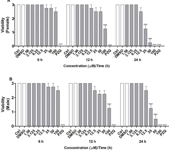
concentra-tions ranging from 1.56 to 100μM or manually. The MTT assay [33] was conducted for 6, 12, or 24 h to assay the viability. Incubation of coupled adult worms with CUR for 6 h did not alter the viability of the female and male worms at any of the tested concentrations (Fig 1A and 1B). After 12 h of incubation, the viability of both female and male worms decreased consider-ably in the presence of CUR at 100μM. After 24 h of incubation, the viability of both female and male worms diminished significantly in the presence of CUR at 25, 50, or 100μM. In con-trast, the couples of adult worms in the negative control groups (RPMI 1640 medium alone or in combination with 0.1% DMSO) exhibited normal viability, whereas the couples of adult worms in the positive control group (PZQ at 1.56μM or heat-killed) were not viable (100% death) (Fig 1A and 1B).
Microscopic observation of couples of adultS.mansoniworms supported the different via-bilities of the worm pairs after exposure to CUR. Viability was assessed on the basis of changes in the motor activity of the worms and of the occurrence of death according to standard proce-dures for the screening of compounds defined by the WHO-TDR [34]. According toFig 2A and 2B, CUR significantly decreased the viability of adult female and male worms at the con-centrations and incubation times described previously. Mortality was 100% for both female and male worms after exposure to CUR at 100μM for 24 h. CUR at 25 or 50μM lowered the motor activity of female and male worms to a minimum or killed them. The LC50values obtained for CUR were 491.0, 90.2, and 32.9 for female worms and 491.0, 90.8, and 43.9 for male worms at 6, 12, and 24 h, respectively. Additionally, CUR at 25μM separated 75% of the couples of adult worms, whilst CUR at 50 or 100μM separated all the couples of adult worms after incubation for 24 h (data not shown). The couples of worms in the negative control groups (RPMI 1640 medium alone or in combination with 0.1% DMSO) exhibited normal via-bility and remained coupled (i.e., the couples of adult worms were not separated), whereas the couples of worms in the positive control group (PZQ at 1.56μM) showed no viability (100% death) and no separation.
CUR induces alterations in the tegument and organelles of adult
S
.
mansoni
worms
control group (RPMI 1640 with 0.1% DMSO), the tegument was intact, the muscular layer had preserved fibers throughout the body, most vitelline cells were normal, the cytoplasm was rich in granular endoplasmic reticulum and mitochondria, and vitelline droplets existed inside vitelline balls (Fig 3A). However, some vitelline cells contained small vacuoles, and some nuclear chromatin was undergoing condensation. After incubation for 6 or 12 h with CUR, the mitochondrial membrane swelled, chromatin condensed, and small vacuoles emerged in femaleS.mansoniworms, but the tegument remained unaltered. Interestingly, drastic changes occurred in the vitelline cells of femaleS.mansoniworms after 24 h of incubation. The intersti-tial tissue underwent lysis, the mitochondria swelled and degenerated, and chromatin con-densed. Additionally, analysis of some parts of the tegument indicated swelling and formation of vacuoles of different sizes (Fig 3A).
Fig 1.In vitroeffect of CUR on the viability of adultS.mansoniworms as measured by MTT assay.Couples of adult worms were incubated with different concentrations of CUR for 6, 12, or 24 h. Adult (A) female and (B) maleS.mansoniworms were separated, and the viability was measured by the MTT assay at 550 nm. In the negative control groups, couples of adult worms were incubated with RMPI 1640 medium or with RPMI 1640 medium with 0.1% DMSO. In the positive control groups, couples of adult worms were incubated with PZQ (1.56μM) or heat-killed at 56˚C. Values are expressed as the mean±S.E.M of three independent experiments. An asterisk indicates statistically significant differences as compared to the negative control group (RPMI 1640 medium with 0.1% DMSO) (***p<0.001).
The maleS.mansoniworms in the control group (RPMI 1640 with 0.1% DMSO) presented intact tegument and spines with regular morphology. The muscular layer exhibited preserved fibers throughout the body, and the mitochondria and cells had normal morphology (Fig 3B). After 6 h of incubation with CUR, there were no structural alterations in maleS.mansoni; at 12 h of incubation, nuclear chromatin began to condense, but the tegument remained unal-tered. However, at 24 h of incubation with CUR, vacuoles of different sizes emerged in the teg-ument, mitochondria swelled and ruptured, small vacuoles arose, and nuclear chromatin condensed (Fig 3B).
Fig 2.In vitroeffect of CUR on the viability of adultS.mansoniworms with emphasis on changes in the motor activity of the worms.Couples of adult worms were incubated with different concentrations of CUR for 6, 12, or 24 h. The viability of separated adult (A) female and (B) maleS.mansoniworms was monitored by using a viability scale of 0–3 (3 = totally vital, normally active, 2 = slowed activity, 1 = minimal activity, 0 = worm death—death was defined as no movement being observed for at least 2 min of examination). In the negative control groups, couples of adult worms were incubated with RMPI 1640 medium or with RPMI 1640 medium with 0.1% DMSO. In the positive control groups, couples of adult worms were incubated with PZQ (1.56μM). Values are expressed as the mean±S.E.M of three independent experiments. An asterisk indicates statistically significant differences as compared to the negative control group (RPMI 1640 medium with 0.1% DMSO) (***p<0.001).
CUR induces DNA fragmentation and damage in adult
S
.
mansoni
worms
We incubated couples of adult worms with CUR at 25 or 50μM for 24 h and then extracted and analyzed the DNA of female and male adult worms (separated either by action of CUR or manually) by electrophoresis on 2% agarose gel. DNA fragmentation slightly increased in the adult female and male worms of the negative control group (worms incubated with RPMI 1640 medium with 0.1% DMSO) (Fig 4A). In contrast, incubation with CUR significantly increased DNA fragmentation in adult female and male worms (Fig 4A). We also evaluated DNA fragmentation by using the Terminal Deoxynucleotidyl Transferase dUTP Nick End Labelling (TUNEL). We incubated couples of adult worms with CUR (50μM). TUNEL-posi-tive cells (dark brown apoptotic nuclei) increased significantly in adult female and male worms as compared to the negative control group (Fig 4B and 4C). Additionally, TUNEL-pos-itive cells increased more significantly in adult female worms as compared to adult male worms.
We also evaluated DNA damage by the comet assay. The comet assay is an alkaline single-cell gel electrophoresis that is used to measure breaks in DNA strands in single-cells that are embed-ded in agarose and submitted to lysis to remove membranes and soluble cell constituents.
Fig 3. CUR induces alterations in the tegument and organelles of adultS.mansoniworms.Couples of adult worms were incubated with CUR at 50μM for 6, 12, or 24 h. After incubation, female and maleS.mansoniworms were separated and processed for Transmission Electron Microscopy (TEM) analysis. In the negative control groups, couples of adult worms were incubated with RPMI 1640 medium with 0.1% DMSO for 24 h. (A) Micrograph of adult female worms and (B) Micrograph of adult male worms. T, tegument; S, spine; L, lipid; N, nucleus, NU, nucleolus; M, Mitochondria; VC, Vitelline cell; VD, Vitelline droplets; Asterisk, vacuolization of the tegument; Arrow, lysis of tissue. A total of 20 adult female and male worms were evaluated at each concentration.
Lesions can be observed because damaged DNA migrates faster than undamaged DNA. In other words, in cells with damaged DNA, the DNA migrates from the nucleus toward the anode, which resembles the shape of a comet [36]. The frequency of DNA damage increased in
Fig 4. CUR induces DNA fragmentation and damage in adultS.mansoniworms.Couples of adult worms were incubated with CUR at the indicated concentrations for 24 h. After incubation, female and maleS.mansoniworms were separated and analyzed. In the negative control groups, couples of adult worms were incubated with RPMI 1640 medium with 0.1% DMSO. (A) Genomic DNA of adult female and male worms was extracted as described in material and methods, and 600 ng of the DNA was run in 2% agarose gel containing 1% GelRed (1:500) (MW Molecular weight marker). The experiments were repeated twice, and ten couples of adult worms were evaluated in each experiment. (B) TUNEL-stained light micrographs of adult female and male worm sections (arrows indicate the dark brown-stained apoptotic nuclei). (C) Histograms indicate the percentage of TUNEL-positive cells. For each experiment, at least 100 cells were analyzed. Values are expressed as the mean±S.E.M of three independent experiments. An asterisk indicates statistically significant differences as compared to the negative control group (RPMI 1640 medium with 0.1% DMSO) or when male and female worms were compared (**p<0.01,***p<0.001).
female and male cells incubated with CUR at 25 or 50μM at 24 h as compared to the negative control group (Table 1).
CUR increases the expression of
SmCASP3/7
transcripts and the
activity of Caspase 3 in adult
S
.
mansoni
worms, but the activity of
Caspase 8 remains unaltered
To test whether CUR induces cell death, we determined the levels of expression ofSmCASP3, 7, and8transcripts by quantitative RT-PCR in adult worms separated either by action of CUR or manually.SmCASP3/7increased significantly in female and male worms incubated with CUR at 25 or 50μM for 24 h as compared to the negative control group (worms incubated with RPMI 1640 medium plus 0.1% DMSO). At higher concentration of CUR (50μM), the
SmCASP3andSmCASP7transcripts were upregulated by about 13- and 11.1-fold in adult
female worms, respectively (Fig 5A). In adult male worms incubated with CUR, theSmCASP3
andSmCASP7transcripts were upregulated by about 7.6- and 5.7-fold, respectively (Fig 5B).
On the other hand, analysis of the transcription levels showedSmCASP8increased only slightly in both female and male worms incubated with CUR as compared to the transcription levels of
SmCASP3andSmCASP7(Fig 5A and 5B). These results resembled the data reported by Morais
et al. [27], who demonstrated about twofold upregulation forSmCASP8.
We also analyzed how the activities of Caspase 3 and 8 changed in adult worms incubated with CUR at 25 or 50μM. The activity of Caspase 3 increased significantly in both adult female and maleS.mansoniworms after incubation with CUR for 24 h as compared to the negative control group. At higher concentration of CUR (50μM), the activity of Caspase 3 increased by more than 80% in adult female and male worms (Fig 5C and 5D). On the other hand, the activity of Caspase 8 remained unaltered in both female and maleS.mansoniworms (Fig 5E and 5F).
CUR induces formation of the superoxide anion and increases the
activity of SOD activity in adult
S
.
mansoni
worms
We incubated pairs of adult worms with CUR at 12.5–100μM for 6, 12, or 24 h and evaluated the level of superoxide anion in adult female and male worms separated either by action of CUR or manually. We employed the colorimetric Nitroblue Tetrazolium (NBT) assay during this analysis [43]. The level of superoxide anion increased significantly in adult female and
maleS.mansoniworms incubated with CUR at 25 to 100μM after 12 or 24 h of incubation as
Table 1. Frequency of DNA damage in the cells of adultS.mansoniworms incubated with CUR.
% Comet class in the cells of female and male adult wormsa Score
0 1 2 3 4
Samples Female Male Female Male Female Male Female Male Female Male Female Male Controlb 67.2±5.2 71.5±6.4 20.1±6.2 18.7±4.3 7.4±4.1 5.3±2.9 6.3±3.0 4.5±2.0 0.0±0.0 0.0±0.0 53.8±23.4 42.8±16.1
25μM 16.3±4.7 23.0±5.2 59.3±8.7 55.1±8.1 14.0±5.9 13.4±4.2 10.4±2.9 8.5±2.6 0.0±0.0 0.0±0.0 118.5±29.2*** 107.4±24.3*** 50μM 13.9±5.8 16.3±6.1 58.1±9.5 53.6±8.3 14.1±6.7 17.5±6.9 13.9±5.4 12.6±4.8 0.0±0.0 0.0±0.0 128.0±39.1*** 126.4±36.5***
aFifty random cells were visually analyzed on each slide. Each comet class had a value that ranged between 0 and 4: (0) undamaged cells (all the DNA was
located in the head) and (4) maximum damage (almost all the DNA was located in the tail). The total score was calculated by multiplying the percentage of damaged nucleoids by the value of the respective comet class (0, 1, 2, 3, or 4). A value of 0 indicates that no damage occurred; a value of 400 corresponds to maximum damage.
bCouples of adult worms incubated with RPMI 1640 with 0.1% DMSO. Values are expressed as the mean±S.E.M of three independent experiments. An
asterisk indicates statistically significant differences as compared to the negative control group (RPMI 1640 medium with 0.1% DMSO) ***p<0.001.
compared to the negative control group (worms incubated with RPMI 1640 medium with 0.1% DMSO). For incubations with CUR at 25, 50, or 100μM for 24 h, the level of superoxide anion increased by more than 44%, 56%, and 66% in adult female worms, respectively, and 34%, 37%, and 44% in adult male worms, respectively. On the other hand, 6 h of incubation with CUR did not alter the level of the superoxide anion as compared to the negative control group (Fig 6A and 6B). Both male and female worms in the positive control group had signifi-cantly increased levels of the superoxide anion at 6 h.
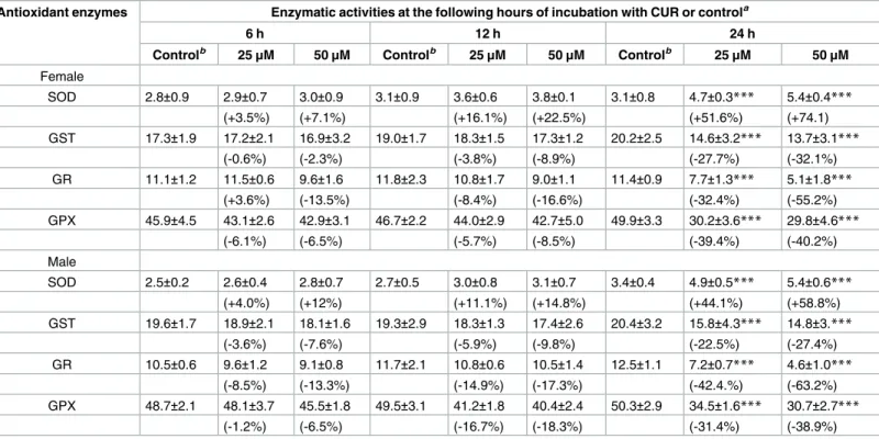
Inside the cells, the superoxide anion (O2-) is converted to hydrogen peroxide (H2O2) in a reaction catalyzed by the enzyme SOD. Here, we determined the activity of SOD in female and male adult worms incubated with CUR at 25 or 50μM for 6, 12, or 24 h. As shown inTable 2, 6 and 12 h of incubation with CUR did not change the activity of SOD significantly in adult female and male worms as compared to the negative control group (worms incubated with RPMI 1640 medium with 0.1% DMSO). In contrast, the activity of SOD increased significantly in adult female and male worms 74% and 58%, respectively at 24 h of incubation (Table 2). The activity of SOD was also significantly higher in the positive control at 6, 12, and 24 h in both male and female worms (data not shown).
CUR alters various oxidative stress parameters in adult
S
.
mansoni
worms
To investigate other oxidative stress parameters, we incubated pairs of adultS.mansoniworms with CUR at 25 or 50μM for 6, 12, or 24 h and evaluated the activities of GST, GR, and GPX and the levels of protein carbonyl in both adult female and male worms separated either by action of CUR or manually.Table 2shows that the activities of GST, GR, and GPX decreased significantly in adult female and male worms after 24 h of incubation with CUR at 25 or 50μM. CUR at 50μM inhibited the activity of GST in adult female and male worms by more than 32% and 27%, respectively. CUR at 50μM inhibited the activity of GR in adult female and male worms by more than 63% and 55%, respectively. Finally, CUR at 50μM inhibited the activity of GPX in adult female and male worms by more than 40% and 38%, respectively. At 6 and 12 h of incubation, the activities of GST, GR, and GPX did not decrease significantly as compared to the negative control group. In the positive control group (100μM hydrogen per-oxide), the activity of GST increased significantly in female and male worms at 6, 12, and 24 h (data not shown). On the other hand, the activities of GR and GPX decreased significantly in both adult female and male worms (data not shown) for all the incubation periods.
The levels of protein carbonyl increased significantly in both adult female and male worms after incubation with CUR at 25 or 50μM for 24 h as compared to the negative control group. At 6 and 12 h, the levels of protein carbonyl did not change significantly (Table 3). Both male and female worms in the positive control group had significantly increased levels of protein carbonyl at 12 and 24 h of incubation (data not shown).
Discussion
The only schistosomicidal drug that is commercially available is Praziquantel, which has been used in monotherapy for several decades. Therefore, widespread emergence of drug resistance
worms incubated with CUR. Expression was calculated according to the comparative Ct method (2-ΔΔCTmethod), and data were normalized relative to an endogenous standard gene (SmGPDH). The activities of Caspase 3 and 8 were measured as described in material and methods. The activity of Caspase 3 referred to adult (C) female and (D) male worms. The activity of caspase 8 referred to adult (E) female and (F) male worms. Values are expressed as the mean±S.E.M of three independent experiments. An asterisk indicates statistically significant differences as compared to the negative control group (RPMI 1640 medium with 0.1% DMSO) (**p<0.01,***p<0.001).
is probable and makes the development of new therapeutic approaches against schistosomiasis mandatory. In the past years, numerous natural products and synthetic drugs have been evalu-ated as potential schistosomicidal agents [18,19], but few studies have shown the effects of these drugs on the induction of apoptosis in this parasite. Curcumin (CUR), a secondary metabolite of turmeric derived fromC.longaL., displays many biological activities [20,48]. Previous studies have suggested that CUR and CUR analogs can generate reactive oxygen
Fig 6. CUR induces formation of the superoxide anion in adultS.mansoniworms.Couples of adult worms were incubated with CUR at 12.5 to 100μM for 6, 12, or 24 h. After incubation, (A) female and (B) maleS.mansoniworms were separated, and the level of superoxide anion was measured by the NBT assay at 620 nm. Couples of adult worms incubated with RPMI 1640 with 0.1% DMSO were used as negative control, and couples of adult worms incubated with RPMI 1640 medium with 100μM hydrogen peroxide were used as positive control. The results represent the mean±SEM of three independent experiments. An asterisk indicates statistically significant differences as compared to the negative control group (RPMI 1640 medium with 0.1% DMSO) (**p<0.01,***p<0.001).
species (ROS) in several cancer cell lines and trigger the apoptotic death of these cells [49–51]. Additionally, CUR exhibits potent anti-filarial effects on a filarial parasite because it induces
Table 2. Effect of CUR on the activity of different antioxidant enzymes in adultS.mansoniworms.
Antioxidant enzymes Enzymatic activities at the following hours of incubation with CUR or controla
6 h 12 h 24 h
Controlb 25μM 50μM Controlb 25μM 50μM Controlb 25μM 50μM
Female
SOD 2.8±0.9 2.9±0.7 3.0±0.9 3.1±0.9 3.6±0.6 3.8±0.1 3.1±0.8 4.7±0.3*** 5.4±0.4***
(+3.5%) (+7.1%) (+16.1%) (+22.5%) (+51.6%) (+74.1)
GST 17.3±1.9 17.2±2.1 16.9±3.2 19.0±1.7 18.3±1.5 17.3±1.2 20.2±2.5 14.6±3.2*** 13.7±3.1***
(-0.6%) (-2.3%) (-3.8%) (-8.9%) (-27.7%) (-32.1%)
GR 11.1±1.2 11.5±0.6 9.6±1.6 11.8±2.3 10.8±1.7 9.0±1.1 11.4±0.9 7.7±1.3*** 5.1±1.8***
(+3.6%) (-13.5%) (-8.4%) (-16.6%) (-32.4%) (-55.2%)
GPX 45.9±4.5 43.1±2.6 42.9±3.1 46.7±2.2 44.0±2.9 42.7±5.0 49.9±3.3 30.2±3.6*** 29.8±4.6***
(-6.1%) (-6.5%) (-5.7%) (-8.5%) (-39.4%) (-40.2%)
Male
SOD 2.5±0.2 2.6±0.4 2.8±0.7 2.7±0.5 3.0±0.8 3.1±0.7 3.4±0.4 4.9±0.5*** 5.4±0.6***
(+4.0%) (+12%) (+11.1%) (+14.8%) (+44.1%) (+58.8%)
GST 19.6±1.7 18.9±2.1 18.1±1.6 19.3±2.9 18.3±1.3 17.4±2.6 20.4±3.2 15.8±4.3*** 14.8±3.***
(-3.6%) (-7.6%) (-5.9%) (-9.8%) (-22.5%) (-27.4%)
GR 10.5±0.6 9.6±1.2 9.1±0.8 11.7±2.1 10.8±0.6 10.5±1.4 12.5±1.1 7.2±0.7*** 4.6±1.0***
(-8.5%) (-13.3%) (-14.9%) (-17.3%) (-42.4.%) (-63.2%)
GPX 48.7±2.1 48.1±3.7 45.5±1.8 49.5±3.1 41.2±1.8 40.4±2.4 50.3±2.9 34.5±1.6*** 30.7±2.7***
(-1.2%) (-6.5%) (-16.7%) (-18.3%) (-31.4%) (-38.9%)
aCouples of adult worms were incubated with CUR at 25 or 50μM for 6, 12, or 24 h. The activities of the antioxidant enzymes were measured as described
the materials and methods section. Values in parentheses indicate the percentage of inhibition/activation as compared to the negative control group.
bPairs of adult worms incubated with RPMI 1640 with 0.1% DMSO. Activities are indicated as U/mg, and the values are expressed as the mean±S.E.M of
three independent experiments. An asterisk indicates statistically significant differences as compared to the negative control group (RPMI 1640 medium with 0.1% DMSO)
***p<0.001.
doi:10.1371/journal.pone.0167135.t002
Table 3. Effects of CUR on the content of protein carbonyl in adultS.mansoniworms.
Samples Content of protein carbonyl at the following hours of incubation with CUR or controla
6 h 12h 24 h
Female Male Female Male Female Male
Controlb 23.3±4.7 26.1±2.8 23.9±2.8 27.0±3.1 25.6±2.8 28.9±3.2
25μM 23.9±1.6 27.5±3.6 27.3±2.0 30.5±4.1 32.7±2.1*** 35.4±4.5***
(+2.5%) (+5.3%) (+14.2%) (+12.9%) (+27.7%) (+22.4%)
50μM 25.8±3.7 28.1±2.1 28.3±3.2 31.2±2.7 36.9±3.4*** 37.8±3.2***
(+7.9%) (+7.6%) (+18.4%) (+15.5%) (+44.1%) (30.7%)
aCouples of adult worms were incubated with CUR at 25 or 50μM for 6, 12, or 24 h. The content of protein carbonyl was measured as described in the
materials and methods section. Values in parentheses indicate the increase in percentage as compared to the control.
bCouples of adult worms incubated with RPMI 1640 with 0.1% DMSO. The content of protein carbonyl is expressed as mmol/mg protein, and the values are
expressed as the mean±S.E.M of three independent experiments. An asterisk indicates statistically significant differences as compared to the negative control group (RPMI 1640 medium with 0.1% DMSO)
***p<0.001.
apoptosis by a mitochondrial pathway [8,31]. Here, we demonstrated that CUR affected oxida-tive stress and induction of apoptosis in adultS.mansoniworms.
Previous investigations by our group have demonstrated that CUR at 50 or 100μM after 24 or 120 h of incubation kills all the couples of adult worms [21]. Based on these results, here we report additional data on the viability of adult worms after conducting the MTT assay and microscopic analyses of pairs of adult worms incubated with CUR for 6, 12, or 24 h. The results suggested potentialin vitroactivity of CUR against female and maleS.mansoniworms at 24 h. However, CUR was more lethal to female worms, LC50values were 32.9μM as compared to 43.9μM in the case of adult male worms.
Ultrastructural analysis showed altered tegument in adult female and male worms after incubation with CUR. The tegument is an essential structure for the survival and maintenance
ofSchistosomaworms because it plays a vital role in evading the host’s immune system,
acquir-ing nutrients, excretacquir-ing catabolic products, and targetacquir-ing drug absorption, among other physi-ological processes [52].
Morphologically, cells killed by apoptosis may exhibit morphological changes such as swelled and ruptured mitochondria, condensed chromatin, and fragmented DNA [53,54]. The ultrastructural analysis also demonstrated alterations such as swelling and degeneration of the mitochondrial membrane, condensation of chromatin, and formation of vacuoles in adult female and male worms. In adult female worms, the alterations occurred mainly in the vitellar-ium, which is a proliferative tissue that occupies the posterior two thirds of the female and pro-duces cells that surround the ovum and provide the precursor proteins that form the eggshell and the nutrients that aid development of the embryo [55]. Additionally, at 24 h, there were some alterations in vitelline cells in adult female worms belonging to the negative control group, as already described by Galanti et al. (2012) [56]. Because the production of eggs is cru-cial to both the transmission and the pathogenesis of schistosomiasis, drug-induced alterations in the reproductive development of schistosomes could constitute a new method to prevent or treat the disease [55,56]. Interestingly, our group has previously shown that CUR reduces the production of eggs by more than 50% as compared to the negative control group underin
vitroconditions [21]. Additionally, Mohapatra et al. (2011) [8] have demonstrated that CUR is
the most effective pharmacological agent to induce apoptosis in embryonic stages of the
nema-todeSetaria digitata, suggesting that blockage of embryogenesis through therapeutic induction
of apoptosis during the embryonic stages of parasites is a promising strategy to develop effec-tive anti-parasitic measures against extracellular parasites.
Considering the alterations observed by TEM, we evaluated different apoptotic parameters in adult worms incubated with CUR. DNA fragmentation is one of the main parameters used to identify cell apoptosis—activated caspases cleave the DNA of the cell, to produce DNA frag-ments [54]. Evaluation of DNA fragmentation by electrophoresis on agarose gel evidenced that CUR fragmented the DNA of adult female and male worms. In addition, TUNEL staining, a method that detects DNA fragmentationin situ(a hallmark of apoptosis), demonstrated that TUNEL-positive cells increased in adult female and male worms as compared to the negative control group. In addition, the number of TUNEL-positive cells was higher in adult female worms as compared to adult male worms. Moreover, the comet assay revealed that CUR dam-aged DNA in adult female and male worms. Studies have suggested that CUR can fragment and damage DNA [31,57–58]. For example CUR inducesin situDNA fragmentation in both embryos and adult females of the parasiteS.servi[31]. Other studies have shown that CUR induces apoptosis in human hepatocellular carcinoma J5 cells [58].
caspases);SmCASP3andSmCASP7, which are homologues of the human effector caspases CASP3 andCASP7, respectively; andSmCASPC, which is a homologue of humanCASP8[16– 17,60–61]. In the present study, we evaluated how CUR affected the expression of transcripts
ofSmCASP3,7, and8as well as the activities of Caspase 3 and 8 in adult female and male
worms.SmCASP3/7transcripts and the activity of caspase 3 were upregulated in female and male adult worms after incubation with CUR at 25 or 50μM. Moreover, the expression of
SmCASP3/7and the activity of Caspase 3 were higher in adult female worms as compared to
adult male worms. The extrinsic pathway that activates caspase starts through binding to death receptors on the cell membrane, to recruit the cytosolic adapter protein FADD and Caspase 8 and to form the death-inducing signaling complex (DISC) that cleaves and activates caspase 3 [9]. Here, the activity of caspase 8 did not increase, suggesting that the activity of caspase 3 could be associated with the induction of apoptosis by the intrinsic pathway. Works by other authors have also suggested that CUR can upregulate the expression ofCASP3, and that this caspase plays a crucial role in CUR-induced apoptosis in filarial parasites by the intrinsic path-way [8,31].
Mitochondria are the major source and target of intracellular reactive oxygen species (ROS) [8,61–63]. During apoptosis, induced by a variety of stimuli, the permeability of the mitochon-drial membrane increases and triggers the release of pro-apoptotic factors including cyto-chrome-c and AIF (Apoptosis-inducing Factor) into the cytosol. The cytosolic cytocyto-chrome-c then interacts with APAF-1 and forms an apoptosome. Activation of the apoptosome can ulti-mately activate effector caspase and apoptosis [63–65]. A wide range of chemicals or natural compounds can stimulate the formation of ROS and trigger apoptosis [66–67]. High concen-trations of CUR (at 25 and 50μM) promote the formation of ROS in different cell lines, whereas low CUR (at 10μM) usually diminishes the formation of ROS [28,68–71]. The results of the present study were consistent with data reported by other authors [8,31,49–51] and showedhat CUR and some of its derivatives induced production of ROS.
In schistosomes and other helminthic parasites, diverse antioxidant systems that depend on antioxidant enzymes, or not, regulate the concentrations of ROS inside the cell. AdultS. man-soniworms have a long life span and constantly face stress conditions within the host, so they have evolved a series of antioxidant enzymes to evade the host’s hostile environment [72]. Among these enzymes are SOD, GST, GR, and GPX, which play an essential part in balancing the production and decomposition of ROS and in protecting the parasite from damage as a result of enhanced production of ROS like superoxide radical anion and hydroxyl radicals [73,74]. The induction of oxidative stress due to abatement of the antioxidant system or to increased production of ROS in adultS.mansonihas been considered an attractive approach to new treatment strategies [74–76].
promoting cell death. Our study evidenced increased content of protein carbonyl content in adult female and male worms incubated with CUR.
In summary, the results of the present work suggest that CUR generates oxidative stress fol-lowed by an apoptotic-like event in adult female and maleS.mansoniworms, which ultimately leads to parasite death. Induction of apoptotic death is a therapeutic approach that needs to be further explored during the development of new drugs with broad spectrum and anthel-minthic activity.
Acknowledgments
The authors are grateful to The Electron Microscopy Laboratory in Ribeirão Preto, University of São Paulo, Brazil, for support with the transmission electron microscopy examinations. The authors are also thankful to Prof. Ma´rcio Luis de Andrade e Silva and Olinda Mara Brigatto for their technical support and to Prof. Conor Caffrey for discussion on the activity of caspase.
Author Contributions
Conceived and designed the experiments:LGM VR.
Performed the experiments:DPA MBMM RGP PMF AA ATO.
Analyzed the data:LGM AA WRC VR ERM CC SB.
Contributed reagents/materials/analysis tools:LGM WRC VR SB CC.
Wrote the paper:LGM AA.
References
1. WHO: Schistosomiasis, Fact sheet Nu 115, Update February 2016. Available:http://www.who.int/ mediacentre/factsheets/fs115/en/. Accessed 01 March, 2016.
2. Cioli D, Pica-Mattoccia L, Basso A, Guidi A. Schistosomiasis control: praziquantel forever? Mol. Bio-chem. Parasitol. 2014; 195: 23–29. doi:10.1016/j.molbiopara.2014.06.002PMID:24955523 3. Caffrey CR. Schistosomiasis and its treatment. Future Med Chem. 2015; 7(6): 675–676. doi:10.4155/
fmc.15.27PMID:25996057
4. Fallon PG, Doenhoff MJ. Drug-resistant schistosomiasis: resistance to praziquantel and oxamniquine induced in Schistosoma mansoni in mice is drug specific. Am. J. Trop. Med. Hyg. 1994; 51: 83–88. PMID:8059919
5. Ismail M, Botros S, Metwally A, William S, Farghally A, Tao LF, et al. Resistance to praziquantel: direct evidence from Schistosoma mansoni isolated from Egyptian villagers. Am J. Trop. Med. Hyg. 1999; 6: 932–935. PMID: 10403323
6. Pica-Mattoccia L, Cioli D. Sex- and stage-related sensitivity of Schistosoma mansoni to in vivo and in vitro praziquantel treatment. Int. J. Parasitol. 2004; 34: 527–533. doi:10.1016/jijpara.2003.12.003 PMID:15013742
7. Yepes E, Varela-M RE, Lo´pez-Aba´n J, Dakir EL, Mollinedo FMA. In vitro and in vivo anti-schistosomal activity of the alkylphospholipid analog edelfosine. PLoS One. 2014; (10): e109431. doi:10.1371/ journal.pone.0109431PMID:25302497
8. Mohapatra AD, Kumar S, Satapathy AK, Ravindran B. Caspase dependent programmed cell death in developing embryos: a potential target for therapeutic intervention against pathogenic nematodes. PLoS Negl Trop Dis. 2011;(9): e1306. doi:10.1371/journal.pntd.0001306PMID:21931872
9. Strasser A, Cory S, Adams JM. Deciphering the rules of programmed cell death to improve therapy of cancer and other diseases. EMBO J. 2011; 30: 3667–3683. doi:10.1038/emboj.2011.307PMID: 21863020
11. Aouacheria A, Brunet F, Gouy M. Phylogenomics of life-or-death switches in multicellular animals: Bcl-2, BH3-Only, and BNip families of apoptotic regulators. Mol. Biol. Evol. 2005; 22: 2395–2416. doi:10. 1093/molbev/msi234PMID:16093567
12. Verjovski-Almeida S, DeMarco R, Martins EA, Guimarães PE, Ojopi EP, Paquola AC, et al. Transcrip-tome analysis of the acoelomate human parasite Schistosoma mansoni. Nat Genet. 2003; 35(2):148– 57. doi:10.1038/ng1237PMID:12973350
13. Berriman M, Haas BJ, Loverde PT, Wilson RA, Dillon GP, Cerqueira GC, et al. The genome of the blood fluke Schistosoma mansoni. Nature. 2009; 460(7253):352–358. doi:10.1038/nature08160PMID: 19606141
14. Protasio AV, Tsai IJ, Babbage A, Nichol S, Hunt M, Aslett MA, et al. A systematically improved high quality genome and transcriptome of the human blood fluke Schistosoma mansoni. PLoS Negl Trop Dis. 2012; 6(1):e1455. doi:10.1371/journal.pntd.0001455PMID:22253936
15. Oliveira KC, Carvalho ML, Venancio TM, Miyasato PA, Kawano T, DeMarco R, Verjovski-Almeida S. PLoS Negl Trop Dis. 2009; 3(12):e556. doi:10.1371/journal.pntd.0000556PMID:19956564
16. Lee EF, Clarke OB, Evangelista M, Feng Z, Speed TP, Tchoubrieva EB, et al. Discovery and molecular characterization of a Bcl-2-regulated cell death pathway in schistosomes. Proc. Natl. Acad. Sci. U.S.A. 2011; 108: 6999–7003. doi:10.1073/pnas.1100652108PMID:21444803
17. Han H. Apoptosis phenomenon in the schistosomulum and adult worm life cycle stages of Schistosoma japonicum. Parasitol. Int. 2013; 62: 100–108. doi:10.1016/j.parint.2012.09.008PMID:23159324 18. Ndjonka D, Rapado LN, Silber AM, Liebau E, Wrenger C. Natural products as a source for treating
neglected parasitic diseases. Int J Mol Sci. 2013; 6: 3395–3439. doi:10.3390/ijms14023395
19. De Moraes J. Natural products with antischistosomal activity. Future Med Chem. 2015; 7: 801–820. doi: 10.4155/fmc.15.23PMID:25996071
20. Sueth SV, Mendes SGP, Decote RD, Lima ME. Curcumina, o po´ dourado do ac¸afrão-da-terra: intro-specc¸ões sobre quı´mica e atividades biolo´gicas. Quim. Nova. 2015; 38(4): 538–552.
21. Magalhães LG, Machado CB, Morais ER, Moreira EB, Soares CS, da Silva SH. In vitro schistosomicidal activity of curcumin against Schistosoma mansoni adult worms. Parasitol Res. 2009; 104: 1197–1201. doi:10.1007/s00436-008-1311-yPMID:19096877
22. Bazh EK, El-Bahy NM. In vitro and in vivo screening of anthelmintic activity of ginger and curcumin on Ascaridia galli. Parasitol Res. 2013; 112(11): 3679–3686. doi:10.1007/s00436-013-3541-xPMID: 24046262
23. Fouladvand MBA, Tahmasebi R. Evaluation of in vitro antileishmanial activity of curcumin and its deriva-tives "gallium curcumin, indium curcumin and diacethyle curcumin. Eur Rev Med Pharmacol Sci. 2013; 17(24): 3306–3308. PMID:24379060
24. Wachter B, Syrowatka M, Obwaller A, Walochnik J. Wien Klin Wochenschr. In vitro efficacy of curcumin on Trichomonas vaginalis. Wien Klin Wochenschr. 2014; 1: S32–S36. doi:10.1007/s00508-014-0522-8 PMID:24619489
25. Goo YK, Yamagishi J, Ueno A, Terkawi MA, Aboge GO, Kwak D, et al. Characterization of Toxoplasma gondii glyoxalase 1 and evaluation of inhibitory effects of curcuminon the enzyme and parasite cultures. Parasit Vectors. 2015; 8(1): 654. doi:10.1186/s13071-015-1268-5PMID:26694921
26. Luz PP, Magalhães LG, Pererira AC, Cunha WR, Rodrigues V, Andrade ESML. Curcumin-loaded into PLGA nanoparticles: preparation and in vitro schistosomicidal activity. Parasitol Res. 2012; 110: 593– 598. doi:10.1007/s00436-011-2527-9PMID:21739309
27. Morais ER, Oliveira KC, Magalhães LG, Moreira EB, Verjovski-Almeida S, Rodrigues V. Effects of cur-cumin on the parasite Schistosoma mansoni: a transcriptomic approach. Mol Biochem Parasitol. 2013; 187(2): 91–97. doi:10.1016/j.molbiopara.2012.11.006PMID:23276630
28. Bhaumik S, Anjum R, Rangaraj N, Pardhasaradhi BVV, Khar A. Curcumin mediated apoptosis in AK-5 tumor cells involves the production of reactive oxygen intermediates. FEBS Lett. 1999; 456(2): 311– 314. doi:10.1016/S0014-5793(99)00969-2PMID:10456330
29. Watson JL, Hill R, Yafee PB, Greenshields A, Walsh M, Lee PW, et al. Curcumin causes superoxide anion production and p53- independent apoptosis in human colon cancer cells. Cancer Lett. 297: 1–8. doi:10.1016/j.canlet.2010.04.018PMID:20472336
30. Thayyullathil F, Chathoth S, Hago A, Patel M, Galadari S. Rapid reactive oxygen species (ROS) gener-ation induced by curcumin leads to caspase-dependent and independent apoptosis in L929 cells. Free Radic Biol Med. 2008; 45: 1403–1412. doi:10.1016/j.freeradbiomed.2008.08.014PMID:18762247 31. Nayak A, Gayen P, Saini P, Mukherjee N, Babu SP. Molecular evidence of curcumin-induced apoptosis
32. Smithers SR, Terry RJ. The infection of laboratory hosts with cercariae of Schistosoma mansoni and the recovery of the adult worms. Parasitology. 1965; 55: 695–700. PMID:4957633
33. Comley JCW, Rees MJ, Turner CH, Jenkins DC. Calorimetric quantitation of filarial viability. Int J Parasi-tol. 1989; 19: 77–83. doi:10.1016/0020-7519(89)90024-6PMID:2707965
34. Ramirez B, Bickle Q, Yousif F, Fakorede F, Mouries MA, Nwaka S. Schistosomes: Challenges in com-pound screening. Expert. Opin. Drug. Discov. 2007; 2: 53–61. doi:10.1517/17460441.2.S1.S53PMID: 23489033
35. Sambrook J, Russel DW. Molecular cloning: a laboratory manual. 3rd ed. Cold Spring Harbour Labora-tory Press. 2001.
36. Azqueta A, Slyskova J, Langie SA, ONeill G, Collins A. Comet assay to measure DNA repair: approach and applications. Front Genet. 2014; 5: 00288. doi:10.3389/fgene.2014.00288PMID:25202323 37. Dubois F, Caby S, Oger F, Cosseau C, Capron M, Grunau C, et al. Histone deacetylase inhibitors
induce apoptosis, histone hyperacetylation and up-regulation of gene transcription in Schistosoma mansoni. Mol Biochem Parasitol. 2009; 68: 7–15. doi:10.1016/j.molbiopara.2009.06.001
38. Mourão MM, Dinguirard N, Franco GR, Yoshino TP. Phenotypic screen of early developing larvae of the blood fluke, Schistosoma mansoni, using RNA interference. PLoS Negl Trop Dis. 2009;(8: ): e502. doi:10.1371/journal.pntd.0000502PMID:19668375
39. De Paula RG, de Magalhães Ornelas AM, de Souza Gomes M, de Paula Aguiar D, Magalhães LG, et al. Proteasome stress responses in Schistosoma mansoni. Parasitol Res. 2015; 14(5): 1747–1760. doi:10.1007/s00436-015-4360-z
40. Livac KJ, Schmittgen TD. Analysis of relative gene expression data using real-time quantitative PCR and the 2(-Delta Delta C(T)). Methods. 2001; 25(4):402–408. doi:10.1006/meth.2001.1262PMID: 11846609
41. Jı´lkova´ A, Reza´cova´ P, Lepsı´k M, Horn M, Va´chova´ J, Fanfrlı´k J, Brynda J, McKerrow JH, Caffrey CR, Mares M. Structural basis for inhibition of cathepsin B drug target from the human blood fluke, Schisto-soma mansoni. J Biol Chem. 2011; 286(41):35770–81. doi:10.1074/jbc.M111.271304PMID: 21832058
42. Abdulla MH, Lim KC, Sajid M, McKerrow JH, Caffrey CR. Schistosomiasis mansoni: novel chemother-apy using a cysteine protease inhibitor. PLoS Med. 2007; 4(1):e14. doi:10.1371/journal.pmed.0040014 PMID:17214506
43. Choi HS, Kim JW, Cha YN. A Quantitative Nitroblue Tetrazolium Assay for Determining Intracellular Superoxide Anion Production in Phagocytic Cells. J Immunoassay and Immunochem. 2006; 27: 31–44. doi:10.1080/15321810500403722PMID:16450867
44. Habig WH, Pabst MJ, Jakoby WB. Glutathione-S-transferases: the first enzymatic step in mercapturic acid formation. J Biol Chem. 1974; 249: 7130–7139. PMID:4436300
45. Carlberg I, Mannervik B. Purification and characterization glutathione reductase from calf liver: An improved procedure for affinity chromatography on 29 59-ADP sepharose 4B. Anal Biochem. 1981; 116:531–536. PMID:7316181
46. Mei H, Arvind T, Schwartz Julie, Lo Verde Philip T. Expression and Characterization of Glutathione Per-oxidase Activity in the Human Blood Fluke Schistosoma mansoni. Infect Immun. 1996; 64: 4299–4306. PMID:8926102
47. Levine RL, Williams JA, Stadtman ER, Shacter E. Carbonyl assay for determination of oxidatively modi-fied proteins. Methods Enzymol. 1994; 233: 346–357. PMID:8015469
48. Oliveira AS, Sousa E, Vasconcelos MH, Pinto M. Curcumin: A Natural Lead for Potential New Drug Candidates. Curr Med Chem. 2015; 22(36): 4196–4232. PMID:26511469
49. Yang ST, Huang AC, Tang NY, Liu HC, Liao CL, Ji BC, et al. Bisdemethoxycurcumin-induced S phase arrest through the inhibition of cyclin A and E and induction of apoptosis via endoplasmic reticulum stress and mitochondria-dependent pathways in human lung cancer NCI H460 cells. Environ Toxicol. 2015. doi:10.1002/tox.22191PMID:26370218
50. Patel PB, Thakkar VR, Patel JS. Cellular Effect of Curcumin and Citral Combination on Breast Cancer Cells: Induction of Apoptosis and Cell Cycle Arrest. J Breast Cancer. 2015; 18(3): 225–234. doi:10. 4048/jbc.2015.18.3.225PMID:26472972
51. Zhang X, Chen M, Zou P, Kanchana K, Weng Q, Chen W, et al. Curcumin analog WZ35 induced cell death via ROS-dependent ER stress and G2/M cell cycle arrest in human prostate cancer cells. BMC Cancer. 2015;6, 15:866. doi:10.1186/s12885-015-1851-3PMID:26546056
52. Wilson RA. The cell biology of schistosomes: a window on the evolution of the early metazoa. Proto-plasma. 2012; 249: 503–518. doi:10.1007/s00709-011-0326-xPMID:21976269