Inductio n o f apo pto sis in cance r
ce lls by tum o r ne cro sis facto r
and butyro lacto ne , an inhibito r
o f cyclin-de pe nde nt kinase s
1Laboratório de Genética, Instituto Butantan, São Paulo, SP, Brasil 2Departamento de Farmacologia, Instituto de Ciências Biomédicas,
Universidade de São Paulo, São Paulo, SP, Brasil
3Genentech, Inc., South San Francisco, CA, USA
J.E. Belizário1,2,
S. Sherwood3 and
W. Beçak1
Abstract
Induction of apoptosis by tumor necrosis factor (TNF) is modulated by changes in the expression and activity of several cell cycle regulatory proteins. We examined the effects of TNF (1-100 ng/ml) and butyro-lactone I (100 µM), a specific inhibitor of cyclin-dependent kinases (CDK) with high selectivity for CDK-1 and CDK-2, on three different cancer cell lines: WEHI, L929 and HeLa S3. Both compounds blocked cell growth, but only TNF induced the common events of apoptosis, i.e., chromatin condensation and ladder pattern of DNA fragmentation in these cell lines. The TNF-induced apoptosis events were increased in the presence of butyrolactone. In vitro phosphorylation assays for exogenous histone H1 and endogenous retinoblastoma protein (pRb) in the total cell lysates showed that treatment with both TNF and butyrolactone inhibited the histone H1 kinase (WEHI, L929 and HeLa) and pRb kinase (WEHI) activities of CDKs, as compared with the controls. The role of proteases in the TNF and butyrolactone-induced apoptosis was evaluated by comparing the number and ex-pression of polypeptides in the cell lysates by gel electrophoresis. TNF and butyrolactone treatment caused the disappearance of several cellular protein bands in the region between 40-200 kDa, and the 110-90- and 50-kDa proteins were identified as the major substrates, whose degradation was remarkably increased by the treatments. Inter-estingly, the loss of several cellular protein bands was associated with the marked accumulation of two proteins apparently of 60 and 70 kDa, which may be cleavage products of one or more proteins. These findings link the decrease of cyclin-dependent kinase activities to the increase of protease activities within the growth arrest and apoptosis pathways induced by TNF.
Co rre spo nde nce
J.E. Belizário
Departamento de Farmacologia ICB, USP
05508-900 São Paulo, SP Brasil
E-mail: jebeliza@ usp.br
Research supported by FAPESP (Nos. 93/0327-4 and 96/0860-6) and CNPq (Nos. 400187-93 and 300786/94-8).
Received May 20, 1998 Accepted December 21, 1998
Ke y wo rds
·Apoptosis ·Cell cycle
·Cyclin-dependent kinases ·Cyclin-dependent kinase
Intro ductio n
Tumor necrosis factor-a (TNF-a) is a 17-kDa protein primarily produced by mac-rophages with a wide range of biological activities (1). TNF exerts cytotoxic or cyto-static effects on a variety of cell types, which may result in cell death by apoptosis (2). Apoptosis is a selective program for cell death controlled by specific genes, which either suppress (bcl-2/CED-9 protein ily) or promote it (CED-3/ICE protein fam-ily) (3,4). The molecular and morphological events triggered by these regulatory proteins lead to the internucleosomal fragmentation of DNA, degeneration of nuclear and cyto-plasmic structures and formation of mem-brane-bound apoptotic bodies, which are engulfed by neighboring cells or tissue mac-rophages (5,6). Recently, a network of genes, including various cell cycle genes, proto-oncogenes, tumor suppressor genes and cell death genes, was shown to play major roles on the regulation of cell growth, differentia-tion and apoptosis as well as in tumor gression (7-9). Two interrelated cellular pro-cesses, the cell cycle and cell death, are involved simultaneously during the cellular response to TNF, with some cell types show-ing that growth arrest was accompanied by apoptosis. The growth inhibitory effects of TNF in normal and cancer cells have been associated with a G1 phase arrest (10) and a
decrease in the activity of cyclin-dependent kinases (CDKs) (11), the inhibition of the expression of cyclin A, cyclin B (11-13) and a concomitant increase of the tumor sup-pressor protein p53 and the CDK inhibitory protein, p21 (14-16). Furthermore, the ex-pression of both cyclin D3 and c-myc can sensitize cancer cells to TNF-induced apop-tosis (17).
Molecular interaction-based screens have revealed molecules derived from microor-ganisms, plants and animals which inhibit the activity of the cyclin-dependent kinases (CDK-1 to 7) and their regulatory catalytic
subunits, cyclins A to H (18,19). Butyrolac-tone I is a microbial alkaloid isolated from an Aspergillus strain (20) which is a competi-tive inhibitor of ATP binding to the ATP-binding pocket of the CDKs. Butyrolactone is a highly selective inhibitor of CDK-1 and CDK-2 in vitro and was shown to arrest the normal and cancer cell cycle progression from the G1 to the S phase and from the G2 to
the M phase (21). Moreover, it suppresses the phosphorylation of pRb at the G1-S phase
and promotes apoptosis of HL-60 cells at doses of 20-50 µM (22). The present data show that the induction by TNF of both chromatin condensation and DNA fragmen-tation in the cancer cell lines WEHI, L929 and HeLa S3 is increased in the presence of butyrolactone. These effects were accompa-nied by the inhibiton of histone H1 and pRb kinase activities as well as by the proteolysis of several cellular proteins in the apoptotic cells compared with untreated control cells.
Mate rial and Me tho ds
Ce ll culture and drug tre atm e nt
The cell lines WEHI, methylcholanthrene-induced mouse fibrosarcoma (CRL 1751), L929, mouse fibrosarcoma (CCL 1), and HeLa S3, human cervix carcinoma (CCL 2.2) were obtained from the Americam Type Culture Collection. The cells were main-tained in DMEM, 10% fetal bovine serum, 2 mM glutamine and antibiotics in a 5% CO2
morpho-logical and biochemical assays. The effects of butyrolactone I, a selective inhibitor of CDK-1 and CDK-2 kinase activity (20,21), on cell growth and apoptosis were deter-mined by incubating the cell lines with 100 µM of the drug for 1 h following the addition of TNF and by further incubation for 1-24 h. Butyrolactone was provided by Dr. Akira Okuyama (Banyu Tsukuba Research Insti-tute and Merck Research Labs, Okubo, Ja-pan).
D e te ctio n o f no rmal and apo pto tic change s
in cance r ce ll nucle i
Changes in nuclear volume during the cell growth and chromatin condensation caused by the different treatments were evalu-ated by fluorescence microscopy after stain-ing with 1 µg/ml of the DNA-bindstain-ing fluoro-chrome Hoechst 33242. Apoptotic nuclei were scored on the basis of the condensation state of chromatin and its marginalization at the edges of nuclear membrane (23). A mini-mum of three fields containing 100 cells per field were counted in three independent ex-periments.
D NA fragm e ntatio n assay
The assay was carried out as previously reported (23), with modifications. A total amount of 1-2 x 10⁶ cells were suspended in
400 µl of the homogenization buffer (0.1 M NaCl, 10 mM EDTA, 0.3 M Tris-HCl, pH 8.0, 0.2 M sucrose, and 0.01% SDS) in Eppendorf tubes and incubated for 1 h in a water bath at 65o
C. Next, 70 µl of 8 M potassium acetate was added and the prepa-ration was incubated on ice for 60 min and centrifuged at 3000 g for 10 min at 4o
C. The upper phase was transferred to a new Eppendorf tube and DNA extraction was performed with 1 volume of phenol:chloro-form:isoamylalcohol solution (25:24:1) and precipitated with 2 volumes of 100% ethanol overnight at -70oC. DNA was quantified by
spectrophotometry at 260/280 nm. Aliquots of DNA containing 5-10 µg were electro-phoresed on 2% agarose gel with ethidium bromide and DNA bands were photographed by UV transillumination.
Histo ne H1 and pRb kinase assays
Culture samples were trypsinized and cen-trifuged and the pellet obtained was lysed by rapid freezing and thawing and sonication for 20 s three times. The cell lysates were cleared by centrifugation at 10,000 g for 30 min. The protein concentration in the super-natants was determined by the Bradford pro-tein assay (BioRad Laboratories, Hercules, CA, USA). The histone H1 kinase reaction (24) was carried out in a total 50-µl volume of 50 mM Tris-HCl, pH 7.4, 10 mM MgCl2,
1 mM DTT, and 50 µM ATP (reaction buf-fer), containing 10 µg of protein from one sample, and 5 µg purified histone H1 and 5 µCi of [g-32
P]ATP (3000 Ci/mmol, Amer-sham, Buckingham, UK). The changes in the expression and phosphorylation status of cellular proteins were analyzed after SDS-PAGE by the method of Laemmli (25). A mixture of protein molecular mass standards (Gibco/BRL, Gaithersburg, MD, USA) was used to determine the apparent molecular masses of proteins. The gels were fixed, stained with 0.1% Coomassie blue, dried and analyzed by autoradiography.
We ste rn blo t analysis
a nitrocellulose membrane. The retinoblas-toma monoclonal antibody RB1(1F8) from Zymed (San Franscisco, CA, USA) was used as the first antibody and a horseradish per-oxidase-conjugated rabbit mouse anti-body was used as second antianti-body, at appro-priate dilutions. Detection of immunoblots was carried out with the ECL Western blot-ting detection system (Amersham, Arlington Heights, IL, USA).
Re sults
Butyro lacto ne e nhance s TNF-induce d
apo pto sis in cance r ce lls
In previous studies (10,23) we have de-termined the doses and times necessary for TNF to induce cell cycle perturbations and cell death in the rodent cell lines WEHI and L929 and the human cell line HeLa. In the present study, we examined the relative changes in size and condensation of nuclear chromatin typical of apoptosis in these cell lines after their growth stimulation with fetal calf serum in the absence or presence of butyrolactone, TNF or both compounds, for various times. Butyrolactone as a single agent, at the dose from 20 to 100 µM, did not induce apoptosis in these three cell lines.
The treatment, however, caused a progres-sive increase in the number of cells with large nuclei (data not shown), which may be related to the cell cycle arrest at the G2/M
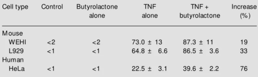
phase induced by the drug (21). Pre-incuba-tion with butyrolactone for 1 h, followed by the addition of TNF and further incubation of WEHI (15 h), L929 (20 h) and HeLa (20 h), caused an increase of 19, 33 and 76%, respectively, of the apoptotic cells, as com-pared with TNF alone (Table 1). Apoptosis was further confirmed by comparing the in-tegrity of isolated cellular DNA by agarose gel electrophoresis. The results (Figure 1) show that the typical ladder pattern of DNA fragmentation was induced for each cell line incubated with TNF and TNF plus butyro-lactone. A higher proportion of fragmented DNA was obtained in cells treated with TNF plus butyrolactone, consistent with the mor-phological data (Table 1).
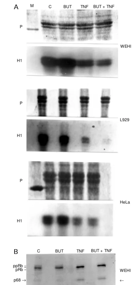
Inhibitio n o f histo ne H1 kinase and pRb
kinase activitie s by TNF and butyro lacto ne
Inhibition of cellular growth and induc-tion of apoptosis by TNF (10) and butyrolac-tone (20-22) in various cell types have been associated with the ability of these compounds to reduce the activity of protein kinases that catalyze the phosphorylation of histone H1 and pRb proteins. Consistent with the previ-ous reports, we noted that the exposure of the three cell lines to TNF reduced the his-tone H1 kinase activities within the cells (Figure 2A). The level of reduction in the histone H1 kinase activity increased with the combination of the TNF and butyrolactone compared to TNF alone. The reduction was more evident with the cell lines WEHI and L929, but not clear with HeLa. We analyzed the phosphorylation status of pRb in the WEHI cell line after the different treatments (Figure 2B). The control and butyrolactone-treated cells exhibited the three phosphoryl-ated forms of pRb whereas the cells exposed to TNF or TNF plus butyrolactone showed
Table 1 - Induction of apoptotic cell death by tumor necrosis factor (TNF) and the combination of TNF plus butyrolactone in mouse and human cell lines.
Cell lines w ere incubated w ithout or w ith butyrolactone (100 µM ) for 1 h and then w ith TNF. The concentration of TNF (mouse or human recombinant protein) added and time of incubation w ere, respectively, 1 ng/ml and 15 h for WEHI, 10 ng/ml and 24 h for L929, and 100 ng/ml and 24 h for HeLa. Apoptotic nuclei w ere scored on the basis of the condensation state of chromatin and its marginalization at the edges of the nuclear membrane. Data are reported as the mean percent of apoptotic cells ± standard deviation of the results of three similar experiments.
Cell type Control Butyrolactone TNF TNF + Increase
alone alone butyrolactone (% )
M ouse
WEHI <2 <2 73.0 ± 13 87.3 ± 11 19
L929 <1 <1 64.8 ± 6.6 86.5 ± 3.6 33
Human
only the fast migrating form of pRb (about 105 kDa). We also observed in the autorad-iographs the presence of a faint band of 68 kDa, which may be a cleavage product from Rb proteins (26). We also detected this 68-kDa product with the Rb monoclonal anti-body (data not shown).
D iffe re ntial e xpre ssio n o f pro te ins in the ce ll
lysate s fro m TNF- and butyro lacto ne -tre ate d
ce lls
We monitored the relative effects of TNF and butyrolactone on the synthesis and degradation of cellular proteins to detect other molecular targets associated with the inhibition of cell growth and induction of apoptosis in WEHI cells in which the extent of cell death was nearly 90%. The content and molecular size of numerous proteins did not change in the cells exposed to butyrolac-tone alone as compared to controls (Figure 3). The intensity of polypeptide bands in the
Coomassie-stained gels showed that the pro-teins of approximately 110, 90 and 50 kDa were drastically reduced in the cell lysates from the cells treated with TNF. This occurred with the simultaneous appear-ance of the two proteins of 60 and 70 kDa. The effect was increased when we used the combination of TNF and butyrolactone. It is interesting to observe that the protein bands between 110-120 kDa (location of pRb hyperphosphorylated forms) were ab-sent in the samples from the TNF and the TNF plus butyrolactone cell lysates. The data suggest that the lack of various protein bands in the 40-200 kDa range and the si-multaneous appearance of new bands of 60 and 70 kDa, as well as of weak bands on the gel, may be the result of proteolysis of cellu-lar proteins and the generation of smaller cleavage products. The data also indicate that the changes may be the result of up or down regulation of gene expression induced by these agents.
Figure 1 - Agarose gel electro-phoresis of genom ic DNA extracted from WEHI, L929 and HeLa cells incubated in the ab-sence (C) and in the preab-sence of 100 µM butyrolactone (BUT), in the presence of tumor necro-sis factor (TNF) at 1, 5 and 100 ng/ml, respectively, or both bu-tyrolactone and TNF for 15 h (WEHI cells) or 24 h (L929 and HeLa cells). The relative mobil-ity of the oligonucleosomal frag-ments from DNA samples re-flects interger multiples of a 180-bp unit (apoptosis ladder pattern) w ith respect to the DNA standard (M ).
WEHI L929 HeLa
C BUT TNF
BUT + TNF M
C BUT TNF
BUT + TNF M
C BUT TNF
D iscussio n
Evidence in a number of studies has sug-gested that oncoproteins, tumor suppressor proteins and the cell cycle regulatory pro-teins play a critical role in the mechanism by which normal and tumor cells respond to TNF and to pharmacological compounds that induce cell differentiation, proliferation or apoptosis (7,8,11,17). Recently, several chemical inhibitors of CDKs have been found on the basis of their ability to inhibit cell cycle proliferation and/or to induce apopto-sis in cancer cells (18,19). The present study shows that TNF inhibits the histone H1 and pRb kinase activities associated with CDKs in the cell lines WEHI, L929 and HeLa undergoing cell cycle arrest and apoptosis, and that these biochemical effects can be sustained with butyrolactone, a specific in-hibitor of protein kinases 1 and CDK-2. These data provide further evidence for
Figure 2 - A, Histone H1 kinase activity in the cell lysates of WEHI, L929 and HeLa cells incubated in the absence (C), in the presence of 100 µM butyrolactone (BUT), tumor necrosis factor (TNF) at 1, 5 and 100 ng/ ml, respectively, or both butyrolactone and TNF for 15 h (WEHI cells) or 24 h (L929 and HeLa cells). Equal amounts of proteins from cell lysates w ere incubated w ith 5 µg histone H1 and [g-32ATP] and the level of
[32P]-phosphate incorporated into histone w as detected
after electrophoresis on 10% SDS/PAGE and autoradi-ography. Upper panel show s the pattern of protein bands (histone H1 and cell lysates) of Coomassie-stained gels, and the low er panel show s the autoradio-graph obtained w ith the dried stained gel after expo-sure to X-ray film. M , Protein molecular mass standard.
B, Phosphorylation by pRb kinases in the cell lysates from WEHI cells in the absence (C) and in the presence of 100 µM butyrolactone (BUT), TNF (1 ng/ml), and both butyrolactone and TNF after 15 h of incubation. Equal amounts of the cell lysate proteins w ere incubated w ith [g-32ATP] and the level of [32P]-phosphate
incorpo-rated into endogenous pRb proteins w as simulta-neously detected in the autoradiograph and nitrocellu-lose membrane containing the phosphorylated proteins after Western blotting w ith RB1(1F8) monoclonal anti-body and the ECL detection method. The presence of a phosphoprotein of approximately 68 kDa of molecular mass is indicated on the gel (arrow ). The results are representative of three similar experiments.
WEHI ppRb
pRb
-p68®
-C BUT TNF BUT + TNF
B
WEHI
L929
HeLa P
H1
P
H1
P
H1
A
M C BUT TNF BUT + TNFthe participation of CDK in the signaling pathway by which TNF induces growth ar-rest and apoptosis in cancer cells.
We found that butyrolactone itself does not induce apoptosis in the cell lines WEHI, L929 and HeLa used in this study, as ob-served for the HL-60 cell line (22). It has been shown that the general inhibitors of multiple protein kinases staurosporine and related compounds (27,28), genistein and tyrphostin (29,30) are potent inducers of apoptosis in some types of cancer cells. For example, staurosporine at 10 nM concentra-tion causes the arrest of cell cycle progres-sion at the G1 phase which occurs
concomi-tantly with the synthesis of the cyclin-de-pendent kinase inhibitors p18 and p27 and consequently the inhibition of 2, CDK-4 and CDK-6 activity (28). More important, apoptosis of various types of cancer cells following treatment with different cytotoxic agents frequently occurs upon the cell cycle arrest and reduction of the CDK activities associated with cyclins A, B, D and E (31-35). Thus, an efficient block of the synthesis and activity of CDK/cyclin in tumor cells appears to be a crucial step toward growth arrest and apoptotic cell death. It has been reported that butyrolactone and other
inhibi-tors of CDKs such as staurosporine, flavo-piridol, suramin and olomoucine inhibit the CDK activity by their direct interaction with the ATP-binding site inside the CDK mol-ecules (18,19). However, we have not yet shown if p21, a CDK protein inhibitor tran-scriptionally regulated by p53, or its family members, p16, p18, p27 and p57, function as mediators of the TNF-induced CDK kinase suppression. The participation of the phos-phatases cdc-25 (36) and retinoblastoma phosphatase (26) has also been considered because of their roles in CDK kinase activity regulation and apoptosis mediated by c-myc and pRb deregulation. It is important to men-tion that various other protein kinase fami-lies with essential roles in the cell survival pathways can also be inhibited by TNF and the competitive inhibitor for ATP binding and, therefore, contribute to the biological effects induced by these factors (37). Taken together, these data further support our hy-pothesis that the apoptosis process is initi-ated after the inhibition of multiple protein cascades which are continually activated by cell survival factors (11).
TNF-induced apoptosis is mediated by the activation of a conserved family of as-partate-specific cysteinyl proteases called
Figure 3 - Relative changes in cellular protein levels in the cell lysates from WEHI cells in the absence (C) and in the presence of 100 µM butyrolactone (BUT), in the presence of tumor necro-sis factor (TNF) (1 ng/ml), and of both butyrolactone and TNF af-ter 15 h of incubation. Equal a-mounts of proteins w ere sepa-rated by 10% SDS/PAGE, fixed and stained w ith Coom assie blue. The location and the appar-ent molecular mass of represen-tative protein bands are indi-cated on the gel.
kDa
150 140 110 90
55 50 45
-- 105 - 90
- 70 - 60
- 50 - 45
- H1 kDa
1 2 3 4
caspases (21,38). Multiple protein substrates of caspases have been found, such as the nuclear proteins PARP, U1-70 and lamins (5,39) and the cytosolic proteins gelsolin and actin (40,41). In present study we have shown that either TNF or TNF plus butyro-lactone provoked the loss of several cellular proteins in WEHI cells when the extent of cell death was nearly 90%. We noted that the level of three major cellular protein bands of approximately 110 (pRb intermediate forms), 90 and 50 kDa and various other weakly stained bands were markedly reduced in re-sponse to TNF. Simultaneously, we observed the appearance of new bands of 60 and 70 kDa, supporting the idea that they may be the cleavage products of one or more proteins. These data suggest that specific modifica-tions, including phosphorylation and pro-teolytic cleavage of the cellular proteins, might be involved in the molecular mecha-nisms behind the morphological and bio-chemical changes occurring during the apoptotic process. Few of the caspase-cleaved substrates are known to have direct physi-ological significance in apoptotic cell death. Recent studies have identified the Rb tumor suppressor proteins as substrates for the caspases during the apoptosis process in-duced by TNF and other death inducers (42-44). In the present report we show that WEHI cells exposed to TNF or both TNF plus butyrolactone exclusively contain the hypo-phosphorylated form of pRb and a product of 68 kDa (Figures 2B and 3), as reported elsewhere (44). It has been demonstrated that pRb functions at a checkpoint that pro-tects cancer cells from entering the S phase
and apoptosis in the absence of serum stim-ulation or deregulated expression of E2F transcription factor (45,46). Therefore, we suggest a mechanism by which the cell cycle-dependent inactivation of CDKs by TNF and CDK chemical inhibitors and an inefficient phosphorylation of Rb would prime the underphosphorylated forms of Rb to caspases for its specific cleavage. Furthermore, the changes in the phosphorylation status and conformation structure of Rb proteins would allow the release of E2F, which in turn promotes S phase entry and induces apopto-sis.
Taken together, the present findings link the decrease of cyclin-dependent kinase ac-tivity and the increase of protease acac-tivity within the growth arrest and apoptotic path-ways induced by TNF. It is hoped that the identification of newer inhibitors of CDKs and the caspase substrates involved in the cell proliferation and apoptosis pathways will be of clinical importance for the therapy of many diseases.
Ackno wle dgm e nts
We thank Edilene M. Lobato, Aicha M. Sarr, Amanda Machion, Branca Trindade and various members of Instituto Butantan and Universidade de São Paulo for valuable assistance and collaboration. We are also grateful to Akira Okuyama (Banyu Tsukuba Research Institute/Merck Research Labs., Okubo, Japan) and Michael A. Palladino Jr. (Genentech, South San Francisco, CA, USA) for providing butyrolactone and murine and human recombinant TNF, respectively.
Re fe re nce s
1. Tracey KJ & Cerami A (1993). Tumor ne-crosis factor, other cytokines and disease.
Annual Review of Cell Biology, 9: 317-343.
2. Wallach D, Boldin M , Varfolomeev M , Beyaert R, Vandenabeele P & Fiers W (1997). Cell death induction by receptors
of the TNF family: tow ards a molecular understanding. FEBS Letters, 410: 96-106.
3. Thompson CB (1995). Apoptosis in the pathogenesis and treatment of disease.
Science, 267: 1458-1462.
4. Vaux DL & Strasser A (1996). The
molecu-lar biology of apoptosis. Proceedings of the National Academy of Sciences, USA, 93: 2239-2244.
5. M iller (1997). The role of the caspase fam-ily proteases in apoptosis. Seminars in Immunology, 9: 35-49.
the Bcl-2 family of apoptosis regulatory proteins in the immune system. Semi-narsinImmunology, 9: 25-33.
7. Sherr CJ (1996). Cancer cell cycle. Sci-ence, 274: 1672-1677.
8. Hunter T (1997). Oncoprotein netw ork.
Cell, 88: 333-346.
9. Evan GL, Brow n L, Whyte M & Harrington E (1995). Apoptosis and the cell cycle. Cur-rent Opinnion in Cell Biology, 7: 825-834. 10. Belizario JE & Dinarello CA (1991).
Inter-leukin-1, interleukin-6, tumor necrosis fac-tor-a and transforming grow th factor-ß in-crease cell resistance to TNF cytotoxicity by grow th arrest in the G1 phase of the
cell cycle. Cancer Research, 51: 2379-2385.
11. Belizário JE, Sherw ood SW & Beçak W (1999). Inhibition of multiple protein ki-nase cascades in cell undergoing cell cycle arrest and apoptosis in response to TNF. Cancer Research (in press). 12. Jeoung D, Tang B & Sonenberg M (1995).
Effects of TNF-a on anti-mitogenicity and cell cycle-related proteins in M CF-7 cells.
Journal of Biological Chem istry, 270: 18367-18373.
13. Vieira KBL, Goldstein DJ & Villa LL (1996). Tumor necrosis factor-a interferes w ith the cell cycle of normal and papillomavi-rus-immortalized human keratinocytes.
Cancer Research,56: 2452-2457. 14. Yin D, Kondo S, Barnett GH, M orimura T
& Takeuchi J (1995). Tumor necrosis fac-tor-ainduce p53-dependent apoptosis in rat glioma cells. Neurosurgery, 37: 758-762.
15. Jacobsen FW, Dubois CM , Rusten LS, Veiby OP & Jacobsen SE (1995). Inhibi-tion of stem cell factor-induced prolifera-tion of primitive murine hematopoietic progenitor cells signalling through the 75-kDa TNFR. Journal of Immunology, 154: 3732-3741.
16. Shiohara M , Akashi M , Gombart AF, Yang R & Koeffler HP (1996). Tumor necrosis factor-a: posttranscriptional stabilization of WAF1 mRNA in p53-deficient human leukemic cells. Journal of Cellular Physiol-ogy, 166: 568-576.
17. Janicke RU, Lin XY & Porter AG (1996). Cyclin D3 sensitizes tumor cells to TNF-induced c-myc dependent apoptosis. M o-lecular and Cellular Biology, 16: 5245-5253.
18. M eijer L (1996). Chemical inhibitors of cyclin-dependent kinases. Trends in Cell Biology, 6: 393-397.
19. M eijer L & Kim S-H (1997). Chemical in-hibit ors of cyclin-dependent kinases.
M ethods in Enzymology, 283: 113-128.
20. Kit agaw a M , Okabe T, Ogino H, M atsumoto H, Takahashi IS, Kokubo T, Higashi H, Saitoh S, Taya Y, Yasuda H, Ohba Y, Nishim ura S, Tanaka N & Okuyama A (1993). Butyrolactone I, a se-lective inhibitor of cdk2 and cdc2 kinase.
Oncogene, 8: 2425-2432.
21. Kitagaw a M , Higashi H, Takahashi IS, Okabe T, Ogino H, Taya Y, Nishimura S & Okuyama A (1994). A cyclin-dependent kinase inhibitor, butyrolactone I, inhibits phosphorylation of RB protein and cell cycle progression. Oncogene, 9: 2549-2557.
22. Shibata Y, Nishimura S, Okuyama A & Nakamura T (1996). p53-independent in-duction of apoptosis by cyclin-dependent kinase inhibition. Cell Grow th and Differ-entiation,7: 887-891.
23. Belizario JE, Tilly J & Sherw ood SW (1993). Caffeine potentiates the lethality of tumor necrosis factor in cancer cells.
British Journalof Cancer, 67: 1229-1235. 24. Simanis V & Nurse P (1986). The cell cycle control gene cdc2+ of fission yeast en-codes a protein kinase potentially regu-lated by phosphorylation. Cell, 45: 261-268.
25. Laemmli UK (1970). Cleavage of struc-tural proteins during assembly of the head of bacteriophage T4. Nature, 227: 22-27. 26. Dou QP, An B & Will PL (1995). Induction
of a retinoblastoma phosphatase activity by anticancer drugs accompanies p53-in-dependent G1 arrest and apoptosis.
Pro-ceedings of the NationalAcademy of Sci-ences, USA, 92: 9019-9023.
27. Wang Q, Worland PJ, Clark JL, Carlson BA & Sausville EA (1995). Apoptosis in 7-hydroxystauropurine-treated T lympho-blasts correlates w ith activation of cyclin-dependent kinases 1 and 2. CellGrow th and Differentiation, 6: 927-936. 28. Kw on TK, Buchholz M A, Chrest FJ &
Nordin AA (1996). Staurosporine-induced G1 arrest is associated w ith the induction
and accumulation of cyclin-dependent ki-nase inhibitors. Cell Grow th and Differen-tiation, 7: 1305-1313.
29. Tallet t A, Chilvers ER, Hannah S, Dransfield I, Law son M F, Haslett C & Sethi T (1996). Inhibition of neuropeptide-stimulated tyrosine phosphorylation and tyrosine kinase activity stimulates apopto-sis in small cell lung cancer cells. Cancer Research, 56: 4255-4263.
30. Gjertsen BT & Doskeland SO (1995). Pro-tein phosphorylation in apoptosis. Bioche-mica et Biophysica Acta, 1269: 187-199. 31. Lock RB (1992). Inhibition of p34/cdc-2
kinase activation, p34/cdc-2 tyrosine
de-phosphorylation, and mitotic progression in Chinese hamster ovary cells exposed to etoposide. Cancer Research, 52: 1817-1822.
32. Dulic V, Kaufmann WK, Wilson SJ, Tisty TD, Lees E, Harper JW, Elledge SJ & Reed SI (1994). p53-dependent inhibition of cyclin-dependent kinase activities in hu-man fibroblasts during radiation-induced G1 arrest. Cell, 76: 1013-1023.
33. Norbury C, M acFarlane M , Fearnhead H & Cohen GM (1994). CDC2 activation is not required for thymocyte apoptosis. Bio-chemical and Biophysical Research Com-munications, 202: 1400-1406.
34. Oberhammer FA, Hochegger K, Froschl G, Tiefenbacher R & Pavelka M (1994). Chromatin condensation during apopto-sis is accompanied by degradation of lamin A + B, w ithout enhanced activation of cdc2 kinase. Journal of Cell Biology, 126: 827-837.
35. Neamati N, Fernandez A, Wright S, Kiefer J & M cConkey DJ (1995). Degradation of lamin B1 precedes oligonucleosomal DNA fragmentation in apoptotic thymocytes and isolated thymocyte nuclei. Journal of Immunology, 154: 3788-3795.
36. Galaktionov K, Chen X & Beach D (1996). Cdc-25 cell cycle phosphatase as a target of c-myc. Nature, 38: 511-517.
37. Am arant e-M endes GP & Green DR (1997). Abl tyrosine kinase and the con-trol of apoptosis. In: M artin SJ (Editor),
Apoptosis and Cancer. R.G. Landes Com-pany, Austin, TX.
38. Boldin M P, Goncharov TM , Goltsev YV & Wallach D (1996). Involvement of M ACH, a novel M ORT1/FADD-interacting pro-tease in Fas/APO-1 and TNF receptor-in-duced cell death. Cell, 85: 803-815. 39. Casciola-Rosen LA, M iller DK, Anhalt GJ
& Rosen A (1994). Specific cleavage of the 70-kDa protein component of U1 small nuclear ribonucleoprotein is a char-acteristic biochemical feature of apoptotic cell death. Journal of Biological Chemis-try, 269: 30757-30760.
40. Kayalar C, Ord T, Testa M P, Zhong L-T & Bredesen DE (1996). Cleavage of actin by interleukin 1ß-converting enzyme to re-verse DNA I inhibition. Proceedings of the National Academy of Sciences,USA, 93: 2234-2238.
42. Janicke RU, Walker PA, Yu X & Porter AG (1996). Specific cleavage of the retino-blastoma protein by an ICE-like protease in apoptosis. EM BO Journal, 15: 6969-6978.
43. Tan X, M artin SJ, Green DR & Wang JYJ (1997). Degradation of retinoblastoma protein in tumor necrosis factor- and CD95-induced cell death. Journal of
Bio-logical Chemistry, 272: 9613-9616. 44. An B & Dou QP (1996). Cleavage of
reti-noblastoma protein during apoptosis: an interleukin 1ß-converting enzyme-like pro-tease as candidate. Cancer Research, 56: 438-442.
45. Haas-Kogan DA, Kogan SC, Levi D, Dazin P, T’Ang T, Fung Y-K, Fung T & Israel M A (1995). Inhibition of apoptosis by the
reti-noblastoma gene product. EM BO Jour-nal, 14: 461-472.