Pancre atic nitric o xide and o xyge n
fre e radicals in the e arly stage s o f
stre pto zo to cin-induce d diabe te s
m e llitus in the rat
1Centro de Estudios Farmacológicos y Botánicos (CEFYBO ),
Consejo Nacional de Investigaciones Científicas y Técnicas, Buenos Aires, Argentina
2Bioanalítica Médica, Instituto de Investigaciones Biomedicas
de Barcelona, Barcelona, España E. González1,
J. Roselló-Catafau2,
A. Jawerbaum1, D. Sinner1,
C. Pustovrh1, J. Vela1,
V. White1,C. Xaus2,
C. Peralta2 and M. Gimeno1
Abstract
The objective of the present study was to explore the regulatory mechanisms of free radicals during streptozotocin (STZ)-induced pancreatic damage, which may involve nitric oxide (NO) production as a modulator of cellular oxidative stress. Removal of oxygen species by incubating pancreatic tissues in the presence of polyethylene glycol-conjugated superoxide dismutase (PEG-SOD) (1 U/ml) pro-duced a decrease in nitrite levels (42%) and NO synthase (NOS) activity (50%) in diabetic but not in control samples. When NO production was blocked by NG
-monomethyl-L-arginine (L-NMMA) (600 µM), SOD activity increased (15.21 ± 1.23 vs 24.40 ± 2.01 U/mg
dry weight). The increase was abolished when the NO donor, spermine nonoate, was added to the incubating medium (13.2 ± 1.32). Lipid peroxidation was lower in diabetic tissues when PEG-SOD was added (0.40 ± 0.02 vs 0.20 ± 0.03 nmol/mg protein), and when L-NMMA
blocked NOS activity in the incubating medium (0.28 ± 0.05); sper-mine nonoate (100 µM) abolished the decrease in lipoperoxide level (0.70 ± 0.02). We conclude that removal of oxygen species produces a decrease in pancreatic NO and NOS levels in STZ-treated rats. Moreover, inhibition of NOS activity produces an increase in SOD activity and a decrease in lipoperoxidation in diabetic pancreatic tissues. Oxidative stress and NO pathway are related and seem to modulate each other in acute STZ-induced diabetic pancreas in the rat.
Co rre spo nde nce
E. González Serrano 669 (1414) Buenos Aires Argentina
Fax: + 54-11-4856-2751 E-mail: elidate@ arnet.com.ar
Research supported by CO NICET (No. PIP 0598/98 (E. González)), PICT (No. 98 03375), Agencia Nacional de Promoción Científica y Tecnológica, and performed under the Interinstitutional Project AR0012 from the CSIC, Spain.
Received November 30, 1999 Accepted July 27, 2000
Ke y wo rds
·Streptozotocin diabetes
·Pancreas
·O xidative stress
·Nitric oxide
Intro ductio n
Streptozotocin (STZ) is an agent widely employed to induce experimental diabetes due to its ability to selectively target and destroy insulin-producing pancreatic islet ß-cells (1). Its diabetogenic action has been ascribed to the enhancement of intracellular
methylation reactions (2) and to production of nitric oxide (NO) (3,4) and free radicals (5). NO has been demonstrated to be the ef-fector molecule responsible for pancreatic islet destruction (6). We have recently shown that the NO synthase (NOS) inhibitors NG
prevent metabolic disorders in tissues and plasma of STZ-induced diabetic rats (7,8).
With respect to oxidative stress, an in-creased free radical generation was reported in diabetic plasma and tissues. Vitamin C levels are lower (9) and altered levels of the antioxidant defense systems are present in diabetic human plasma compared to control (10). Elevated plasma lipid leads to changes in lipid oxidation (11). Lipid peroxides are higher (12), an early sign of oxygen activa-tion (13). Superoxide dismutase (SOD) cata-lyzes the removal of the superoxide radical, and the addition of exogenous SOD in vitro
also protects cellular membranes from chem-ical damage attributable to superoxide pro-duction (14). The use of a covalent conju-gated form of SOD, such as polyethylene glycol-SOD (PEG-SOD), increases the half-life of the enzyme in the circulation and the efficacy of its protective effect (15,16).
The fact that ß-cells contain much more SOD than a-cells (17) suggests that this enzyme may play an important role in ß-cell homeostasis.
As stated, NO is significantly involved in pancreatic destruction, and seems to be closely related to the events leading to oxida-tive stress. The interaction between NO and O2·- may be biologically significant. NO is able to combine with reactive oxygen spe-cies (ROS) (18) and to modulate oxidative damage (19). This study aims at exploring the regulatory mechanisms of free radicals during STZ-induced pancreatic damage, which may involve NO production as a modu-lator of cellular oxidative stress.
Mate rial and Me tho ds
Che micals
Citrate buffer, STZ, nitroblue tetrazo-lium, xanthine, xanthine-oxidase, polyethyl-ene glycol-superoxide dismutase, sodium thiobarbiturate, malondialdehyde (1,1,3,3 tetraethoxypropane), L-NMMA, L-NAME,
NADPH, L-valine and HEPES were obtained from Sigma Chemical Co., St. Louis, MO, USA. Glucostix reagent strips were obtained from Bayer Diagnostics, Buenos Aires, Ar-gentina.
The nitrate/nitrite assay kit and spermine nonoate (SN) were purchased from Cayman Chemical Co., Ann Arbor, MI, USA. Dowex
AG50W-X8 columns (Na+
form) were pur-chased from BioRad Laboratories, Rich-mond, CA, USA. [14
C]-L-Arginine was from New England Nuclear, Boston, MA, USA.
Anim al pre paratio ns
Female albino Wistar rats bred in the laboratory, weighing 200-230 g were used. The animals were kept in a temperature- (20-23o
C) and illumination- (12-h light/12-h dark) controlled room, and were made diabetic by a single injection of STZ (55 mg/kg body weight, ip) dissolved in 0.1 M citrate buffer, pH 4.5 (day 1). Blood glucose from the tail vein was assayed 5 days later (day 5) using glucostix reagent strips and a refractance meter. Control animals showed glucose lev-els between 80 and 110 mg/dl, and STZ-injected animals between 300 and 500 mg/ dl. Animals were killed by cervical disloca-tion on day 5 after STZ injecdisloca-tion, and their pancreas was removed and placed on Petri dishes containing Krebs-Ringer bicarbonate solution for subsequent assays. The ionic composition of this medium was described previously (20). The guidelines for the care and use of animals approved by the local institution were followed and they conformed to the standards for use of laboratory animals established by the Institute of Laboratory Animals Resources, U.S. National Academy of Sciences.
Nitrate /nitrite assay
min, with the addition of L-NMMA (600 µM) or PEG-SOD (1 U/ml), were homog-enized and deproteinized. Nitrates in the supernatant were reduced to nitrites using nitrate reductase, and total nitrites were meas-ured by the Griess method (21), employing an assay kit for nitrate/nitrite determination. Absorbance was measured at 540 nm in a microliter plate using NaNO3 and NaNO2 as standard. Results are reported as nmol/mg protein.
Assay o f NO S activity
NOS enzyme activity was quantified in pancreatic tissues from control and diabetic rats previously incubated in a metabolic shak-ing bath under an atmosphere of 95% O2 and 5% CO2 for 60 min with the addition of the NOS blocker L-NMMA (600 µM) or the ROS scavenger PEG-SOD (1 U/ml), by the [14
C]-arginine to [14
C]-citrulline conversion assay as described by Bredt et al. (22), modi-fied by Salter et al. (23). Briefly, tissues were homogenized in 1.5 ml 20 mM HEPES buf-fer, pH 7.4. The homogenates of each sample were fractionated in 3 tubes, with 0.45 mM Ca2+
(control tube), and 1 mM EGTA + 2 mM L-NAME (nonspecific tube) added, re-spectively. Each fraction was incubated at 37o
C with 0.1 µCi of [14
C]-arginine and 0.5 mM NADPH. L-Valine (50 mM) was added to the reaction buffer to minimize any inter-ference from arginase. After 15 min of incu-bation, samples were centrifuged for a pe-riod of 10 min at 10,000 g and the superna-tant was applied to a 1-ml Dowex AG50W-X8 column (Na+
form) balanced with 20 nM HEPES, pH 7.4, and [14
C]-citrulline was eluted with 3 ml of water. Radioactivity was measured by liquid scintillation counting. Total NOS activity was calculated as the difference between the [14C]-citrulline pro-duced by samples with Ca2+
and by samples containing both EGTA + L-NAME (nonspe-cific). Ca2+-independent enzyme activity was calculated from the difference between
samples containing EGTA and samples con-taining both EGTA + L-NAME (nonspe-cific). Ca2+
-dependent activity was calcu-lated by subtracting Ca2+
-independent activ-ity from total activactiv-ity. As formation of L-citrulline from L-arginine is stoichiometric with respect to the formation of NO, we could assume that an equal amount of NO was formed. Enzymatic activity is expressed as pmol NO min-1
100 mg of wet tissue-1 . Intra- and interassay variations were less than 10%.
Supe ro xide dism utase activity
To perform SOD determination, tissue samples were previously incubated in a meta-bolic shaking bath under an atmosphere of 95% O2 and 5% CO2 for 60 min with the addition of the NO donor SN (100 µM) or the NOS blocker L-NMMA (600 µM). Tis-sues were homogenized in 100 mM Tris (hydroxymethyl) aminomethane (Tris-HCl) buffer. SOD activity was assayed according to the method of Yamanaka et al. (24) modi-fied by Sun et al. (25). The method consists of determining the ability of the enzyme to inhibit the superoxide anion-mediated re-duction of nitroblue tetrazolium (25 µM) to formazan. The latter was determined spec-trophotometrically at 560 nm. The superox-ide anion necessary for this reaction is gen-erated by xanthine (100 µM) and xanthine oxidase (200 U/l). Enzymatic activity is ex-pressed as units (U) per mg of dry tissue.
Lipid pe ro xidatio n
were homogenized in 100 mM Tris (hy-droxymethyl) aminomethane (Tris-HCl) buf-fer, the chemical reaction was performed and peroxidation was quantified spectropho-tometrically at 530 nm (26). Lipoperoxides are expressed as nmol per mg of protein.
Statistical analysis
Results are reported as mean ± SEM, and were analyzed by ANOVA and the Tukey-Kramer multiple comparisons test. Differ-ences between means were considered sig-nificant if P<0.05.
Re sults
Influe nce o f SO D o n pancre atic nitrate /nitrite
le ve ls
Nitrate/nitrite levels were evaluated after incubation of pancreatic tissues in the pres-ence of the NOS blocker L-NMMA (600 µM) and the ROS scavenger PEG-SOD (1 U/ml). Figure 1 shows that the blockade of NO synthesis is reflected by a decrease in tissue nitrate/nitrite levels in control (P<0.05) and diabetic rats (P<0.05). Nitrate/nitrite values were higher in diabetic than in control pancreas (P<0.05). When PEG-SOD was added to the incubating bath, no effects could be detected in non-diabetic tissues, but the ROS scavenger decreased the nitrate/nitrite increase due to a diabetic state (P<0.05).
Pancre atic NO S activity: influe nce o f SO D
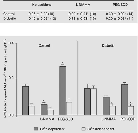
NOS activity from pancreatic tissues was evaluated in the presence of L-NMMA (600 µM) and PEG-SOD (1 U/ml) added to the incubating bath (Table 1 and Figure 2). The Ca2+
-dependent isoform was predominant in control tissues, while the Ca2+
-independent isoform was increased in diabetic tissues compared to control. L-NMMA inhibited NOS activity (300 µM and up to 1 mM); the maximum effect was demonstrable from 600 Table 1 - Nitric oxide synthase (NOS) activity: influence of ROS removal. NOS activity in
pmol NO min-1 100 mg w et w eight-1 in isolated control and diabetic rat pancreas incubated alone or in the presence of 600 µM L-NM M A or 1 U/ml PEG-SOD.
Results are reported as means ± SEM . The number of animals is given in parentheses. * P<0.05 vs control (no additions). +P<0.05 vs diabetic (no additions) (ANOVA and Tukey-Kramer multiple comparisons test).
No additions L-NM M A PEG-SOD
Control 0.25 ± 0.02 (10) 0.09 ± 0.01* (10) 0.30 ± 0.02* (14)
Diabetic 0.40 ± 0.05* (12) 0.15 ± 0.03+ (10) 0.20 ± 0.06+ (11)
Figure 2 - Influence of PEG-SOD on pancreatic nitric oxide synthase (NOS) activity. NOS activity (Ca2+ dependent and Ca2+ independent) in pmol nitric oxide (NO) min-1 100 mg w et tissue-1, from isolated control (N = 6) and diabetic (N = 8) rat pancreas incubated alone or in the presence of 600 µM L-NM M A or 1 U/ml PEG-SOD. Conditions and details as in legend to Figure 1. * P<0.05 vs basal levels from tissues incubated alone (ANOVA and Tukey-Kramer multiple comparisons test).
Figure 1 - Influence of PEG-SOD on pancreatic nitrate/nitrite lev-els. Nitrate/nitrite production by isolated control and diabetic rat pancreas incubated alone or in t he presence of 600 µM L-NM M A or 1 U/ml PEG-SOD. N: Number of experiments. Data are reported as mean ± SEM . * P< 0.05 vs control (N = 6); +P< 0.05 vs diabetic (N = 7) (ANOVA and Tukey-Kramer
mul-tiple comparisons test). N
it
ra
te
s
/n
it
ri
te
s
(
n
m
o
l/
m
g
p
ro
te
in
)
5
4
3
2
1
0
*
* +
* +
Control Diabetic
L-NM M A PEG-SOD
N
O
S
a
c
ti
v
it
y
(
p
m
o
l
N
O
m
in
-1 1
0
0
m
g
w
e
t
w
e
ig
h
t
-1) 0.4
0.2
0.0
L-NM M A PEG-SOD
L-NM M A PEG-SOD
Ca2+ dependent Ca2+ independent
*
*
* *
µM on (data not shown). The influence of the inactive stereoisomer D-NMMA was also tested, but NOS activity was unaffected, showing a specific inhibitory action of L-NMMA (data not shown). The presence of 600 µM L-NMMA significantly reduced the enzymatic activity in control and diabetic tissues (P<0.05). The addition of PEG-SOD improved NOS activity in control animals (P<0.05). It seems that the increase occurred due to a Ca2+
-dependent isoform. Decreased Ca2+
-independent NOS levels were observed when PEG-SOD was added to the incubat-ing medium in pancreas from diabetic rats (P<0.05), showing a different modulatory profile compared to that of control animals.
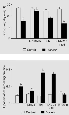
SO D activity in pancre atic tissue s fro m
co ntro l and STZ-diabe tic rats: influe nce o f
L-NMMA and SN
Figure 3 shows that SOD activity was lower in diabetic than in control rats (P<0.05). We can observe that the NOS blocker L-NMMA (600 µM) or L-L-NMMA plus the NO donor SN (100 µM) could not modify pan-creatic SOD activity in control animals.
When L-NMMA was added to the incubat-ing bath of diabetic tissues, an increase in SOD level was detected (P<0.01). The addition of L-NMMA and SN abolished the increase of SOD activity due to the presence of L-NMMA in the incubating medium (P<0.05). Similar experiments were carried out with the addition of L-NAME (1 mM), showing an increase in SOD levels, and were also carried out with L-NAME plus SN (600 µM), showing a de-crease of enzyme activity (data not shown). When NO production was blocked, the anti-oxidative defense seemed to improve in pan-creatic tissue from diabetic rats.
Lipo pe ro xide le ve ls in pancre atic tissue s
fro m co ntro l and STZ-diabe tic rats: influe nce
o f NO S blo ckade and NO do no rs
Lipoperoxidation was higher in diabetic
S
O
D
(
U
/m
g
d
ry
w
e
ig
h
t) 30
20
10
0
*
+
*
Control Diabetic
L-NM M A L-NM M A
+ SN
L
ip
o
p
e
ro
x
id
e
s
(
n
m
o
l/
m
g
p
ro
te
in
)
0.8
0.4
0
L-NM M A PEG-SOD
than in control tissues (P<0.05), as shown in Figure 4.
Lipoperoxide levels were evaluated in the presence of the NOS blocker L-NMMA (600 µM), L-NMMA plus the NO donor SN (100 µM) or the ROS scavenger PEG-SOD (1 U/ml). L-NMMA, SN or PEG-SOD could not modify pancreatic lipoperoxidation lev-els of control animals. The addition of PEG-SOD in the incubating medium of diabetic tissues caused a decrease of lipoperoxides (P<0.05). In diabetic pancreas, NOS inhibi-tion decreased lipoperoxidainhibi-tion (P<0.05), and NO generation abolished L-NMMA ef-fects and increased pancreatic lipoperoxide levels (P<0.05). Similar experiments were carried out with the addition of the NOS blocker L-NAME, which decreased lipoper-oxide levels, and L-NAME plus SN (100 µM), which increased lipid peroxidation (data not shown). The presence of NO increased lipid peroxidation in pancreatic tissues from STZ-treated animals.
Figure 3 - Pancreatic superoxide dismutase (SOD) activity: influ-ence of nit ridergic pat hw ay. SOD activity in U/mg dry w eight in isolated control and diabetic rat pancreas incubated alone or in the presence of 600 µM L-NM M A, 100 µM sperm ine nonoate (SN) or L-NM M A plus SN. Conditions and details as in Figure 1. * P<0.05 vs control (N = 6); +P<0.05 vs diabetic (N = 7) (ANOVA and Tukey-Kramer mul-tiple comparisons test).
SN L-NM M A
+ SN *
*
+
Figure 4 - Influence of NO on pancreatic lipoperoxide levels. Lipid peroxidation (nmol/mg pro-tein) from isolated control and diabetic rat pancreas incubated alone or in the presence of 600 µM L-NM M A, 100 µM spermine nonoate (SN), L-NM M A plus SN or PEG-SOD (1 U/ml). Condi-tions and details as in Figure 1. * P< 0.05 vs control (N = 6); +P< 0.05 vs diabetic (N = 8) (ANOVA and Tukey-Kramer mul-tiple comparisons test). SN
D iscussio n
STZ is widely used in studies of experi-mental diabetes because it selectively de-stroys the pancreatic ß-cell. A considerable body of evidence indicates that the genera-tion of superoxide radicals may mediate the cytotoxic effect. NO overproduction has also been pointed out as a main instrument of destruction in STZ-damaged pancreatic is-lets. Our hypothesis is that both mechanisms are interconnected, since NO acts as a modu-lator of pancreatic oxidative damage in STZ diabetes mellitus.
We performed our experiments 5 days after STZ treatment, considering that this is an adequate interval to address the time point of cell necrosis when STZ is used. In this sense, Doi (27) has determined that 48 h after injection, rats were completely diabetic and microscopic examinations showed pyk-nosis, degranulation and marked degenera-tion of ß-cells. Buchanan and Mawhinney (28) observed similar degeneration and ne-crosis, indicating that only a small number of
a-cells were affected. Metabolic alterations were usually found in animals which re-ceived STZ injected 3 to 5 days earlier (29,30). Our own studies indicate that pan-creatic endothelin and prostanoid levels are altered at day 5 after STZ treatment (31). Over longer periods of time, the extent of damage is higher. Gruber et al. (32) have pointed out that islets of rats that were sacri-ficed 10 days after STZ injection were smaller and much less frequent than those of control animals, and endocrine functions were highly affected, but some islet cells retained fea-tures of ß-cells.
Pancreatic nitrate/nitrite levels from dia-betic animals were higher than in control rats. NO has been proposed as a mechanism mediating the diabetogenic effects of STZ (3), and our previous studies have indicated that there is an overproduction of NO when animals are injected with STZ and diabetes mellitus develops (7,8).
The metabolic process leading to pancre-atic NO generation in STZ-diabetic rats is not fully explained in the literature, and may involve the metabolism or decay of STZ (33).
The decrease in pancreatic nitrite pro-duction in diabetic animals when the tissues were incubated with PEG-SOD is unclear. We suggest that SOD might be blocking NO oxidation to nitrites/nitrates and peroxyni-trites (34), scavenging STZ-generated su-peroxides. Our observations agree with re-ports about the protective role of SOD against diabetogenic drugs in vitro (35) and in vivo
(5).
Our data confirm that the excess of NO production in STZ pancreatic damage in-volves an increased activity of NOS, whose level is higher in diabetic than in control tissues. The increase in NOS activity in dia-betic tissues is the result of the induction of the Ca2+
-independent isoform. This agrees with McDaniel et al. (36), who stated that ß-cells, selectively destroyed in diabetes, seem to express the inducible isoform of NOS and to overproduce NO, which exerts deleteri-ous effects on their function.
Previous studies from our laboratory (8) have shown that NOS inhibition ameliorates diabetic signals in STZ-diabetic rats. The presence of the ROS scavenger PEG-SOD increases Ca2+-dependent NOS activity in control tissues, while it seems to decrease Ca2+
-independent NOS in diabetic pancreas, showing a different modulatory profile for the two enzymatic isoforms, and resulting in an inhibition of total NOS activity in STZ-induced diabetic pancreas.
contri-bute to ß-cell injury. On the other hand, when diabetic tissues are assayed, inhibition of NO synthesis and the presence of NO donors increase and decrease SOD activity, respectively. In this sense, the antioxidant capacity of the diabetic pancreas seems to be related to NO levels in the tissue.
NO exerts a deleterious effect on ß-cells through the inactivation of enzymes that are specifically protective against oxidative stress damage. ROS has toxic effects on membrane phospholipids, resulting in the formation of malondialdehyde. Membrane peroxidation alters membrane fluidity and permeability, and leads to a loss of membrane integrity
(38). We found here that NO level and ROS scavenging also modulate lipid peroxides in pancreatic tissues from STZ-diabetic ani-mals, increasing lipid peroxides and lower-ing lipoperoxidation levels, respectively.
In the present study we found that oxida-tive stress and NO pathway are related and seem to modulate each other, leading to ß-cell destruction after STZ administration.
Ackno wle dgm e nts
We want to thank Maria Ester Castro for excellent technical assistance and Ignacio Burak for helpful suggestions.
Re fe re nce s
1. Johansson EB & Tjalve H (1969). Studies on the tissue-deposition and fate of [14 C]-streptozotocin w ith special reference to the pancreatic islets. Acta Endocrinolo-gica, 89: 339-347.
2. W ilson GL, Pat t on NJ, M cCord JM , M ullins DW & M ossm an BT (1984). M echanisms of streptozotocin and al-loxan-induced damage in rat ß-cells. Dia-betologia, 27: 587-596.
3. Kw on NS, Lee SH, Choi CS, Kho T & Lee HS (1994). Nitric oxide generation from streptozotocin. FASEB Journal, 8: 529-536.
4. Corbett JA, Lancaster Jr JR, Sw eetland M A & M cDaniel M L (1991). Interleukin-1ß induced formation of EPR-detectable iron-nitrosyl complexes in islets of Lan-gerhans. Journal of Biological Chemistry, 266: 21351-21355.
5. Gandy SE, Buse M G & Crouch RK (1982). Protective role of superoxide dismutase against diabetogenic drugs. Journal of Clinical Investigation, 70: 650-659. 6. Kroncke KD, Kolb-Bachofen V, Berschick
B, Burkart V & Kolb H (1991). Activated macrophages kill pancreatic syngeneic is-let cells via arginine-dependent nitric ox-ide generation. Biochemical and Biophysi-cal Research Communications, 175: 752-758.
7. González E, Roselló-Catafau J, Subro A, Jaw erbaum A, Novaro V, Gelpi E & Gimeno M (1997). Effect of HOE 140 and L-NAM E in metabolic impairment due to streptozotocin diabetes in the rat. M edi-cal Science Research, 25: 133-136.
8. González E, Roselló-Catafau J, Xaus C, Jaw erbaum A, Novaro V, Gómez G, Gelpí E & Gimeno M AF (1997). Influence of NOS and kinin antagonists on metabolic parameters in chronic streptozotocin-in-duced diabetes mellitus. Prostaglandins, 53: 321-336.
9. Hunt JV (1995). Ascorbic acid and diabe-tes mellitus. In: Harris RJ (Editor), Subcel-lular Biochemistry. Ascorbic Acid: Bio-chemistry and Biomedical Cell Biology. Vol. 25. Plenum Press, New York, 369-405.
10. Ernster L & Forksmark AP (1993). Ubiqui-nol: an endogenous antioxidant in aerobic organism s. Clinical Investigations, 71 (Suppl 8): S60-S65.
11. Cow PB, Gross RW & Sobel BE (1984). Amphipathic metabolites and membrane dysfunction in ischemic myocardium. Cir-culation Research, 55: 135-154. 12. Wohaieb SA & Godin DV (1987).
Alter-ations in free radical tissues defense mechanisms in streptozotocin-induced diabetes in rat. Effect of insulin treatment.
Diabetes, 36: 1014-1018.
13. Kakkar R, Kaira J, M antha SV & Prasad K (1995). Lipid peroxidation and activity of antioxidant enzymes in diabetic rats. M o-lecular and Cellular Biochemistry, 151: 113-119.
14. M ichelson AM (1978). Biological aspects of superoxide dismutase. In:Pullman B (Editor), Frontiers in Physicochemical Bi-ology. Academic Press, New York, 309-355.
15. Beckman JS, M inor Jr RL, White CW,
Repine JE, Rosen GM & Freeman BA (1988). Superoxide dismutase and cata-lase conjugated to polyethylene glycol in-creases endothelial enzyme activity and oxidant resistance. Journal of Biological Chemistry, 263: 6884-6892.
16. Viau AT, Abuchoski A, Greenspan S & Davis FF (1986). Safety evaluation of free radical scavengers catalase and PEG-superoxide dismutase. Free Radical Biol-ogy and M edicine, 2: 283-288.
17. Gandy SE, Galbraith RH, Crouch RK, Buse M G & Galbraith GM P (1981). Superoxide dismutase in human islets of Langerhans.
New England Journal of M edicine, 304: 1547-1548.
18. M oncada S & Higgs EA (1995). M olecular mechanisms and therapeutic strategies related to NO. FASEB Journal, 9: 1319-1330.
19. Rubbo H (1995). Inhibition of lipoxygen-ase-dependent liposome and LDL oxida-tion: termination of radical chain propaga-tion reacpropaga-tions and formapropaga-tion of nitrogen-containing oxidized lipids. Archives of Bio-chemistry and Biophysics, 324: 15-23. 20. Jaw erbaum A, Roselló-Catafau J,
Gonzá-lez ET, Novaro V, Gómez G, Gelpi E, Gimeno AL & Gimeno M AF (1994). Glu-cose metabolism, triglyceride and glyco-gen levels, as w ell as eicosanoid produc-tion in isolated uterine strips and in em-bryos in a rat model of non-insulin-de-pendent diabetes mellitus during preg-nancy. Prostaglandins, 47: 81-95. 21. Green LC, Wagner DA & Glogow ski J
nitrate in biological fluids. Analytical Bio-chemistry, 126: 131-138.
22. Bredt DS & Snyder SH (1989). Nitric oxide mediates glutamate-linked enhancement of cGM P levels in the cerebellum. Pro-ceedings of the National Academy of Sci-ences, USA, 86: 9030-9033.
23. Salter M , Know les RG & M oncada S (1991). Widespread tissue distribution, species distribution and changes in activ-ity of Ca2+-dependent and Ca2+ -independ-ent nitric oxide synthases. FEBS Letters, 291: 145-149.
24. Yamanaka N, Nishida K & Ota R (1979). Increase of superoxide dismutase activity in various human leukemia cells. Physi-ological Chemistry and Physics, 11: 253-256.
25. Sun Y, Oberley LW & Li Y (1988). A simple method for clinical assay of superoxide dismutase. Clinical Chemistry, 34: 497-500.
26. Jha HV, Recklinghausen G & Zilliken F (1985). Inhibition of in vitro microsomal lipid peroxidation by isoflavonoids. Bio-chemical Pharmacology, 34: 1367-1369. 27. Doi K (1975). Studies on the mechanism
of the diabetogenic activity of streptozo-tocin and on the ability of compounds to block the diabetogenic activity of strepto-zot ocin (aut hors' t ranslat ion). Nippon Naibunpi Gakkai Zasshi, 51: 129-147.
28. Buchanan KD & M aw hinney WA (1973). Glucagon release from isolated pancreas in streptozotocin-treated rats. Diabetes, 22: 797-800.
29. Catanzaro OL, M arina-Prendes M G, Hope SI, Zuccollo A & Dominguez AL (1994). Streptozotocin-induced hyperglycemia is decreased by nitric oxide inhibition. Bra-zilian Journal of M edical and Biological Research, 27: 2043-2047.
30. Ortiz M A, Roselló J, Xaus C, Peralta C, M ato E & Pou JM (1999). M odulation of vasoconstrictor and dilator pancreatic me-tabolites in streptozotocin diabetic rats: effect of bradykinin blockage and NO inhi-bition. Prostaglandins and other Lipid M e-diators, 57: 281-290.
31. González E, Jaw erbaum A, Sinner D, Pustovrh C, Xaus C, Gómez G, Peralta C, Roselló-Catafau J & Gimeno M (1999). Evolution of streptozotocin-pancreatic damage in the rat: modulatory effect of endothelins on nitridergic and prostanoid pathw ay. Nitric Oxide, 3: 459-466. 32. Gruber HE, Schedl HP & Osborne JW
(1980). Alterations in islet cell ultrastruc-ture follow ing streptozotocin-induced dia-betes in the rat. Acta Diabetologica Latina, 17: 213-223.
33. Turk J, Corbet t JA, Ram anadham S, Borher A & M cDaniel M L (1993). Bio-chemical evidence for nitric oxide
forma-tion from streptozotocin in isolated pan-creatic islets. Biochemical and Biophysi-cal Research Communications, 197: 1468-1474.
34. Tanaka Y, Shimizu H, Sato N, M ori M & Shim om ura Y (1995). Involvem ent of spontaneous nitric oxide production in the diabetogenic action of streptozotocin.
Pharmacology, 50: 69-74.
35. Uchigata Y, Hiroshi Y, Akinori K & Hiroshi O (1982). Protection by superoxide dis-mutase, catalase and poly (ADP-ribose) synthetase inhibitors against alloxan- and streptozotocin-induced islet DNA strand breaks and against the inhibition of proin-sulin synthesis. Journal of Biological Chemistry, 257: 6084-6088.
36. M cDaniel M , Kw on G, Hill JR, M arshall C & Corbett J (1996). Cytokines and nitric oxide in islet inflammation and diabetes.
Proceedings of the Society for Experimen-tal Biology and M edicine, 211: 24-32. 37. Kakkar R, M antha SV, Radhi J, Prasad K &
Kalra J (1997). Antioxidant defense sys-tem in diabetic kidney: a time course study. Life Sciences, 60: 667-679. 38. Baynes JW (1995). Reactive oxygen in
the aetiology and complications of diabe-tes. In: Ioannides C & Flatt PR (Editors),