Distribution of microglial cells in the
cerebral hemispheres of embryonic
and neonatal chicks
1Departamento de Ciências Biológicas, Instituto de Ciências Naturais e Tecnológicas,
Universidade Estadual do Mato Grosso, Cáceres, MT, Brasil
2Departamento de BiologiaCelular, Embriologia e Genética, Centro de Ciências
Biológicas, Universidade Federal de Santa Catarina, Florianópolis, SC, Brasil A.R. Ignácio1,
Y.M.R. Müller2,
M.S.L. Carvalho2
and E.M. Nazari2
Abstract
The distribution, morphology and morphometry of microglial cells in the chick cerebral hemispheres from embryonic day 4 (E4) to the first neonatal day (P1) were studied by histochemical labeling with a tomato (Lycopersicon esculentum) lectin. The histochemical analysis revealed lectin-reactive cells in the nervous parenchyma on day E4. Between E4 (5.7 ± 1.35 mm length) and E17 (8.25 ± 1.2 mm length), the lectin-reactive cells were identified as ameboid microglia and observed starting from the subventricular layer, distributed through-out the mantle layer and in the proximity of the blood vessels. After day E13, the lectin-reactive cells exhibited elongated forms with small branched processes, and were considered primitive ramified micro-glia. Later, between E18 (5.85 ± 1.5 mm cell body length) and P1 (3.25 ± 0.6 mm cell body length), cells with more elongated branched processes were observed, constituting the ramified microglia. Our findings provide additional information on the migration and differen-tiation of microglial cells, whose ramified form is observed at the end of embryonic development. The present paper focused on the arrange-ment of microglial cells in developing cerebral hemispheres of embry-onic and neonatal chicks, which are little studied in the literature. Details of morphology, morphometry and spatial distribution of mi-croglial cells contributed to the understanding of bird and mammal central nervous system ontogeny. Furthermore, the identification and localization of microglial cells during the normal development could be used as a morphological guide for embryonic brain injury re-searches.
Correspondence
Y.M.R. Müller
Laboratório de Reprodução e Desenvolvimento Animal BEG, CCB, UFSC 88010-970 Florianópolis, SC Brasil
Fax: +55-48-331-5148 E-mail: [email protected]
Presented at the XI Congresso Brasileiro de Biologia Celular, Campinas, SP, Brazil, July 15-18, 2004.
Received July 16, 2004 Accepted May 10, 2005
Key words
•Microglia
•Cerebral hemispheres •Embryos
•Neonatal chicks •Gallus domesticus •Tomato lectin
Introduction
In birds, the cerebral hemispheres de-velop from the telencephalon by a process of evagination, and there is a gradual thicken-ing of the telencephalic walls as neuroblasts migrate radially from the neuroepithelium lining the ventricular surface of the spheres (1). In chicks the cerebral
The complex organization of the nervous system has been shown to arise from cell proliferation and migration during early his-togenesis. Structures in the telencephalon are identified in relatively mature chick brains at 16 days of embryonic development (3), and the histological features observed in the cerebral hemispheres of newly hatched chicks are similar to those seen in 24-day-old and adult chicks (2).
From the initial neurogenesis, the neu-rons become intimately related to the glial cells, which in the central nervous system (CNS) of vertebrates carry out specialized functions in close interaction with surround-ing neurons and blood vessels (4). In 16-day-old embryos the differentiation of neu-rons is sufficiently advanced to enable the distinction between neurons and glial cells by tritiated thymidine autoradiography (5,6). The neurons, astrocytes and oligodendro-cytes arise from the neuroectoderm (7); how-ever, the origin of microglial cells is still controversial (8-10).
The presence of transiently ameboid cells is a typical feature in the developing CNS (8,11). Del Río-Hortega (12) first referred to these ameboid cells as possible precursors of microglial cells, implying that the rounded ameboid cells were giving rise to more dif-ferentiated or ramified forms. It is well docu-mented that microglial cells exhibit two dif-ferent morphological forms: ameboid mi-croglia, which exist transiently in the devel-oping brain, and ramified microglia (13), which also occur in the perinatal brain and represent most of the microglia in the avian and mammalian adult brain (14).
The roles of microglia can be related to many of the complex morphogenetic and histogenetic processes occurring during CNS development to establish the complex net-work of connections present in the adult (8,15). Besides promoting axonal growth, the microglia also exhibit phagocytic activi-ties, specifically in the elimination of transi-tory or aberrant axons and the removal of
apoptotic bodies, which are abundantly pres-ent in the normal developing CNS, and also promote axonal growth and stimulate the vascularization of the CNS (7,8,10).
The distribution and morphology of mi-croglial cells in the brain of mammalian and bird embryos and adults and in the optic nerve, retina and cerebellum have been stud-ied with the aid of markers and histochemi-cal procedures (2,9). Specific labels, such as the lectins, are able to recognize the micro-glial phenotypes with greater precision (16-20). The localization of reactive microglial cells during the normal development of the nervous system could be used as an acces-sory procedure for delineating areas of neu-rotoxicant-induced brain injury (8,21).
The aim of the present study was to char-acterize the distribution and morphological and morphometric features of the microglial cells present in the developing cerebral hemi-spheres of embryos and neonatal chicks by labeling with the lectin Lycopersicon escu-lentum.
Material and Methods
Gallus domesticus eggs were incubated at a temperature of 38ºC and 65% humidity. Seventy-six embryos were analyzed, rang-ing in age between the 4th embryonic day (E4) and the 1st neonatal day (P1). On each embryonic day, four embryos were desensi-tized and sacrificed, and the brains were quickly removed and fixed in 4% parafor-maldehyde in 0.1 M phosphate-buffered sa-line (PBS), pH 7.4, at 4ºC and then trans-ferred to 30% sucrose in 0.1 M PBS at room temperature.
(Sigma, St. Louis, MO, USA) at 4ºC. Sec-tions were then incubated with the avidin-peroxidase complex (Sigma) for 1 h at room temperature and washed three times in PBS. The reaction was visualized by application of a 3,3'-diaminobenzidine solution (1 mg/ ml; Sigma) and H2O2 (1 ml/60 µl), before counterstaining with Harris’ hematoxylin. Sections of E7 (4 embryos) and E18 (4 em-bryos) were incubated without lectin as a negative control.
In this study, the labeled microglial cells were classified into three groups: ameboid cells, primitive ramified cells, and ramified cells (22). The regions of the cerebral hemi-spheres were described according to a new terminology for the avian telencephalon (23). Morphometric analysis of microglial cells was performed by cell diameter measure-ment using a light microscope with an eye-piece scale (10X). The diameter of micro-glial cells was obtained from the average of the longitudinal and transverse axes. From E18 to P1 the cellular processes were also measured. The number of cells needed to establish the average size was determined by the equation n = 1.96 s/l, where s = standard deviation of the first five cell measurements and l = 10% of the first average (24).
Stereological analysis of the sections was performed with the Weibel graticule (40X) (25) to determine the percentage of micro-glial cells. The number of these cells was obtained in five random visual fields, in which 42 cells were counted in each field to give a total of 210 cells in each section. The frequency of microglial cells was taken as a percentage of the total number of cells (N = 210).
Results
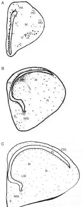
Serial sections of the cerebral hemi-spheres of chick embryos between the ages of E4 and P1 were analyzed and lectin-labeled microglial cells were identified. Fig-ure 1 shows in rostral and intermediate
sec-Figure 1. Schematic drawing of coronal sections of the chick embryo telencephalon showing the location of lectin-reactive cells (points). A, (E7) rostral sec-tion; B,(E12); C, (P1) intermedi-ate section. CDL = dorsolintermedi-ateral corticoid area; HA = apical part of the hyperpallium; HD = den-socellular part of the hyperpal-lium; Hp = hippocampus; LV = lateral ventricle; LSt = lateral striatum; M = mesopallium; MD = dorsal mesopallium; MSt = me-dial striatum; N = nidopallium; S = septum. Scale bar = 200 µm.
tions the main locations of lectin-reactive cells at three representative developmental ages. The microglial cells showed a diver-sity of shape and size (Table 1) and were distributed in the different areas of the cere-bral hemispheres (Table 2).
with a short process. Some cell clusters were visualized towards the dorsal pallial region. During the period between E18 and E21 the first ramified microglial cells (Figure 2D) with long processes (Table 1) were ob-served mainly in the dorsal pallial region, although these were also present in the sub-ventricular layer and meningeal tissue. After hatching (P1), only the ramified microglia were labeled, with most of the cells distrib-uted in the area of the nidopallium, mesopal-lium, dorsal corticoid area, from the subven-tricular layer and pial surface to the area of the lateral striatum and medial striatum, where there was a smaller number of micro-glial cells.
Discussion
In the present study, microglial cells dis-played intense labeling with L. esculentum lectin and during the CNS development of G. domesticus ameboid microglia, primitive ramified microglia and ramified microglia were all identified. These results are similar to those described for the quail cerebellum and for the cerebral hemispheres of chick embryos and chicks (2,26).
The distribution of the ameboid micro-glial cells in the subventricular layer sug-gests that these cells are the microglial pre-cursors, since they are present from the ear-liest ages studied and they morphologically resemble the cells described for the cerebral hemispheres of chick embryos and chicks (2,9).
During development, the distribution of microglial cells probably depends on the functions that they perform, but this distri-bution may be a consequence of their migra-tion to their final locamigra-tions in the adult CNS. This is similar to the situation in neuroblasts, which play no specific role before they reach their final location (7).
The temporal-spatial distribution of lec-tin-reactive cells observed here shows that the microglial cells are present in the
cere-Table 1. Cell diameter and cellular process measurements of ameboid and ramified microglial cells in chick embryos and neonatal chicks from embryonic day 4 (E4) to the first neonatal day (P1).
Diameter of Size of branched Total number of microglial cells (µm) process (µm) cells counted
Ameboid cells
E4 5.7 ± 1.35 - 8
E5 7.1 ± 1.5 - 20
E6 6.85 ± 1.2 - 20
E7 7.1 ± 1.0 - 46
E8 6.2 ± 0.95 - 45
E9 5.95 ± 0.5 - 33
E10 6.6 ± 1.55 - 36
E11 8.95 ± 1.1 - 30
E12 9.7 ± 1.1 - 29
E13 8.9 ± 1.1 - 34
E14 10.45 ± 1.7 - 29
E15 7.45 ± 1.0 - 27
E16 7.85 ± 0.8 - 27
E17 8.25 ± 1.2 - 23
Ramified cells
E18 5.85 ± 1.5 11.5 ± 6.55 21
E19 4.7 ± 0.95 9.3 ± 1.85 14
E20 4.7 ± 0.9 8.55 ± 2.2 20
E21 4.15 ± 0.75 8.15 ± 2.1 13
P1 3.25 ± 0.6 4.75 ± 2.25 13
Data are reported as means ± SD.
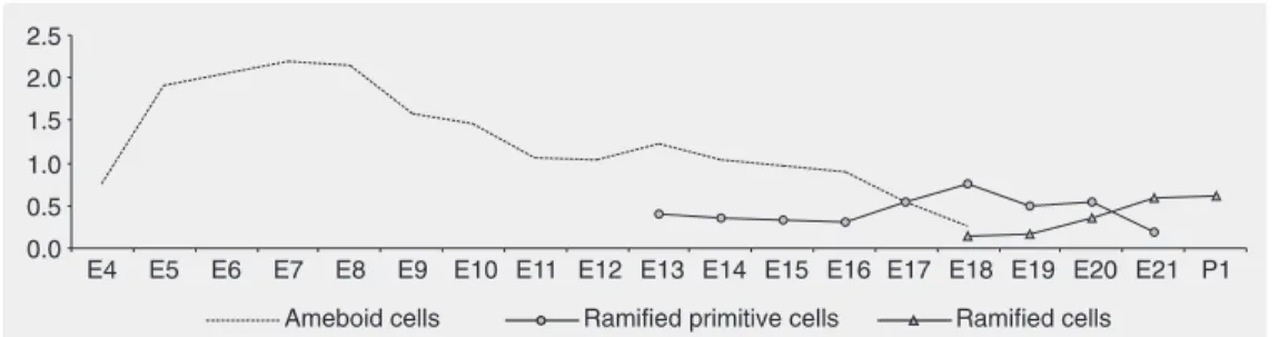
enlarged in all directions and the ventricular layer could be recognized. There was a marked increase in the number of microglial ameboid cells at this age, distributed through-out the ventricular layer and in close ap-proximation to blood vessels (Figure 2A). From E5 to E8 there was no increase in the number of ameboid microglial cells. At E9 there was a decrease in the number of ame-boid cells (Figure 2B), and after E18 these cells were no longer observed (Figure 3).
A B
C D
BV
AM LV
BV
AM
RM
LV PRM
Figure 2. Coronal sections of the cerebral hemispheres of chick embryos of different ages (A, E5; B, E12; C, E19; D, E19) show-ing lectin-reactive cells. LV = lateral ven-tricle; AM = ameboid microglial cell; PRM = primitive ramified microglial cell; RM = ramified microglial cell; BV = blood vessel. Scale bars: A, B, C = 22 µm; D = 10 µm. Table 2. Distribution and lifespan of microglial cells in different areas of the cerebral hemispheres of chick embryos and neonatal chicks
Embryonic Categories of microglial cells
day
Ameboid Primitive ramified Ramified
E4 CNS parenchyma and leptomeninges -
-E5 Ventricular layer -
-E6-E9 Subventricular and dorsal pallial structures -
-E10 Subventricular layer and region next to the -
-blood vessels in the pallial regions
E11 Subventricular layer and pallial regions -
-E12 Subventricular layer and pallial regions -
-E13 Subventricular layer, nidopallium, Subventricular layer, nidopallium, -mesopallium, and dorsal mesopallium mesopallium, and dorsal mesopallium
E14-E16 Nidopallium, mesopallium and Nidopallium, mesopallium, and
-hyperpallium regions hyperpallium regions
E17-E18 Subventricular layer and mesopallium and Uniformly distributed in the pallium and Subventricular layer, pallium and
hyperpallium regions striatum regions striatum
E19 - Dorsal pallial region and striatum regions Dorsal pallial region, medial striatum and lateral striatum
E20-E21 - Dorsal corticoid area Striatum regions, subventricular layer
and in parallel to the choroid plexus of the lateral ventricles
P1 - - Nidopallium, mesopallium dorsal
bral hemispheres from early neurogenesis and tend to increase in number during CNS differentiation. These observations corrobo-rate the hypothesis presented by others (10) and are compatible with the descriptions of different areas of the CNS including the quail cerebellum (26), quail retina (22) and the prenatal rat hippocampus (27). Micro-glia constitute a significant part of the Micro-glial cell population, between 5 and 12% of the total number of CNS cells (28), and are distributed unevenly in the regions of the CNS (29,30). Our data show the percentage of microglial cells in a particular area of the chick CNS, the cerebral hemispheres, that explains the lower percentage of microglial cells present in Figure 3 compared to the literature.
During development, the differentiation of the ameboid lectin-reactive cells into primitive ramified and ramified microglia suggests that these cells may belong to a single glial population, and that during neu-rogenesis they develop into morphologically distinct types characterized by a decreased volume of the cell body, a decrease in the number of vacuoles and the growth of pro-cesses leading to the final differentiated, resident ramified form (2,9,27).
Therefore, our results suggest that in the cerebral hemispheres apparently not all the ameboid cells develop into ramified resident microglia (Figure 2). Other studies (2,27) have shown that many ameboid cells carry out a phagocytic function and in specific conditions in the neuronal environment they can undergo cell death. Other cells can reach the immature stage, characterized by the poorly ramified microglia form and they can be activated by adverse conditions to the nervous system, returning to the phagocytic form. Only a part of the microglial precursor population would pass through all the stages reaching the mature ramified form during the late embryonic development and the in-itial postnatal phase, a hypothesis supported by the small number of ramified lectin-reac-tive cells in comparison to ameboid cells.
Microglial cells are considered to be the most plastic cell population in the CNS and exhibit a primordial activity in normal em-bryonic development and adult brain func-tion. They have also assumed an important role in various neurodegenerative diseases and neurological disorders (31) and this will be the focus of study in microglia research in the next decades.
Figure 3. Ameboid, primitive ramified and ramified microglial cell percentage from embryonic day 4 (E4) to embryonic day 21 (E21) embryos and the first neo-natal day (P1) chicks.
References
1. Ulinski PS & Margoliash D (1990). Neurobiology of the reptile-bird transition. In: Jones EG & Peters A (Editors), Cerebral Cortex. Plenum Press, New York, 217-265.
2. Fujimoto E, Miki A & Mizoguti H (1987). Histochemical studies of the differentiation of microglia cells in the cerebral hemispheres of chick embryos and chicks. Histochemistry, 87: 209-216.
3. Kallen B (1962). Embryogenesis of brain nuclei in the chick telen-cephalon. Ergebnisse Anatomie Entwicklung Geschichte, 36: 62-82.
5. Tsai HM, Garber BB & Larramendi MH (1981). 3H-Thymidine
auto-radiographic analysis of telencephalic histogenesis in the chick embryo: I. Neuronal birthdates of telencephalic compartments in situ. Journal of Comparative Neurology, 198: 275-292.
6. Tsai HM, Garber BB & Larramendi MH (1981). 3H-Thymidine
auto-radiographic analysis of telencephalic histogenesis in the chick embryo: II. Dynamics of neuronal migration, displacement, and ag-gregation. Journal of Comparative Neurology, 198: 293-306. 7. Cuadros MA & Navascués J (1998). The origin and differentiation of
microglial cells during development. Progress in Neurobiology, 56: 173-189.
8. Streit WJ (2001). Microglia and macrophages in the developing CNS. Neurotoxicology, 22: 619-624.
9. Kaur C, Hao AJ, Wu CH et al. (2001). Origin of microglia. Micros-copy Research and Technique, 54: 2-9.
10. Navascués J, Calvente R, Marin-Teva JL et al. (2000). Entry, disper-sion and differentiation of microglia in the developing central ner-vous system. Anais da Academia Brasileira de Ciências, 72: 91-102.
11. Fujimoto E, Miki A & Mizoguti H (1989). Histochemical study of the differentiation of microglial cells in the developing human cerebral hemispheres. Journal of Anatomy,166: 253-264.
12. Del Río-Hortega P (1932). Microglia. In: Penfield W (Editor), Cytol-ogy and Cellular PatholCytol-ogy of the Nervous System. Paul B. Hoeber, New York, 482-534.
13. Nakajima K & Kosaka S (1993). Functional roles of microglia in the brain. Neuroscience Research,17: 187-203.
14. Ling EA & Wong WC (1993). The origin and nature of ramified and ameboid microglia: a historical review and current concepts. Glia, 7: 9-18.
15. Ferrer I, Bernet E, Soriano E et al. (1990). Naturally occurring cell death in the cerebral cortex of the rat and removal of dead cells by transitory phagocytes. Neuroscience, 39: 451-458.
16. Acarin L, Vela JM, González B et al. (1994). Demonstration of poly-N-acetyl lactosamine residues in ameboid and ramified microglial cells in rat brain by tomato lectin binding. Journal of Histochemistry and Cytochemistry, 42: 1033-1041.
17. Barradas PC & Cavalcante LA (1998). Proliferation of differentiated glial cells in the brain stem. Brazilian Journal of Medical and Biologi-cal Research, 31: 257-270.
18. Boya J, Calvo JL, Carbonell AL et al. (1991). A lectin histochemistry study on the development of rat microglial cells. Journal of Anatomy, 175: 229-236.
19. Boya J, Carbonell AL, Calvo JL et al. (1991). Microglial cells in the nervous system of the rabbit and rat: cytochemical identification of two different lectins. Acta Anatomica, 140: 250-253.
20. Cavalcante LA, Santoro GF, Barradas PC et al. (1995). Lectin histochemistry of microglia in superior colliculus of the developing opossum. Ciência e Cultura, 4: 240-245.
21. Aschner M, Allen JW, Kimelberg HK et al. (1999). Glial cells in neurotoxicity development. Annual Review of Pharmacology and Toxicology,39: 151-173.
22. Navascués J, Moujahid A, Almendros A et al. (1995). Origin of microglia in the quail retina: central-to-peripheral and vitreal-to-scleral migration of microglial precursors during development. Jour-nal of Comparative Neurology, 354: 209-228.
23. Reiner A, Perkel DJ, Bruce LL et al. (2004). Revised nomenclature for avian telencephalon and some related brainstem nuclei. Journal of Comparative Neurology, 473: 377-414.
24. Bussab WO & Morettin PA (2004). Estatística Básica. Editora Saraiva, São Paulo, SP, Brazil.
25. Freere RH & Weibel ER (1967). Stereologic techniques in micros-copy. Journal of the Royal Microscopical Society,87: 25-34. 26. Cuadros MA, Rodríguez-Ruiz J, Valvente R et al. (1997). Microglia
development in the quail cerebellum. Journal of Comparative Neu-rology, 389: 390-401.
27. Dalmau I, Finsen B, Tonder N et al. (1997). Development of micro-glia in the prenatal rat hippocampus. Journal of Comparative Neu-rology, 377: 70-84.
28. Fedoroff S (1995). Development of microglia. In: Kettenmann H & Ransom BR (Editors), Neuroglia. Oxford University Press, New York, 162-181.
29. Lawson LJ, Perry VH, Dri P et al. (1990). Heterogeneity in the distribution and morphology of microglia in the normal adult mouse brain. Neuroscience, 39: 151-170.
30. Cuadros MA & Navascués J (2001). Early origin and colonization of the developing central nervous system by microglial precursors. Progress in Brain Research, 132: 51-59.