Phytohemagglutinin improves the development
and ultrastructure of
in vitro
-cultured goat
(
Capra hircus
) preantral follicles
E.V. Cunha
1, J.J.N. Costa
1, R.O.D.S. Rossi
1, A.W.B. Silva
1, J.R.S. Passos
1, A.M.L.R. Portela
1,
D.C.S.T. Pereira
1, M.A.M. Donato
3, C.C. Campello
2, M.V.A. Saraiva
1, C.A. Peixoto
3,
J.R.V. Silva
1and R.P. Santos
1 1Nu´cleo de Biotecnologia de Sobral, NUBIS, Universidade Federal do Ceara´, Sobral, CE, Brasil 2Laborato´rio de Manipulac¸a˜o de Oo´citos e Folı´culos Pre´-Antrais, Faculdade de Medicina Veterina´ria, Universidade Estadual do Ceara´,Fortaleza, CE, Brasil
3Laborato´rio de Ultraestrutura, CPqAM/FIOCRUZ, Universidade Federal de Pernambuco, Recife, PE, Brasil
Abstract
The objective this study was to determine the effect of phytohemagglutinin (PHA) on survival, growth and gene expression in caprine secondary follicles culturedin vitro. Secondary follicles (,0.2 mm) were isolated from the cortex of caprine ovaries and
cultured individually for 6 days ina-MEM+
supplemented with PHA (0, 1, 10, 50, 100, or 200mg/mL). After 6 days of culture, follicle diameter and survival, antrum formation, ultrastructure and expression of mRNA for FSH receptors (FSH-R), proliferating cell nuclear antigen (PCNA), and neuronal nitric oxide synthase were determined. All treatments maintained follicular survival [a-MEM+
(94.59%); 1mg/mL PHA (96.43%); 10mg/mL PHA (84.85%); 50mg/mL PHA (85.29%); 100mg/mL PHA (88.57%), and 200mg/mL PHA (87.50)], but the presence of 10mg/mL PHA in the culture medium increased the antrum formation rate (21.21%) when compared with control (5.41%, P,0.05) and ensured the maintenance of oocyte and granulosa cell ultrastructures after 6 days of culture. The expression of mRNA for FSH-R (2.7 ± 0.1) and PCNA (4.4 ± 0.2) was also significantly increased in follicles cultured with 10mg/mL PHA in relation to those cultured in a-MEM+
(1.0 ± 0.1). In conclusion, supplementation of culture medium with 10mg/mL PHA maintains the follicular viability and ultrastructure, and promotes the formation of antral cavity after 6 days of culturein vitro.
Key words: Caprine; Antrum formation; FSH-R; PCNA; Ultrastructure
Introduction
The development of efficient culture systems is extremely important both to understand the mechanisms that regulate follicular development and to assure thein vitrofollicular growth up to the stage at which the oocytes are capable of being matured and fertilized in vitro. In different species, great advances have been made in culturing preantral follicles. In human, bovine and canine species, secondary follicles have grownin vitrountil the stage of antral follicles (1-3). More satisfactory results were obtained in porcine (4), bubaline (5), ovine (6), and caprine (7) species, since a small number of embryos have been obtained after fertilization of oocytes fromin vitro-grown secondary follicles. To improve this technique in domestic species it is necessary to study substances that may contribute to promote the oocyte growth and
proliferation of granulosa cells. Several hormones and growth factors, such as follicle-stimulating hormone (FSH) (8,9), growth and differentiation factor-9 (10) and kit ligand (11), have been tested during the culture of caprine preantral follicles. Among them, FSH is the main regulator of ovarian function and its receptor has been demon-strated in goat preantral follicles (7). However, the effects of substances not produced by the ovaries, such as lectins, have not been described.
Lectins have been isolated from various sources and have a wide spectrum of biological activities (12). Most of them are proteins or glycoproteins that specifically bind to carbohydrates. In addition to recognizing sugars, some lectins promote mitogenic stimulation of lymphocytes (13), leukocytes (14) and fibroblasts (15). Phytohemagglutinin
Correspondence: J.R.V. Silva, Nu´cleo de Biotecnologia de Sobral (NUBIS), UFCE, Av. Comandante Mauroce´lio Rocha Ponte, 100, 62041-040 Sobral, CE, Brasil. Fax:+55-88-3611-8000. E-mail: jrvsilva@ufc.br
(PHA) is a lectin extracted fromPhaseolus vulgaris, which bind to complex oligosaccharide containing N-acetylga-lactosamine/galactose residues (15). Due to its ability to stimulate subpopulations of T cells, the PHA has been used as a mitogen to increase proliferation of various cell types in vitro (16). Fagbohun and Downs (17) demon-strated that mitogenic lectins, such as PHA, promote oocyte maturation and cumulus cell expansion in rats. On the other hand, Wang et al. (18) showed that the PHA has no apparent effect during maturation of bovine oocytes. Furthermore, PHA has been used to improve the efficiency of the nuclear transfer of somatic cells for oocytes from diverse species (19), because its capacity to induce closer contacts between adjacent cell membranes. In addition, PHA can stimulate the production of nitric oxide (NO) in different cells (20,21). Some studies have shown that neuronal NO synthase (nNOS) is expressed in ovarian follicles (22) and is involved in the control of folliculogenesis, steroidogenesis, oocyte maturation, and ovulation through the production of NO (23).
The aim of this study was to evaluate the effect of different concentrations of PHA on survival, growth, antrum formation, and ultrastructure of caprine secondary follicles culturedin vitro. In addition, the effect of PHA on the expression of mRNA for proliferating cell nuclear antigen (PCNA), FSH, FSH receptors (FSH-R), and nNOS was analyzed.
Material and Methods
Chemicals
Unless otherwise stated, the culture media, the lectin PHA (Cat. No. L-1668) and other chemicals were purchased from Sigma-Aldrich Corp. (USA). The stock solution of the lectin PHA was prepared in phosphate-buffered saline, pH 7.2, and stored at -206C.
Source of ovaries
Ovaries (n = 42) from 21 adult (1 to 3 years old) crossbreed goats (Capra hircus) were collected at a local slaughterhouse. Immediately postmortem, the surround-ing fat tissue and ligaments were removed and the ovaries were washed in 70% alcohol followed by two washes in sterile saline solution. The ovaries were placed into tubes containing 20 mL alpha minimum essential medium (a-MEM), supplemented with 200 IU/mL penicil-lin and 150mg/mL streptomycin and then transported to
the laboratory at 46C within 1 h.
Isolation andin vitroculture of preantral follicles
Ovarian cortical slices (1 mm) were cut from the ovarian surface, using a surgical blade under sterile conditions. The slices were subsequently placed in fragmentation medium, consisting ofa-MEM supplemented with 150 IU/mL penicillin and 150mg/mL streptomycin.
Secondary follicles (,0.2 mm) were visualized under a
stereomicroscope (SMZ 645 Nikon, Japan) and manually dissected from strips of ovarian cortex using 26 gauge (26 G) needles. Follicles with a visible oocyte, surrounded by two or more granulosa cell layers, an intact basement membrane and no antral cavity were selected for culture. After selection, follicles were cultured individually in 100mL
drops of culture medium under mineral oil on Petri dishes (60615 mm, Corning, USA). The basic culture medium (a
-MEM+) consisted ofa-MEM, pH 7.2-7.4, supplemented with
3.0 mg/mL bovine serum albumin, 1% ITS (10mg/mL
insulin, 5.5mg/mL transferrin, and 5 ng/mL selenium),
2 mM glutamine, 2 mM hypoxanthine, 50mg/mL ascorbic
acid, 150 IU/mL penicillin, 150 IU/mL streptomycin, and 100 ng/mL FSH (from sheep pituitary, Sigma). Follicles were randomly distributed in each of the following treat-ments: a-MEM+ alone (control) and
a-MEM+ associated
with lectin PHA at concentrations of 1, 10, 50, 100, or 200mg/mL. Incubation was carried out at 396C and 5% CO2
in air for 6 days. Previous studies have shown that goat preantral follicles begin their growth from 24 h of in vitro
culture and antrum formation occurs after 6 days (24). Fresh media were prepared before use and incubated for 2 h prior to use. Every other day, 60mL of the culture media was
replaced with fresh medium. The culture was replicated four times, and at least 28 follicles were cultured per treatment.
Morphological evaluation of follicular development
After 6 days of culture, follicles were classified according to their morphology. A follicle was considered to be normal when presented a centrally located spherical and homogeneous oocyte, surrounded by compact layers of granulosa cells, and without apparent damage to the basement membrane in the beginning of culture. Those follicles that showed morphological signs of degeneration, such as darkness of oocytes and surrounding granulosa cells or those with misshapen oocytes, were considered to be degenerated. The follicular growth, survival and the presence/absence of the antral cavity were evaluated every 2 days of culture. Antral cavity formation was defined as a visible translucent cavity within the granulosa cell mass. Follicular diameter was measured only in healthy follicles by calculating two perpendicular dia-meters using Motic Images Plus 2.0. In order to better examine follicular morphology, transmission electron microscopy was performed to analyze the ultrastructure of caprine secondary follicles grown in control medium and in the treatment that provided the best.
Ultrastructural analysis of follicles culturedin vitro
Transmission electron microscopy was used to ana-lyze the ultrastructure of preantral follicles cultured witha -MEM+ alone or supplemented with 10
were embedded in drops of 4% low melting agarose, and kept in sodium cacodylate buffer. Specimens were post-fixed in 1% osmium tetroxide, 0.8% potassium ferricya-nide and 5 mM calcium chloride in 0.1 M sodium cacodylate buffer for 1 h at room temperature, washed in sodium cacodylate buffer and counterstained with 5% uranyl acetate. The samples were then dehydrated through a gradient of acetone solutions and thereafter embedded in epoxy resin (Epoxy-Embedding Kit, Fluka Chemika-BioChemika, Switzerland). Afterwards, semi-thin sections (2mm) were cut, stained with toluidine blue
and analyzed by light microscopy at a 400X magnification. Ultra-thin sections (70 nm) were obtained from caprine preantral follicles classified as morphologically normal in semi-thin sections. Subsequently, ultra-thin sections were counterstained with uranyl acetate and lead citrate, and examined under a Morgani-FEI transmission electron microscope (FEI, The Netherlands).
Expression of mRNA for FSH-R, PCNA and nNOS in cultured follicles
To evaluate RNA expression, three groups of 8 follicles, from three different replicates, cultured either in control medium or in medium supplemented with 10mg/ mL PHA were collected and stored in microcentrifuge tubes at -806C.
Total RNA was extracted using the TRIzolH reagent (Invitrogen, Brazil). According to manufacturer instruc-tions, 1 mL Trizol solution was added to each frozen samples and the lysate was aspirated through a 20-gauge needle before centrifugation at 10,000gfor 3 min at room temperature. Thereafter, all lysates were diluted 1:1 with 70% ethanol and subjected to a mini-column. After binding of the RNA to the column, DNA digestion was performed using RNAse-free DNAse (340 Kunitz U/mL) for 15 min at room temperature. After washing the column
three times, the RNA was eluted with 30mL RNAse-free water. The RNA concentration was estimated by reading the absorbance at 260 nm and was checked for purity at 280 nm in a spectrophotometer (Amersham Biosciences, England). Before the reverse transcription reaction, samples of RNA were incubated for 5 min at 706C and then cooled in ice. From 2mg of total RNA, the reverse transcription was performed in a total volume of 20mL
composed of 10mL sample RNA, 4mL reverse
transcrip-tase buffer (Invitrogen), 8 U RNAsin, 150 U reverse transcriptase Superscript III, 0.036 U random primers, 10 mM DTT and 0.5 mM of each dNTP (Invitrogen). The mixture was incubated at 426C for 1 h, subsequently at 806C for 5 min, and finally stored at -206C. The negative control was prepared under the same conditions, but without addition of reverse transcriptase.
Quantification of the mRNA for FSH-R, PCNA, and nNOS was performed by using SYBR Green. Each reaction in real-time (20mL) containing 10mL SYBR Green Master MixH (Applied Biosystems, UK), 7.3mL
ultrapure water, 1mL cDNA and 5mM of each primer.
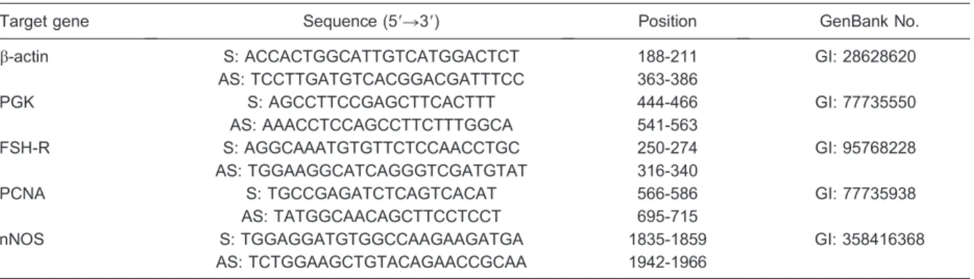
Real-time PCR was performed in a thermocycler (MastercyclerH ep Realplex, Eppendorf, Germany). The primers designed to perform amplification of mRNA for FSH-R, PCNA and nNOS are shown in Table 1. This Table also shows the primers for b-actin and phospho-glycerate kinase, which were used as endogenous controls for normalization of gene expression. The specificity of each primer pair was confirmed by melting curve analysis of PCR products. The thermal cycling profile for the first round of PCR was: initial denaturation and activation of the polymerase for 10 min at 956C, followed by 50 cycles of 15 s at 956C, 30 s at 586C, and 30 s at 726C. The final extension was for 10 min at 726C. The delta-delta-CT method was used to transform CT values into normalized relative expression levels (25).
Table 1.Primer pairs used in real-time PCR for quantification of FSH-R, PCNA and nNOS in cultured caprine follicles with 10mg/mL phytohemagglutinin.
Target gene Sequence (59R39) Position GenBank No.
b-actin S: ACCACTGGCATTGTCATGGACTCT 188-211 GI: 28628620
AS: TCCTTGATGTCACGGACGATTTCC 363-386
PGK S: AGCCTTCCGAGCTTCACTTT 444-466 GI: 77735550
AS: AAACCTCCAGCCTTCTTTGGCA 541-563
FSH-R S: AGGCAAATGTGTTCTCCAACCTGC 250-274 GI: 95768228
AS: TGGAAGGCATCAGGGTCGATGTAT 316-340
PCNA S: TGCCGAGATCTCAGTCACAT 566-586 GI: 77735938
AS: TATGGCAACAGCTTCCTCCT 695-715
nNOS S: TGGAGGATGTGGCCAAGAAGATGA 1835-1859 GI: 358416368
AS: TCTGGAAGCTGTACAGAACCGCAA 1942-1966
Statistical analyses
The percent of follicular survival and antrum formation afterin vitroculture were compared by the Fisher exact test and the results are reported as percent. The data corresponding to the follicular diameter were subjected to the Shapiro-Wilk test and the Bartlett test for verification of normal distribution and homoscedasticity, respectively. Follicular diameters show homogeneity of variance and were compared by the paired t-test. The results are reported as means ± SD and differences were consid-ered to be significant when P,0.05. The levels of mRNA for FSH-R, PCNA, and nNOS in follicles cultured in the treatments were examined by the Mann-Whitney U-test. The results are reported as means ± SE and differences were considered to be significant when P,0.05.
Results
Effect of PHA on survival and growth of goat secondary follicles
At the end of the period of culture, all treatments were capable of maintaining follicular survival and no significant difference (P . 0.05) among them were observed [a -MEM+(94.59%); 1
mg/mL PHA (96.43%); 10mg/mL PHA
(84.85%); 50mg/mL PHA (85.29%); 100mg/mL PHA (88.57%), and 200mg/mL PHA (87.50%)]. Regarding
the follicular diameter, from day 0 to 6 of culture, a significant increase in follicular diameter was observed in all treatments (P,0.05), but no differences were found among them (Table 2).
Regarding the antrum formation, at the end of culture, follicles cultured in the presence of 10mg/mL PHA showed a significant increase in antrum formation when compared to the control medium (a-MEM+). On the other
hand, the percent of antrum formation in follicles cultured with 1, 50, 100, and 200mg/mL PHA did not differ either
from the control treatment or among each other (Table 3).
Ultrastructural analysis of follicles culturedin vitro
The ultrastructural analysis showed that follicles cultured in a-MEM+had an abnormal profile, the oocyte
cytoplasm was extremely vacuolated, having a greater open area and organelles were no longer recognizable in ooplasm (Figure 1A). Despite a regular zona pellucida and well-organized granulosa cells surrounding the oocyte, these follicles had reduced number of microvilli (Figure 1A). On the other hand, follicles cultured in medium supplemented with 10mg/mL PHA (Figure 1B
and C) presented a well-preserved oocyte, with visible organelles, such as Golgi complex, endoplasmic reticu-lum and lipid droplets. Mitochondria had some swollen-ness, but this feature is common in cultured cells. Granulosa cells were present with normal organelles and regular chromatin. Zona pellucida was preserved, and microvilli were evident.
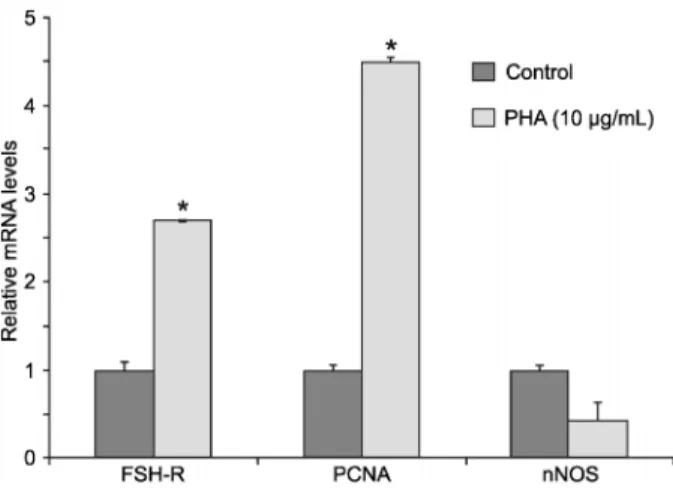
Levels of mRNA for FSH-R, PCNA and nNOS in cultured follicles
Levels of mRNA for FSH-R, PCNA and nNOS in follicles cultured ina-MEM+alone or supplemented with
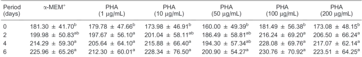
Table 2.Follicular diameters of caprine secondary follicles cultured for 6 days ina-MEM+supplemented with different concentrations of PHA.
Period (days)
a-MEM+ PHA (1mg/mL)
PHA (10mg/mL)
PHA (50mg/mL)
PHA (100mg/mL)
PHA (200mg/mL)
0 181.30 ± 41.70b 179.78 ± 47.66b 173.98 ± 46.91b 160.00 ± 49.39b 181.49 ± 56.38b 173.08 ± 48.15b
2 199.98 ± 50.83ab 197.67 ± 56.10a 201.04 ± 58.11ab 186.49 ± 58.81ab 216.24 ± 69.20a 206.50 ± 66.24a 4 214.29 ± 59.30a 205.64 ± 64.10a 215.88 ± 66.40a 194.30 ± 57.34ab 228.08 ± 69.76a 217.07 ± 62.14a
6 225.96 ± 65.26a 212.30 ± 60.01a 228.34 ± 76.50a 200.90 ± 54.27a 230.76 ± 70.92a 223.51 ± 64.25a
Data are reported as means ± SD.a-MEM+= alpha minimum essential medium; PHA = phytohemagglutinin. Different superscript letters indicate statistically significant differences within a column (P,0.05, pairedt-test).
Table 3.Percentages of follicles showing signs of antrum formation after culture of secondary follicles ina-MEM+supplemented with different concentrations of PHA.
Period (days)
a-MEM+ PHA (1
mg/mL) PHA (10mg/mL) PHA (50mg/mL) PHA (100mg/mL) PHA (200mg/mL)
0 0.00% (00/37) 0.00% (00/28) 0.00% (00/33) 0.00% (00/34) 0.00% (00/35) 0.00% (00/00) 2 0.00% (00/37) 0.00% (00/28) 6.06% (02/33) 0.00% (00/34) 0.00% (00/35) 0.00% (00/00) 4 0.00% (00/37) 3.57% (01/28)A 12.12% (04/33)A 0.00% (00/34) 5.71% (02/35)A 0.00% (00/00)
6 5.41% (02/37)B 7.14% (02/28)AB 21.21% (07/33)A 5.88% (02/34)AB 5.71% (02/35)AB 0.00% (00/00)
10mg/mL PHA are shown in Figure 2. No significant difference in the levels of mRNA for nNOS between follicles cultured in control medium or medium supple-mented with PHA was observed. However, the presence of PHA significantly increased the levels of mRNA for both PCNA and FSH-R, when compared with the control medium (Figure 2).
Discussion
The present study demonstrated that PHA (10mg/mL)
stimulates antrum formation, helps to keep ultrastructure integrity and increases the expression of FSH-R and PCNA in caprine preantral follicles culturedin vitro.
Morphological analysis showed that after 6 days of culture, high survival rates were observed in all the treatments. This is probably due to the use of a culture
medium rich of nutrients such as vitamins, amino acids and minerals that have been successfully used to culture preantral follicles (26). FSH is known to support follicle viability by inhibiting the expression of anti-apoptotic proteins (27) and its presence in the culture medium certainly helped to maintain follicles viable. Other studies have shown that FSH is very important for preantral follicle survival in different species (2,28). On the other hand, despite the lack of a dose/response relationship, the presence of 10mg/mL PHA increased the rate of formation of antral cavity. This may be associated with increased expression of the FSH receptors stimulated by PHA at this concentration. Previousin vitrostudies have shown that this gonadotropin improves formation of antrum in cultured secondary follicles in caprine (9) and mouse (29) species.
The ultrastructural analysis confirmed the integrity of
follicles cultured in medium supplement with 10mg/mL PHA. Previous dose response studies have shown that high concentrations of PHA (.80mg/mL) have toxic effects on cultured fibroblasts, while low concentrations (0.1-10mg/mL) have no positive effects (30). In agree-ment with these data, the present study has demonstrated that the optimal concentration of PHA to stimulate gene expression and antrum formation in cultured preantral follicles is 10mg/mL. The increased expression of the
FSH receptors stimulated by PHA may have contributed to maintain follicular ultrastructure. Previous in vitro
studies have shown that FSH kept ultrastructural char-acteristics of caprine-cultured follicles (9,28). Studies using mice with deficiencies in FSH-R expression have allowed further elucidation of the role of FSH in ovarian follicles and it has been reported that mice lacking the FSH-R gene have structural alterations in the ovary (31). Furthermore, there is evidence to suggest that FSH-R deletion results in changes in oocyte structure and function, and disruption of oocyte-granulosa cell commu-nication (32). In addition, PHA facilitates close cell contact by binding to the N-linked carbohydrate core structure (beta 1-6 branching) of glycoproteins on the cell mem-brane (33). In this study, this lectin may have recognized and mediated adhesion between carbohydrates present in granulosa cells and oocyte, maintaining the follicular ultrastructure. The communication between granulosa cells during the preantral and early antral stages is necessary to ensure subsequent oocyte developmental competence (34). The oocyte is the central regulator of follicular cell functions through the secretion of soluble growth factors such as BMP-15 and GDF-9, which act in
the surrounding follicular cells to promote their prolifera-tion and survival (35).
The addition of 10mg/mL PHA to the culture medium increased the expression of mRNA for PCNA, indicating an influence of this lectin on granulosa cell proliferation, but a dose/response relationship was not demonstrated. PCNA performs the essential function of providing replicative polymerases with the high processivity required to duplicate the entire genome (36) and has been used as a marker of granulosa cell proliferation in various species (8,37,38). Although lectin PHA has a known role in the mitogenesis of various cell types (14-16), the increase in PCNA expression did not reflect an increase in follicular diameter during the culture, probably because of the short culture period. Although extensively studied, the mechanism of stimulation of mitosis by lectins is still not understood. It has been suggested that mitogenic lectins interact with components of cell mem-branes to stimulate cell proliferation (13).
The present study demonstrates the expression of nNOS in goat ovarian follicles, but no increase in the levels of mRNA for nNOS was observed in response to PHA stimulation during in vitroculture. The nNOS is an enzyme responsible for the synthesis of NO that was recently demonstrated to be expressed in bubaline granulosa cells and oocytes in different stages of development, indicating its role in the control of follicular growth (24). Nitric oxide can act as a pro- or anti-apoptotic agent in a variety of structures, including ovarian follicles (39). At low concentrations, NO has anti-apoptotic action, but with the increase of its production, it causes DNA damage and induces cell death by apoptosis (39). The role of NO in regulating ovarian function and reproductive systems has been described by Rosselli et al. (40), who observed correlations between ovarian hormones and NO production, since an increase in NO is correlated with increased steroidogenesis. Previous studies have shown that PHA can stimulate the production of NO in different cellular types (22,23), but this was not observed in cultured preantral follicles.
Addition of 10mg/mL PHA duringin vitroculture caprine
secondary follicles stimulates antrum formation, increases the expression of FSH-R and PCNA, and helps to keep ultrastructural integrity of cultured follicles. These data may be useful for the development of an efficient culture system to promote oocyte growth and maturationin vitro.
Acknowledgments
Research supported by CNPq (#562686/2010-0 and
#501221/2009-3) and Fundac¸a˜o Cearense de Apoio ao Desenvolvimento Cientı´fico e Tecnolo´gico (FUNCAP,
#PRN-0040-00053.01.00/10). J.R.V. Silva and R.P. Santos are investigators of CNPq.
References
1. Roy SK, Treacy BJ. Isolation and long-term culture of human preantral follicles.Fertil Steril1993; 59: 783-790. 2. Gutierrez CG, Ralph JH, Telfer EE, Wilmut I, Webb R.
Growth and antrum formation of bovine preantral follicles in long-term culturein vitro.Biol Reprod2000; 62: 1322-1328, doi: 10.1095/biolreprod62.5.1322.
3. Serafim MK, Arau´jo VR, Silva GM, Duarte AB, Almeida AP, Chaves RN, et al. Canine preantral follicles cultured with various concentrations of follicle-stimulating hormone (FSH). Theriogenology 2010; 74: 749-755, doi: 10.1016/ j.theriogenology.2010.03.028.
4. Wu J, Tian Q. Role of follicle stimulating hormone and epidermal growth factor in the development of porcine preantral follicle in vitro. Zygote 2007; 15: 233-240, doi: 10.1017/S0967199407004194.
5. Gupta PS, Ramesh HS, Manjunatha BM, Nandi S, Ravindra JP. Production of buffalo embryos using oocytes from in vitrogrown preantral follicles.Zygote2008; 16: 57-63, doi: 10.1017/S096719940700442X.
6. Arunakumari G, Shanmugasundaram N, Rao VH. Development of morulae from the oocytes of cultured sheep preantral follicles.Theriogenology2010; 74: 884-894, doi: 10.1016/j.theriogenology.2010.04.013.
7. Saraiva MV, Celestino JJ, Araujo VR, Chaves RN, Almeida AP, Lima-Verde IB, et al. Expression of follicle-stimulating hormone receptor (FSHR) in goat ovarian follicles and the impact of sequential culture medium onin vitrodevelopment of caprine preantral follicles.Zygote2011; 19: 205-214, doi: 10.1017/S0967199410000511.
8. Silva JR, van den Hurk R, Costa SH, Andrade ER, Nunes AP, Ferreira FV, et al. Survival and growth of goat primordial follicles after in vitro culture of ovarian cortical slices in media containing coconut water.Anim Reprod Sci2004; 81: 273-286, doi: 10.1016/j.anireprosci.2003.09.006.
9. Saraiva MV, Rossetto R, Brito IR, Celestino JJ, Silva CM, Faustino LR, et al. Dynamic medium produces caprine embryo from preantral follicles grownin vitro. Reprod Sci 2010; 17: 1135-1143, doi: 10.1177/1933719110379269. 10. Martins FS, Celestino JJ, Saraiva MV, Chaves RN,
Rossetto R, Silva CM, et al. Interaction between growth differentiation factor 9, insulin-like growth factor I and growth hormone on thein vitrodevelopment and survival of goat preantral follicles.Braz J Med Biol Res2010; 43: 728-736, doi: 10.1590/S0100-879X2010007500066.
11. Celestino JJ, Bruno JB, Lima-Verde IB, Matos MH, Saraiva MV, Chaves RN, et al. Steady-state level of kit ligand mRNA in goat ovaries and the role of kit ligand in preantral follicle survival and growthin vitro.Mol Reprod Dev2010; 77: 231-240.
12. Lis H, Sharon N. Biological properties of lectins. In: Anonymous,The lectins: properties, functions and applica-tions in biology and medicine. London: Academic Press; 1986. p 265-285.
13. Nowell PC. Phytohemagglutinin: an initiator of mitosis in cultures of normal human leukocytes.Cancer Res1960; 20: 462-466.
14. Sheng Y, Pero RW, Wagner H. Treatment of chemother-apy-induced leukopenia in a rat model with aqueous extract fromUncaria tomentosa.Phytomedicine2000; 7: 137-143,
doi: 10.1016/S0944-7113(00)80086-0.
15. Sell AM, Costa CP. PHA: a lectin that enhanced wound healing in the skin rats. Proceedings of the International Meeting on Vaccines. Salvador: 1998. p 124.
16. Myers RL.Immunology - a laboractory manual. Dubuque: WCB Publishers; 1995.
17. Fagbohun CF, Downs SM. Maturation of the mouse oocyte-cumulus cell complex: stimulation by lectins. Biol Reprod 1990; 42: 413-423, doi: 10.1095/biolreprod42.3.413. 18. Wang S, Panter KE, Evans RC, Bunch TD. The effects of
pokeweed mitogen (PWM) and phytohemagglutinin (PHA) on bovine oocyte maturation and embryo developmentin vitro. Anim Reprod Sci2001; 67: 215-220, doi: 10.1016/ S0378-4320(01)00122-1.
19. Tesarik J, Nagy ZP, Mendoza C, Greco E. Chemically and mechanically induced membrane fusion: non-activating methods for nuclear transfer in mature human oocytes. Hum Reprod2000; 15: 1149-1154, doi: 10.1093/humrep/ 15.5.1149.
20. Kesherwani V, Sodhi A. Differential activation of macro-phagesin vitroby lectin concanavalin A, phytohemaggluti-nin and wheat germ agglutiphytohemaggluti-nin: production and regulation of nitric oxide.Nitric Oxide2007; 16: 294-305, doi: 10.1016/ j.niox.2006.11.001.
21. Kim D, Yamasaki Y, Jiang Z, Nakayama Y, Yamanishi T, Yamaguchi K, et al. Comparative study on modeccin- and phytohemagglutinin (PHA)-induced secretion of cytokines and nitric oxide (NO) in RAW264.7 cells. Acta Biochim Biophys Sin2011; 43: 52-60, doi: 10.1093/abbs/gmq105. 22. Dubey PK, Tripathi V, Singh RP, Saikumar G, Nath A,
Pratheesh, et al. Expression of nitric oxide synthase isoforms in different stages of buffalo (Bubalus bubalis) ovarian follicles: effect of nitric oxide onin vitrodevelopment of preantral follicle.Theriogenology2012; 77: 280-291, doi: 10.1016/j.theriogenology.2011.08.002.
23. Thaler CD, Epel D. Nitric oxide in oocyte maturation, ovulation, fertilization, cleavage and implantation: a little dab’ll do ya. Curr Pharm Des 2003; 9: 399-409, doi: 10.2174/1381612033391748.
24. Huanmin Z, Yong Z.In vitrodevelopment of caprine ovarian preantral follicles.Theriogenology2000; 54: 641-650, doi: 10.1016/S0093-691X(00)00379-4.
25. Livak KJ, Schmittgen TD. Analysis of relative gene expression data using real-time quantitative PCR and the -2DDCT method.Methods2001; 25: 402-408, doi: 10.1006/ meth.2001.1262.
26. Rossetto R, Saraiva MV, Dos Santos RR, da Silva CM, Faustino LR, Chaves RN, et al. Effect of medium composi-tion on the in vitro culture of bovine pre-antral follicles: morphology and viability do not guarantee functionality. Zygote2012; 2: 1-4, doi: 10.1017/S0967199412000044. 27. Markstrom E, Svensson EC, Shao R, Svanberg B, Billig H.
Survival factors regulating ovarian apoptosis - dependence on follicle differentiation.Reproduction 2002; 123: 23-30, doi: 10.1530/rep.0.1230023.
S0967199407004169.
29. Cortvrindt R, Hu Y, Smitz J. Recombinant luteinizing hormone as a survival and differentiation factor increases oocyte maturation in recombinant follicle stimulating hor-mone-supplemented mouse preantral follicle culture.Hum Reprod 1998; 13: 1292-1302, doi: 10.1093/humrep/ 13.5.1292.
30. Sell AM, Costa CP. Effects of plant lectins on in vitro fibroblast proliferation. Braz Arch Biol Technol 2003; 46: 349-454, doi: 10.1590/S1516-89132003000300006. 31. Balla A, Danilovich N, Yang Y, Sairam MR. Dynamics of
ovarian development in the FORKO immature mouse: structural and functional implications for ovarian reserve. Biol Reprod 2003; 69: 1281-1293, doi: 10.1095/biolre-prod.103.015552.
32. Yang Y, Balla A, Danilovich N, Sairam MR. Developmental and molecular aberrations associated with deterioration of oogenesis during complete or partial follicle-stimulating hormone receptor deficiency in mice. Biol Reprod 2003; 69: 1294-1302, doi: 10.1095/biolreprod.103.015610. 33. Sharma V, Surolia A. Analyses of carbohydrate recognition
by legume lectins: size of the combining site loops and their primary specificity. J Mol Biol 1997; 267: 433-445, doi: 10.1006/jmbi.1996.0863.
34. Albertini DF, Combelles CM, Benecchi E, Carabatsos MJ. Cellular basis for paracrine regulation of ovarian follicle
development. Reproduction 2001; 121: 647-653, doi: 10.1530/rep.0.1210647.
35. Gilchrist RB, Ritter LJ, Armstrong DT. Oocyte-somatic cell interactions during follicle development in mammals.Anim Reprod Sci 2004; 82-83: 431-446, doi: 10.1016/j.anire-prosci.2004.05.017.
36. Maga G, Hubscher U. Proliferating cell nuclear antigen (PCNA): a dancer with many partners.J Cell Sci2003; 116: 3051-3060, doi: 10.1242/jcs.00653.
37. Wandji SA, Srsen V, Voss AK, Eppig JJ, Fortune JE. Initiationin vitroof growth of bovine primordial follicles.Biol Reprod 1996; 55: 942-948, doi: 10.1095/biolre-prod55.5.942.
38. Muskhelishvili L, Wingard SK, Latendresse JR. Proliferating cell nuclear antigen - a marker for ovarian follicle counts. Toxicol Pathol 2005; 33: 365-368, doi: 10.1080/ 01926230590930164.
39. Goud AP, Goud PT, Diamond MP, Gonik B, Abu-Soud HM. Reactive oxygen species and oocyte aging: role of super-oxide, hydrogen persuper-oxide, and hypochlorous acid. Free Radic Biol Med2008; 44: 1295-1304, doi: 10.1016/j.free-radbiomed.2007.11.014.