Use of biological Glue (Bioglue®) in laparoscopic partial
nephrectomy: a study in pigs
_______________________________________________
Luis Felipe Brandão
1, Fabio Cesar Miranda Torricelli
2, Glauco Melo
1, Luiz Fernando Takano
1, Anuar
Ibrahim Mitre
1,2, Marco Antonio Arap
1,21 Instituto de Ensino e Pesquisa do Hospital Sirio Libanes - Sao Paulo, SP – Brazil; 2 Division of Urology,
Hospital das Clinicas, University of Sao Paulo Medical School - Sao Paulo, SP - Brazil
ABSTRACT
ARTICLE
INFO
______________________________________________________________ ______________________
Introduction: Partial nephrectomy is the standard of care for localized renal tumors. However, bleeding and warm ischemia time are still controversial when laparoscopic surgeries are carried out. Herein, we aim to compare the outcomes from laparoscopic partial nephrectomy with and without the use of biological glue with purified bovine albumin and glutaraldehyde (BioGlue®).
Materials and Methods: Twenty-four kidneys of 12 pigs were used in this study. A pre--determined lower pole segment was resected (3 cm x 1 cm) and one of two different hemostatic techniques was performed. In one kidney, hemostatic “U suture” (poligleca-prone 3.0) was performed and in the contra-lateral kidney, only the biological glue was applied. Data recorded was comprised of warm ischemia time (seconds) and estimated blood loss (mL) for each procedure. In cases of bleeding after glue administration, a complementary suture was done.
Results: Mean warm ischemia time was 492.9±113.1 (351-665) seconds and 746±185.3 (409-1125) seconds for biological glue and suture groups, respectively. There was a positive significant difference in terms of warm ischemia favoring the biological glue group over the suture group (p<0.001). Mean blood loss was 39.4 (0-115) mL for the biological glue group and 39.1 (5-120) mL for the suture group (p=0.62).
Conclusion: Biological glue is an important tool for laparoscopic partial nephrecto-mies. It is effective for hemostatic control in selected cases, and it can be used in com-bination with the traditional suture techniques.
Key words:
Bio-glue [Supplementary Concept]; Kidney; Nephrectomy; Laparoscopy
Int Braz J Urol. 2015; 41: 252-7
_____________________ Submitted for publication: January 06, 2014 _____________________ Accepted after revision: May 03, 2014
INTRODUCTION
During the last few decades, ultrasound and computed tomography have increased the de-tection of renal masses smaller than 4 cm. Because of the high incidence of small renal masses and based on the fact that studies have shown that partial resection has the same oncological efficacy as radical surgery, partial nephrectomy has been increasingly used for the treatment of these
pa-tients (1). Several studies evaluating laparoscopic surgery showed overall and cancer-specific sur-vival rates, as well as complication rates, compa-rable to those of open partial nephrectomy (2, 3).
as the upper limit (3, 5). The major concerns with the laparoscopic partial nephrectomy technique are the difficulty to perform kidney cooling (cold ischemia) and the longer time required to perform the parenchyma suture after the resection of the tumor. Therefore, measures should be taken so that the clamping time can be as short as possible in order to preserve as much glomerular function as possible. The shorter the ischemia time, the gre-ater the number of nephrons spared after the pro-cedure in both short and long term outcomes (6).
In order to improve homeostasis and re-duce warm ischemia time, several tissue sealants have been developed (7-11). Here we evaluate the potential role of glutaraldehyde glue with puri-fied bovine serum albumin (Bioglue ® - Cryolife inc. Nennesaw, GA, USA) in the homeostasis on renal surface, comparing it to traditional laparos-copic suturing. Estimated blood loss and warm ischemia time of the kidney were used to assess this comparison.
MATERIALS AND METHODS
Study design: Twenty-four kidneys from 12 swine (minipig BR) were used for the compa-rison. All animals were acquired from the same facility and all surgeries were performed in the Institute of Education and Research of the Sirio--Libanês Hospital in São Paulo, after approval from the ethics committee.
Pigs underwent general anesthesia with endotracheal intubation. Anesthesia was induced with intramuscular ketamine (5 mg/kg), intramus-cular midazolam (0.5 mg/kg), and maintained with continuous intravenous propofol (8mg/kg) and inhalatory Isoflurane (2%). For analgesia, con-tinuous infusion of intravenous fentanyl (30µg/ kg/h) was taken. At the end of the procedures, all animals were euthanized.
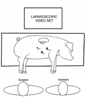
Each procedure was initiated with the cre-ation of pneumoperitoneum using Veress needle (Ethicon inc. Somerville, NJ, USA), followed by trocar insertion according to Figure-1. Then, lapa-roscopic dissection and clamping of renal hilum (both renal artery and renal vein) were performed, followed by partial resection of a pre-determined lower pole segment of kidney (3 cm in longer
dia-meter and 1 cm deep, Figure-2). The 1-cm deep resection was enough to extract the parenchyma segment without injuring the collecting system. Both surgeons confirmed it visually and in none of the cases, the collecting system was opened. In all procedures, we pre-established a double “U” poliglecaprone 3.0 suture using a 1 cm Hem’o’lok clip (Weck Surgical Instruments, Teleflex Medi-cal, Durham, NC, USA) in each end of each thread as the standard. A third “U” suture was used in case of persistent bleeding. When using biological glue, the renal pedicle was unclamped two minu-tes after the end of application of the material (ac-cording to the company´s instructions). Ac(ac-cording to the ethics committee protocol, a poliglecaprone 3.0 suture should be added to control eventual re-sidual bleeding after the use of biological glue.
We chose to test biological glue in one kidney and suture in the contralateral side, in or-der to avoid selection bias. After the first side was completed, all the blood was aspirated, the cavity
Figure 1 - Trocar placement - 3 ports were placed and a small puncture (“X”) was performed for the passage of the cannula for the glue application.
was reviewed and all the trocar incision were clo-sed using 2.0 poligalactine simple sutures. For the following side, the animal was repositioned, the pneumoperitoneum was re-gained and the trocars were placed in same fashion, however in the con-tralateral side.
Warm ischemia time (in seconds) and esti-mated blood loss (in milliliters) were recorded for each procedure. Warm ischemia time was measu-red from the clamp placement in the renal vessels to its removal. Extra care was taken in order to have all the blood out of the cavity after the fi rst side was completed, avoiding inaccuracy in the contralateral measurement. All procedures were performed by two surgeons (LF and GM) with si-milar intermediate laparoscopic experience, pre-senting the same level of knowledge and practice. Both of them were participants of the Minimally Invasive Urologic Surgery Post-Graduation An-nual Course. Each surgeon performed exactly the
same procedures in terms of number, side and type (using either the glue or the suture).
Statistical analysis
Results were described as mean, standard deviation and range values. Mann-Whitney U test was used to compare continuous variables betwe-en the groups. Statistical analysis was performed using SPSS version 20.0 (SPSS Inc., Chicago, IL) and the level of signifi cance was set at p<0.05.
RESULTS
The results are shown in the table-1. The mean warm ischemia time was 492.9±113.1 (351-665) seconds and 746±185.3 (409-1125) secon-ds for bioglue and suture groups, respectively (p<0.001). The mean estimated blood loss was 39.4 (0-115) mL for the group using only the biological glue and 39.1 (5-120) mL for the group that used the suture. In the biological glue group, 6 cases required one “U” suture for strict control of ble-eding and one case required two additional sutu-res. There were no intra-operative complications, there were no collecting system injuries, and all procedures were successfully fi nished according to the protocol. There was no signifi cant differen-ce between surgeons LF and GM regarding their respective outcomes: mean warm ischemia time using the biological glue (523±98 vs. 495±116, p=0.68); mean warm ischemia time using the su-ture (733±179 vs. 758±206, p=0.74); estimated blood loss using the biological glue (38.1±41.8 vs. 27±25.4, p=0.93); estimated blood loss using the suture (33.3±43.6 vs. 45.0±48.8, p=0.68).
DISCUSSION
Open partial nephrectomy was considered the standard of care for renal tumors smaller than 4 cm. However, recent publications have shown that laparoscopy can be used to tackle small renal masses (12). Indeed, laparoscopic partial nephrec-tomy has been increasingly used for this type of lesions, despite being a challenging procedure. Hemostasis control and collecting system sutu-ring are the most diffi cult parts dusutu-ring a laparos-Figure 2 - Partial nephrectomy: A. Lower pole resected. B.
copic procedure, increasing warm ischemia time. For best outcomes with nephron sparing surgery, several tissue sealants were developed to be asso-ciated with, or even replace sutures of the renal parenchyma (7-11). The BioGlue ® is a mixture of bovine serum albumin (45% wt / vol) and gluta-raldehyde (10% wt / vol) in a 4:1 ratio. To avoid any contamination, the bovine serum is purified by precipitation heat, gamma irradiation and chromatography.
Our study showed a significant shorter warm ischemia time for the biological glue group. Although we were able to remove the laparosco-pic clamp in a significantly shorter period of time when using only the biological glue, in half of times we were not satisfied with the achieved he-mostasis. Thus, in 50% of our procedures, an extra suture was taken in order to stop any sort of active bleeding. For this reason, we highlight here that biological glue can be helpful in bleeding control,
but it still cannot be considered a replacement for renal sutures, especially because extra sutures of renal parenchyma are frequently necessary despi-te its use.
Nadler et al (9) showed a significantly lower blood loss in partial nephrectomies for renal tumors with biological glue (BioGlue ®) as a complementary armamentarium. The authors described it as highly effective in stopping surgical oozing. Moreover, they highlighted that due to its initially liquid consistency, biological glue can be easily maneuvered along the resected area or over a Surgicel bolster. In their se-ries, they demonstrated the safety and efficacy of the BioGlue, when used to form a protective covering that can stabilize the bolster, after laparoscopic par-tial nephrectomy.
It is important to remember that the glue cannot enter the urinary tract because it may cau-se adhesions in the collecting system or over ves-sels with relevant active bleeding because adhe-Table 1 - Warm ischemia time (WIT) and estimated blood loss (EBL) for the use of bioglue and suture.
Case Bioglue WIT (sec) Suture WIT (sec) p value Bioglue EBL (mL) Suture EBL (mL) p value
1 552A 596B
p<0.001
50 120
p=0.62
2 363B 748A 0 5
3 474A 773B 27 5
4* 351A 828A 32 15
5* 592A 841B 115 35
6* 567B 873A 10 20
7 627A 584B 5 15
8* 449B 884A 63 100
9* 542B 1125B 52 115
10 348A 660A 0 10
11 665B 633B 10 15
12* 385B 409A 30 15
Mean (stdev**) 492.9 (113.1) 746 (185.3) 39.4 (33.4) 39.1 (44.6)
sion efficacy is reduced in such situation (11). In order to avoid the contact of the glue with the collecting system, we pre-determined a deep leng-th of 1cm of leng-the resected segment. This was an important maneuver, due to the possibility of local occlusion by the serum albumin glutaraldehyde, as it was previously described (13).
Our study has some limitations that should be pointed. We have a small number of cases in each arm. However, even with such numbers, we were able to report a significant shorter warm ischemia time in the bioglue group. Second, our study was made in a porcine model of partial ne-phrectomy, which is known to be an easier pro-cedure when compared to partial nephrectomies in humans. The position of the kidney and easier visualization of the renal pedicle make the proce-dure less complex. Although our results may not be the same in humans due to these differences, the porcine model facilitated standardization of the procedure, therefore allowing our results to be better controlled. Third, as an acute experiment, some other important outcomes, including posto-perative bleeding, rate of urinary extravasation, and the postoperative kidney function could not be assessed. Lastly, we did not evaluate costs. Ho-wever, Dalpiaz et al (14) reported in a review that the use of sealants in the renal parenchyma may decrease the rate of bleeding during and after sur-gery, resulting in reduced costs for blood trans-fusions and blood products in perioperative time and also reducing costs with complications, ope-rative time, length of hospital stay and intensive care unit indications.
CONCLUSIONS
The biological glue is an important tool in laparoscopic partial nephrectomies. In our series, the group that used the glue sealant had a significant shorter warm ischemia time, allowing an earlier un-clamping. However complementary suture were re-quired in half of cases.
Human clinical trials with larger numbers are needed to confirm our results in patients with small renal tumors that could lead us to better outcomes, by decreasing warm ischemia time and bleeding, when performing minimally invasive partial nephrectomy.
CONFLICT OF INTEREST
None declared.
REFERENCES
1. Celia A, Zeccolini G, Guazzoni G, Pansadoro V, Disanto V, Porpiglia F, et al.: Laparoscopic nephron sparing surgery: a multi-institutional European survey of 592 cases. Arch Ital Urol Androl. 2008; 80: 85-91.
2. Lucas SM, Mellon MJ, Erntsberger L, Sundaram CP: A comparison of robotic, laparoscopic and open partial nephrectomy. JSLS. 2012; 16: 581-7.
3. Porpiglia F, Volpe A, Billia M, Scarpa RM: Laparoscopic versus open partial nephrectomy: analysis of the current literature. Eur Urol. 2008; 53: 732-42; discussion 742-3. 4. Carlos AS, Tobias-Machado M, Starling ES, Corrêa de
Araujo FB, Faria EF, Nogueira L, et al.: Alternative techniques to reduce warm ischemia time in laparoscopic partial nephrectomy. Int Braz J Urol. 2013; 39: 145, discussion 146. 5. Hung AJ, Tsai S, Gill IS: Does eliminating global renal
ischemia during partial nephrectomy improve functional outcomes? Curr Opin Urol. 2013; 23: 112-7.
6. Simmons MN, Lieser GC, Fergany AF, Kaouk J, Campbell SC: Association between warm ischemia time and renal parenchymal atrophy after partial nephrectomy. J Urol. 2013; 189: 1638-42.
7. Bernie JE, Ng J, Bargman V, Gardner T, Cheng L, Sundaram CP: Evaluation of hydrogel tissue sealant in porcine laparoscopic partial-nephrectomy model. J Endourol. 2005; 19: 1122-6.
8. Hidas G, Kastin A, Mullerad M, Shental J, Moskovitz B, Nativ O: Sutureless nephron-sparing surgery: use of albumin glutaraldehyde tissue adhesive (BioGlue). Urology. 2006; 67: 697-700; discussion 700.
9. Nadler RB, Loeb S, Rubenstein RA, Vardi IY: Use of BioGlue in laparoscopic partial nephrectomy. Urology. 2006; 68: 416-8.
10. Stojkovic I, Savic V, Djokic M, Balint B, Ljubenovic S, Ignjatovic I: Possibilities and limitations of fibrin glue usage in nephron-sparing surgery: experimental study. Urol Int. 2005; 74: 355-60.
11. Johnston WK 3rd, Kelel KM, Hollenbeck BK, Daignault S, Wolf JS Jr.: Acute integrity of closure for partial nephrectomy: comparison of 7 agents in a hypertensive porcine model. J Urol. 2006; 175: 2307-11.
13. Kim IY, Eichel L, Edwards R, Uribe C, Chou DS, Abdelshehid C, et al.: Effects of commonly used hemostatic agents on the porcine collecting system. J Endourol. 2007; 21: 652-4.
14. Dalpiaz O, Neururer R, Bartsch G, Peschel R: Haemostatic sealants in nephron-sparing surgery: what surgeons need to know. BJU Int. 2008; 102: 1502-8
_______________________ Correspondence address: