14
1. Ph.D. Pediatrician, Hospital de Reabilitação de Anomalias Craniofaciais,Universidade de São Paulo (HRAC/USP), São Paulo, SP, Brazil. 2. Plastic surgeon, HRAC/USP, São Paulo, SP, Brazil.
3. Othorinolaryngologist, HRAC/USP, São Paulo, SP, Brazil. 4. Ph.D. Nutritionist, HRAC/USP, São Paulo, SP, Brazil.
5. Full professor, Department of Well-child Care and Pediatrics, School of Medicine of Ribeirão Preto, Universidade de São Paulo (USP), Ribeirão Preto, SP, Brazil.
6. Ph.D. Assistant professor, Department of Well-child Care and Pediatrics, School of Medicine of Ribeirão Preto, USP, Ribeirão Preto, SP, Brazil.
Manuscript received Feb 26 2004, accepted for publication Mar 31 2004.
Suggested citation: Marques IL, de Sousa TV, Carneiro AF, Peres SP, Barbieri MA, Bettiol H. Robin sequence: a single treatment protocol. J Pediatr (Rio J). 2005;81:14-22.
Abstract
Objective: To present a single protocol that might cover both the respiratory and feeding difficulties of neonates and infants with Robin sequence.
Sources of data: The article was prepared on the basis of the most recent publications available in bibliographic databases and in books that discuss the treatment of Robin sequence, especially the studies conducted at the Hospital for Rehabilitation of Craniofacial Anomalies of Universidade de São Paulo (HRAC/USP).
Summary of the findings: We present the morphological and genetic aspects of Robin sequence and concepts about nasopharyngoscopy and its clinical implications; we discuss the treatment of respiratory and feeding difficulties, and we present a single protocol for the treatment of all Robin sequence cases regardless of their severity and complexity.
Conclusions: Robin sequence is not only an anatomic obstructive disorder to be treated with surgical procedures, but knowledge about childrens growth and development must be applied by a multidisciplinary team, since this permits the maintenance of airway permeability and of the ability to feed orally, often without the need of surgical procedures and their risks, especially when applied to neonates and small infants.
J Pediatr (Rio J). 2005;81(1):14-22: Pierre Robin syndrome, cleft palate, airway obstruction, micrognathia. Copyright © 2005 by Sociedade Brasileira de Pediatria
R
EVIEWA
RTICLERobin sequence: a single treatment protocol
Ilza L. Marques,1 Telma V. de Sousa,2 Arakem F. Carneiro,3 Suely P. de B. A. Peres,4 Marco A. Barbieri,5 Heloisa Bettiol6
Introduction
Robin sequence (RS) is described in the literature as a triad of anomalies characterized by micrognathia, glossoptosis and cleft palate. Even though cleft palate is observed in most cases, it may be absent in some of them.1 Clinically, the triad consists of airway obstruction and feeding difficulty, which are more frequent and more
severe in the neonatal period. The various clinical manifestations also are a characteristic, ranging from slight breathing and feeding difficulties to severe asphyxia, which may result in death if not promptly treated.
Several treatment options are described in the literature: prone positioning (the infant is nursed prone), nasopharyngeal intubation, glossopexy, tracheostomy and, more recently, mandibular distraction. No consensus exists in the literature about the treatment of RS. Most studies are related to the field of otolaryngology and craniofacial surgery; several surgical techniques have been developed to alleviate airway obstruction, as if the anomaly were nothing but an anatomic obstructive respiratory disorder. Few studies are concerned with treating feeding difficulty, characterized by low intake of milk, lengthy oral feeding (usually longer than 30 minutes), fatigue, coughing, gagging, vomiting and regurgitation during or after breastfeeding. These difficulties may cause protein-energy malnutrition or require prolonged use of tube feeding and its severe consequences.
Several studies were conducted at the Hospital for Rehabilitation of Craniofacial Anomalies of Universidade de São Paulo (HRAC/USP) with the aim of resolving breathing and feeding difficulties in newborn infants,4-6 which resulted in a different perspective of RS problems compared to most studies published in the international literature: RS is not only an anatomic obstructive disorder, but also a developmental disorder, and therefore it should be dealt with by a multidisciplinary team. Thus, the aim of the present article is to propose a single protocol for the treatment of breathing and feeding difficulties in newborns and infants with RS.
Figure 4 - Type 4 of airway obstruction A - Pharynx.
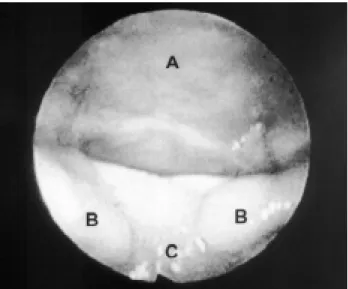
Figure 1 - Type 1 of airway obstruction revealed through nasopharyngoscopy
A - Pharynx; B - Bifid uvula; C - Tongue.
Figure 2 - Type 2 of airway obstruction A - Pharynx; D - Palate.
Methods
The present article is a review of the most recent publications about RS available from databases such as MEDLINE, Lilacs, and SciELO, from textbooks, and mainly from articles published by researchers from HRAC-USP, dealing with its etiopathogenesis and treatment.4-9 The article concludes by proposing a single treatment protocol, which can be applied to all RS cases.
Morphological and genetic aspects
In 1934,10 Pierre Robin described the failure of the tongue to descend, which caused airway obstruction; he also described a cleft palate, which is an aggravating factor in infants and children. In 1976, it was agreed that it was not a specific syndrome (Pierre Robin syndrome, as it was called for many years), but a non-specific combination of symptoms that may occur in several situations: in isolation, associated with a known syndrome, or associated with other developmental disabilities, which conjointly do not correspond to a specific syndrome.11 Other authors suggested the name Robin sequence since they believe manifestations occur sequentially.12 It is currently known as isolated Robin sequence (IRS) when it occurs alone.
In 1999, the use of the term Robin sequence for airway obstruction types 3 and 4 was questioned, since in these cases, glossoptosis does not cause airway obstruction. The term Robin complex was considered more appropriate for these cases.13
The etiology of IRS has been discussed by many authors throughout the years. Some posited theories about abnormal intrauterine positions,11,14-16 which caused micrognathia and posterior displacement of the tongue with consequent obstruction of palatal closure; other authors, although they do not support the theories above, do not regard inheritability as a relevant determining factor for the disease.17 Some isolated RS cases with family history have been described in the literature.18,19 A study carried out at HRAC-USP including 36 infants with IRS suggested inheritability as a relevant factor for the etiopathogenesis of the triad of anomalies.8 Some authors suggest cleft palate as a primary event in the etiopathogenesis of IRS instead of micrognathia.8,20
The genetic syndrome that most frequently co-occurs with RS is Stickler syndrome, also known as hereditary arthroophthalmopathy.4,5,21 In this syndrome, RS results from an intrinsic mandibular hypoplasia caused by poor penetration of the connective tissue through the palate.22,23
A study conducted at HRAC-USP with 159 infants with RS reported the most frequent syndromes associated with this anomaly4 (Table 1).
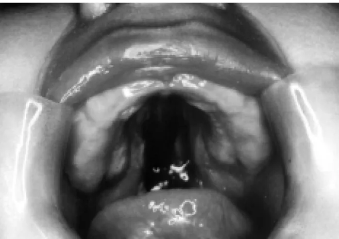
Ninety percent of RS cases revealed a cleft palate; in 70% of these cases, the clefts were complete, wide and U-shaped (Figure 5), and in 30%, they were complete or incomplete, narrow and V-shaped7 (Figure 6).
The literature reports an RS incidence between 1:2,00016 and 1:30,00024 live births. A more controlled study conducted in England suggests an incidence of 1:8,500 live births.25
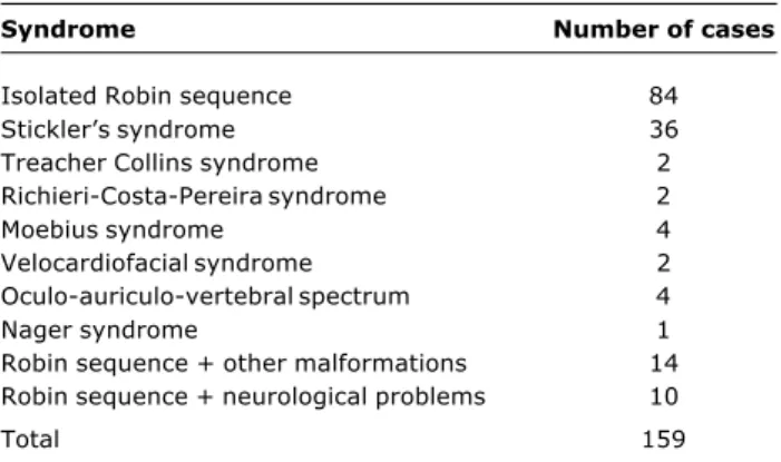
Table 1 - Syndromes associated with RS in a study4 conducted at HRAC-USP with 159 infants
Syndrome Number of cases
Isolated Robin sequence 84
Sticklers syndrome 36
Treacher Collins syndrome 2
Richieri-Costa-Pereira syndrome 2
Moebius syndrome 4
Velocardiofacial syndrome 2
Oculo-auriculo-vertebral spectrum 4
Nager syndrome 1
Robin sequence + other malformations 14 Robin sequence + neurological problems 10
Total 159
Nasopharyngoscopy: clinical implications
The importance of nasopharyngoscopy for the treatment of RS is highlighted in the literature. The different types of airway obstruction were classified into 1, 2, 3 and 4 (Figures 1, 2, 3 and 4), as previously described. Nasopharyngoscopic findings were correlated with prognosis and type of treatment.2,9
Type 1, which truly represents glossoptosis as the cause for airway obstruction, is the most frequent type observed in RS.2,4,5 Studies carried out at HRAC-USP revealed that this type of obstruction occurs in 80% of the cases.4,5. The literature describes its best prognosis, in which 61% of the cases present with IRS.2,4,5 Despite the heterogeneous clinical manifestations in this patient group, prone positioning and long-term nasopharyngeal intubation may reduce respiratory discomfort in 83%, without the need for surgical intervention.4,5 All cases with type 3 or 4 and most cases with type 2 obstruction revealed genetic syndromes, neurological disorders or other malformations associated with RS. Among type 2 cases, 50% required tracheostomy for alleviation of respiratory discomfort, and among type 3 and 4 cases, tracheostomy was the only treatment that allowed the relief of severe respiratory discomfort; attempts to use other types of treatment for the latter cases would only prolong hospital stay, causing problems to infants and their families.4,5 Glossopexy, for reduction of respiratory discomfort, is only recommended for type 1 cases that do not resolve after nasopharyngeal intubation after a maximum period of 15 days.4,5
In order to study the correlation between the severity of glossoptosis assessed by way of nasopharyngoscopy (following the criteria described above), and the severity of clinical manifestations, clinical criteria were established, as follows: mild slight breathing difficulty, absence of intercostal or furcular retraction, without cyanotic or apneic spells, oxygen saturation (SatO2) measured by continuous pulse oximetry greater than 90% and slight feeding difficulty (oral feeding only); moderate presence of respiratory effort with intercostal or furcular retraction, without cyanotic or apneic spells, SatO2 greater than 90% and remarkable feeding difficulty (use of feeding tubes); severe presence of cyanotic or apneic spells, SatO2 less than or equal to 90% and remarkable feeding difficulty. However, no statistically significant correlation was observed between severity of glossoptosis and severity of clinical manifestations in the neonatal period.9 The same patients prospectively followed up until the end of their first year of life by way of nasopharyngoscopy and clinical examination showed less severe glossoptosis and less severe clinical manifestations with the advance of age; the correlation between advanced age and reduction of glossoptosis was statistically significant. These infants were virtually asymptomatic at six months of
age. Despite reduction of the posterior displacement of the tongue, at the age of one year, some infants had moderate or severe glossoptosis, but were asymptomatic.9
With regard to other types of obstruction, infants with type 2 obstruction in the neonatal period presented with type 1 obstruction at the end of the first year, with mild clinical manifestations, whereas infants with type 3 or 4 showed the same type of obstruction throughout their first year of life, having to live with a tracheostomy, with no possibility of decannulation.9
Figure 5 - U-shaped cleft palate
Figure 6 - V-shaped cleft palate
During the neonatal period, nasopharyngoscopy was an important procedure for the diagnosis of the type of airway obstruction and for the planning of RS treatment; therefore, to define the prognosis of the severity of airway obstruction and of clinical outcome, nasopharyngoscopy is important only when the different types of obstruction are compared, with a worse prognosis for types 3 and 4. When the intention is to establish the prognosis of the severity of type 1 obstruction, nasopharyngoscopy is not a good method, probably due to the intrinsic activity of the genioglossus muscle, which pulls the tongue forward; since it is a static test, nasopharyngoscopy is not able to measure this parameter.
At HRAC-USP, infants with a cleft palate often are submitted to palatoplasty at 12 months of life. Edema of the palate and tongue secondary to surgical manipulation are commonly observed after this procedure; consequently, respiratory discomfort right after surgery is a frequent clinical manifestation, especially among infants. Postoperative consequences may be severe in infants who still have glossoptosis, causing remarkable respiratory discomfort.
The follow-up of infants by means of serial nasopharyngoscopy to determine the position of the tongue in relation to the hypopharynx and to the velopharyngeal region, can provide information about the risks of postoperative respiratory complications. Indication of palatoplasty in RS should be mandatorily preceded by nasopharyngoscopy in order to determine the presence of remarkable glossoptosis, since some infants still have moderate or severe glossoptosis at the end of their first year of life. In these cases, it would be sensible to await growth and development, which is quick during the first two years of life, so that surgical closure of the palate can be made more safely. For tracheostomized infants, palatal closure should always be performed before decannulation.
Treatment of respiratory difficulties
The clinical expression of RS is quite heterogeneous, ranging from mild breathing difficulty to severe episodes of asphyxia; the cases can occur in isolation or be associated with different genetic syndromes, showing variable expression, but airway obstruction is a factor that is common to all cases. Obstruction should be alleviated in order to improve respiratory conditions, but also growth, development, feeding and nutritional conditions, as respiratory discomfort becomes more severe the younger the infant is, and the younger the infant, the more susceptible he/she is to respiratory and nutritional complications.
The priority in RS treatment should be the maintenance of airway permeability. Without proper treatment, chronic hypoxia with CO2 retention and increase of pulmonary vascular resistance may lead to cor pulmonale. Moreover, recurrent episodes of cyanosis may result in cerebral hypoxia.
Feeding difficulty is of the consequences of breathing difficulty; the necessity of tube feeding is frequent among these patients. However, by improving respiratory
conditions, one may minimize feeding difficulty, allowing for oral feeding.
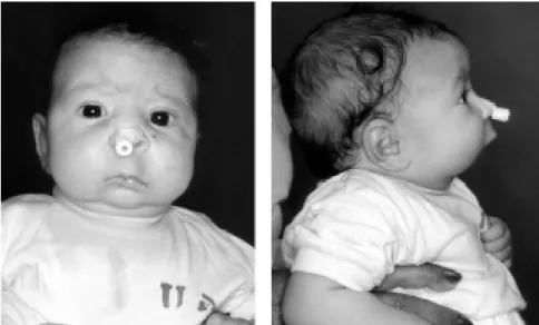
Several types of treatment are described in the literature, but a consensus about their use and efficacy has not been reached yet. HRAC-USP has developed studies whose aim is to standardize the indication and use of these different types of treatment.4,5 The most commonly used types of treatment are: prone positioning (PP) the infant is placed in ventral decubitus; nasopharyngeal intubation (NPI) placement of a silicone orotracheal tube measuring 3 to 3.5 mm in diameter, inserted from 7 to 8 cm through the nostril into the pharynx and cut 1 cm outside the nostril (Figures 8A and 8B); surgical procedures glossopexy, tracheostomy and mandibular distraction, as described next.
At HRAC-USP, PP is used only when the infant has mild breathing and feeding difficulties.4,5 Videofluoroscopic evaluation demonstrated that infants improved their ability to control tongue and jaw movements in this position; improvement in respiratory condition was not only attributed to this position, but mainly to neck extension.26
Long-term NPI is used at HRAC-USP in severe cases, with episodes of cyanosis, apnea or pallor and/or remarkable respiratory effort and/or decrease in SatO2 to a value less than or equal to 90%, with remarkable feeding difficulty.4,5 This type of treatment is advocated by several authors, due to its simplicity.2,5,27,28 Infants treated with NPI revealed greater weight gain than those treated with PP,28 and NPI is more efficient in infants younger than 30 days of life.2,5 A prospective study carried out at HRAC-USP with 159 RS cases showed that NPI resolved airway obstruction in 25% of all cases and in 50% of severe cases. Seventy-three percent of RS patients and 83% of IRS patients improved with PP or NPI, without the need of surgical procedures.4
After insertion of the tube for NPI, only clinical evaluation is necessary in order to observe the adequacy of this procedure. The feeding tube should allow airflow (noted during expiration), but should not allow the backflow of milk or saliva, especially during feeding; if fluids flow back, the tube should be repositioned 0.5 to 1 cm outwards, since it may be located too close to the esophagus. Using this technique, it is possible to keep the infant comfortable without the necessity of x-rays in order to reposition the tube. The aims of NPI are to maintain a good respiratory pattern by reducing respiratory effort, to maintain SatO2 greater than 90%, to reinforce the acceptance of oral foods, consequently reducing the duration of tube feeding, and to promote weight gain. If these aims are not attained within 15 days, glossopexy is indicated only for those cases with type 1 airway obstruction; cases of type 2 obstruction should be submitted to tracheostomy. NPI is not indicated for type 3 cases, in which tracheostomy should be immediately performed. A waiting period of 15 days is important, as this may avoid surgical procedures, but longer periods may lengthen hospital stay and the use of tube feeding and its severe consequences.5
running water, cotton-tipped swabs, and soap) does not imply cyanotic or apneic episodes, due to minimization of airway obstruction with neuromotor development.5
Airway obstruction in RS does not depend only upon the abnormal anatomy of the jaw and/or tongue position, but also upon the intrinsic effectiveness of the parapharyngeal muscles. Such effectiveness depends on individual maturation during the neonatal period.3 The level of neuromuscular dysfunction and the speed of maturation of this function varies among patients and plays a key role in the recovery of airway permeability.2 The development of the function of parapharyngeal muscles is faster than mandibular development.26 All previously described factors allowed for the temporary use of NPI as single treatment option in a large number of RS cases.4,5
The simplicity of NPI allows it to be managed at home by the parents, who are previously trained by nurses at HRAC-USP during the hospitalization period. After hospital discharge, a monthly one-day hospitalization is necessary to monitor respiratory and feeding patterns, when the feeding tube is removed until it is possible to discontinue its use, but it should be noted that there may be some deterioration of the respiratory pattern during sleep, due to relaxation of the tongue and of the parapharyngeal muscles.5 The nasopharyngeal tube should be permanently removed when discontinuation of its use for a long period (24 hours) does not result in cyanotic or apneic episodes, a decrease in SatO2 to values less than or equal to 90% or refusal of oral feeding. Weight gain should be monitored on a monthly basis.5 At HRAC-USP, the average time of NPI use was 60 days, resulting in exclusively oral feeding and satisfactory growth.5
Efficacy of NPI is higher in IRS cases with type 1 airway obstruction; NPI was efficient in only 50% of the cases with
type 2 obstruction, as these were more severe cases, often associated with genetic syndromes.4,5
Glossopexy currently used at HRAC consists of the attachment of the tongue to the lower lip and to the jaw.29 This surgery is indicated only for type 1 cases that do not improve with NPI, being a definitive treatment in 10.7% of the cases.4
At HRAC-USP, tracheostomy is indicated only for cases with type 1 airway obstruction that do not improve with glossopexy, type 2 cases that do not improve with NPI, and is totally indicated for type 3 or type 4 cases, which usually are extremely severe, 100% of them being associated with genetic syndromes, neurological disorders or other malformations.4 Tracheostomy was the definitive treatment in 15.7% of the cases.4
Mandibular distraction is not performed at HRAC-USP, and for now, it is not included in its treatment protocol. In this surgical procedure, the placement of an appropriate distractor at the mandibular angle allows forward positioning of the jaw and, consequently, of the tongue, in an attempt to keep the airways open. Several studies have been conducted to improve this technique in newborns.13,30-36 However, there is no consensus about its risks and benefits in newborns. Given the extensive experience of HRAC-USP in NPI and the number of studies carried out at this institution,4 only 26.4% of RS cases require surgical procedures. Considering the surgical and anesthetic risks for newborns and young infants, mandibular distraction is believed to have a restricted indication, being dependent on previous nasopharyngoscopic evaluation. It is only indicated for cases with type 1 airway obstruction that do not improve with NPI as an alternative to glossopexy and/or before indication of tracheostomy, as the latter procedure is not free from complications and sequelae.37-41
Despite the heterogeneity of RS, its prognosis is usually good. Mortality corresponds to approximately 7%; however, by analyzing these cases, it was observed that they were extremely severe, and that all of them were submitted to tracheostomy due to the severity of airway obstruction and that 50% showed other malformations associated with RS, which were incompatible with life and resulted in death.4,5
Treatment of feeding difficulties
Infants with RS do not present with only breathing difficulty, but also with feeding difficulty. Breathing difficulty leads to incoordination of sucking, swallowing and breathing. Besides this type of incoordination, glossoptosis hinders the forward positioning of the tongue, which is necessary for adequate suction, and cleft palate causes a deficit in negative intraoral pressure, resulting in inefficient suction as well as in nasal regurgitation, thus favoring bronchial aspiration.42
Feeding difficulty in infants with RS often hampers oral feeding, consequently resulting in the necessity for tube feeding which, in its turn, increases the risk of pathological gastroesophageal reflux.43-45 These infants already show a predisposition for such pathology due to the increase in negative intrathoracic pressure resulting from respiratory effort.45,46
Some authors reported that 37% of RS patients required the use of feeding tubes for at least 12 weeks.47 At HRAC-USP, some techniques that help the oral feeding of infants with IRS have been developed. These techniques are known as Speech Therapy Techniques for Facilitating Feeding (STTFF), and are called so because they are applied by a speech therapist. A study conducted at our hospital showed that by applying these techniques on a daily and gradual basis, it is possible to promote oral feeding in a short period of time (7 days) and to discontinue the use of the nasogastric tube.48 These techniques consist of the stimulation of non-nutritive sucking by the use of a pacifier, massage to relax and pull the tongue forward, manual support to sustain the jaw, long and soft bottle nipple with a 1-mm hole, placement of the bottle nipple exactly on the tongue, symmetric global posture and rhythmic movements of the nipple in the oral cavity during nutritive sucking. The nasogastric tube is removed when the infant accepts oral feeding, with a mean volume of milk per feeding of approximately 70% of the volume recommended for the age, ingested at a mean time less than 30 minutes, without intercurrent events such as gagging, cyanosis or coughing.48
It has been demonstrated that the average volume of milk ingested orally by infants with RS is lower than the volume of milk ingested by healthy infants belonging to the same age group.49 The low ingested volume and high energy expenditure during oral feeding indicate the need for calorie supplementation in order to provide a proper weight gain.5,49
The study of growth (weight and length) from birth to six months of life of 15 infants with RS, exclusively treated with NPI, STTFF and an age-appropriate diet, showed that these infants exhibited a catch-up growth for length reaching the
50th percentile (P50) of the NCHS curve50 in the fifth month of life. Nevertheless, weight remained below the 10th percentile (P10) throughout the study period, showing appropriate protein intake and insufficient calorie intake, suggesting calorie supplementation for improvement of the nutritional status.5
Besides STTFF, another strategy used at HRAC-USP to help the oral feeding of infants with RS is the use of a special diet for newborns and infants, which requires lower intake of milk for weight gain, allowing for early removal of the nasogastric tube.6,49 This hypercaloric diet consists of a milk-based formula or, whenever possible, breastmilk, to which 5 to 8% of glucose polymers and 3 to 5% of medium-chain triglycerides, with essential fatty acids, are added.
A study conducted at HRAC-USP with two groups of orally fed infants treated with NPI and STTFF, one receiving an age-appropriate diet and the other one receiving a hypercaloric diet, revealed a faster growth speed in the latter group, which in its turn, had an average time of NPI use significantly lower (25 days) compared to the former group (60 days), showing that nutritional recovery was important for improvement of respiratory capacity.6 Maturation and improvement of neuromuscular dysfunction are reliant on nutritional recovery during the first months of life, suggesting that the improved intrinsic activity of the genioglossus and parapharyngeal muscles obtained with dietary intervention through hypercaloric diet is responsible for the improved respiratory pattern of infants with RS.6
Hypercaloric diet and STTFF are not beneficial only to patients treated with NPI or PP, but also to infants submitted to surgical treatments, as the promotion of oral feeding should be one of the main objectives of RS treatment.
When the posterior displacement of the tongue is very pronounced, without quick resolution after STTFF, and when this situation is not always accompanied by respiratory discomfort indicating the need for surgical procedures to keep the airways open, glossopexy may be indicated, not to improve breathing but to facilitate suction, by allowing infants to retract their tongue and press it up against the bottle nipple, thus enabling them to feed orally.
Single treatment protocol
The following single treatment protocol, applied to all RS cases, regardless of their severity, was designed at HRAC-USP.
Nasopharyngoscopy performed on the first days of hospitalization in all cases for the diagnosis of the type of airway obstruction and planning of the treatment.
Prone positioning (PP) for cases with type 1 or type 2 airway obstruction with breathing difficulty.
Nasopharyngeal intubation (NPI) for cases with type 1 or type 2 airway obstruction that have cyanotic episodes, apnea, pallor, remarkable respiratory effort and/or decrease in que SatO2 measured by continuous pulse oximetry for values less than or equal to 90%.
not improve with NPI for at most 15 days and for those cases with mild respiratory discomfort and severe posterior displacement of the tongue that are not able to feed orally after STTFF for at most 30 days.
Tracheostomy for all cases with type 3 or 4 obstruction, for cases with type 2 obstruction that show no improvement with NPI for at most 15 days, and for type 1 cases that show no improvement with glossopexy.
Speech therapy techniques for facilitating feeding (STTFF) for all cases with type 1 or type 2 obstruction treated with PP or NPI, and for more complex cases only after maintenance of airway permeability by surgical treatment (glossopexy or tracheostomy).
Hypercaloric diet for all orally fed cases with or without supplementation of the prescribed volume by means of tube feeding, regardless of the type of airway obstruction.
Growth monitoring all cases.
Anti-GER (gastroesophageal reflux) drug all cases with long-term use of nasogastric tube (longer than 30 days).
Feeding gastrostomy all cases with severe dysphagia and use of nasogastric tube for more than three months without improvement after STTFF, associated or not with fundoplication, depending on the presence and severity of gastroesophageal reflux.
Serial nasopharyngoscopy performed every six months after 12 months of life until it is possible to perform palatoplasty.
Palatoplasty after 12 months of life, depending on previous nasopharyngoscopic assessment, indicated only for well-nourished infants with or without mild glossoptosis. In tracheostomized infants, palatoplasty should be performed after 12 months of life before decannulation.
Conclusions
Nasopharyngoscopy should be performed in the first months of life in all RS cases for the diagnosis of the type of airway obstruction and for planning of the treatment; it should be regularly performed after 12 months of life for determining the best moment for palatoplasty, in order to prevent postoperative respiratory complications.
The great novelty in RS treatment was the large experience acquired by HRAC-USP in long-term NPI. This procedure, when properly indicated, can alleviate respiratory discomfort in newborns and young infants, reducing the indication of surgical procedures for maintenance of airway permeability. STTFF and hypercaloric diet are also innovative techniques for RS treatment, allowing for oral feeding and nutritional recovery, and also for a decrease in the necessity for surgical procedures.
Despite the heterogeneity of clinical manifestations and complexity of the cases, the studies performed showed that a multidisciplinary team (pediatrician, surgeon, otolaryngologist, nutritionist, speech therapist, nurse, among others) should be involved in giving assistance to infants with RS. By following a single treatment protocol, it is possible to meet the needs of all cases, considering that RS
is not only an anatomic obstructive disorder to be resolved with surgical procedures. Knowledge about growth and development should be applied, as it allows for the quick recovery of airway permeability and of oral feeding, often avoiding surgical procedures and their risks, especially in newborns and young infants.
References
1. Elliott MA, Studen-Pavlovich DA, Ranalli DN. Prevalence of selected pediatric conditions in children with Pierre Robin sequence. Pediatr Dent. 1995;17:106-11.
2. Sher AE. Mechanisms of airway obstruction in Robin sequence: implications for treatment. Cleft Palate Craniofac J. 1992;29: 224-31.
3. Sher AE, Shprintzen RJ, Thorpy MJ. Endoscopic observations of obstructive sleep apnea in children with anomalous upper airways: predictive and therapeutic value. Int J Pediatr Otorhinolaryngol. 1986;11:135-46.
4. Marques IL, Sousa TV, Carneiro AF. Large experience with infants with Robin sequence: a prospective study on 159 cases. In: Lilja J, editor. Transactions 9th International Congress on Cleft Palate and Related Craniofacial Anomalies; 2001 25-29 June; Goteborg, Sweden. Goteborg: s.n.; 2001. p. 81-7. 5. Marques IL, Sousa TV, Carneiro AF, Barbieri MA, Bettiol H,
Gutierrez MR. Clinical experience with infants with Robin sequence: a prospective study. Cleft Palate Craniofac J. 2001;38:171-8.
6. Marques IL, Peres SP, Bettiol H, Barbieri MA, Andrea M, Souza L. Growth of children with isolated Robin sequence treated by nasopharyngeal intubation: importance of a hypercaloric diet. Cleft Palate Craniofac. 2004;41:53-8.
7. Marques IL. Crescimento de crianças portadoras de seqüência de Robin isolada de zero a 1 ano de idade [tese]. Ribeirão Preto: Faculdade de Medicina de Ribeirão Preto, Universidade de São Paulo; 1995.
8. Marques IL, Barbieri MA, Bettiol H. Etiopathogenesis of isolated Robin sequence. Cleft Palate Craniofac J. 1998;35:517-25. 9. Sousa TV, Marques IL, Carneiro AF, Bettiol H, Freitas JA.
Nasopharyngoscopy in Robin sequence: clinical and predictive value. Cleft Palate Craniofac J. 2003;40:618-23.
10. Robin P. Glossoptosis due to atresia and hypotrophy of the mandible. Am J Dis Child. 1934;48:541-7.
11. Cohen Jr MM. The Robin anomalad: its nonspecificity and associated syndromes. J Oral Surg. 1976;34:587-93. 12. Pasyayan HM, Lewis MB. Clinical experience with the Robin
sequence. Cleft Palate J. 1984;21:270-6.
13. Williams JK, Maull D, Grayson BH, Longaker MT, McCarthy JG. Early decannulation with bilateral mandibular distraction for tracheostomy-dependent patients. Plast Reconstr Surg. 1999;103:48-59.
14. Cocke Jr W. Experimental production of micrognathia and glossoptosis associated with cleft palate (Pierre Robin syndrome). Plast Reconstr Surg. 1966;38:395-403.
15. Latham RA. The pathogenesis of cleft palate associated with the Pierre Robin syndrome. An analysis of a seventeen-week human foetus. Br J Plast Surg. 1966;19:205-14.
16. Poswillo D. The aetiology and surgery of cleft palate with micrognathia. Ann R Coll Surg Engl. 1968;43:61-88.
17. Edwards JR, Newall DR. The Pierre Robin syndrome reassessed in the light of recent research. Br J Plast Surg. 1985;38:339-42. 18. Becker R, Palm D. Zur kausalen und formalen genese des Pierre
Robin syndrome. Dtsch Zahnarztl Z. 1966;21:1321-38. 19. Tan KL. The Pierre-Robin syndrome. J Singapore Paediatr Soc.
1968;10:88-94.
20. Cohen SR, Chen LL, Burdi AR, Trotman CA. Patterns of abnormal myogenesis in human cleft palates. Cleft Palate Craniofac J. 1994;31:345-50.
21. Shprintzen RJ. The implications of the diagnosis of Robin sequence. Cleft Palate Craniofac J. 1992;29:205-9.
Corresponding author: Ilza Lazarini Marques
Unidade de Ensino e Pesquisa do HRAC-USP Rua Silvio Marchione, 3/20
CEP 17043-900 Bauru, SP Brazil
Fax: +55 (14) 3235.8162/3265.1306 E-mail: ilza_marques@uol.com.br 23. Turner G. The Stickler syndrome in a family with the Pierre Robin
syndrome and severe myopia. Aust Paediatr J. 1974;10:103-8. 24. Salmon MA. Developmental defects and syndromes. Aylesbury:
HMM; 1978.
25. Bush PG, Williams AJ. Incidence of the Robin anomalad (Pierre Robin syndrome). Br J Plast Surg. 1983;36:434-7.
26. Takagi Y, Bosma JF. Disability of oral function in an infant associated with displacement of the tongue: therapy by feeding in prone position. Acta Paediatr Scand. 1960;49(Suppl):62-9. 27. Freeman MK, Manners JM. Cor pulmonale and the Pierre Robin anomaly: airway management with a nasopharyngeal tube. Anaesthesia. 1980;35:282-6.
28. Heaf DP, Helms PJ, Dinwiddie R, Matthew DJ. Nasopharyngeal airways in Pierre Robin syndrome. J Pediatr. 1982;100:698-703.
29. Argamaso RV. Glossopexy for upper airway obstruction in Robin sequence. Cleft Palate Craniofac J. 1992;29:232-8.
30. Denny A, Kalantarian B. Mandibular distraction in neonates: a strategy to avoid tracheostomy. Plast Reconstr Surg. 2002;109:896-904.
31. Denny AD, Talisman R, Hanson PR, Recinos RF. Mandibular distraction osteogenesis in very young patients to correct airway obstruction. Plast Reconstr Surg. 2001;108:302-11. 32. Dogliotti PL, Nadal E. Distracción de mandíbula em la asociación
retrognatia glosoptosis (síndrome de Pierre Robin). Rev Cir Infant. 1999;9:94-6.
33. Judge B, Hamlar D, Rimell FL. Mandibular distraction osteogenesis in a neonate. Arch Otolaryngol Head Neck Surg. 1999;125: 1029-32.
34. McCarthy JG. The role of distraction osteogenesis in the reconstruction of the mandible in unilateral craniofacial microsomia. Clin Plast Surg. 1994;21:625-31.
35. Morovic CG, Monasterio L. Distraction osteogenesis for obstructive apneas in patients with congenital craniofacial malformations. Plast Reconstr Surg. 2000;105:2324-30. 36. Sidman JD, Sampson D, Templeton B. Distraction osteogenesis
of the mandible for airway obstruction in children. Laryngoscope. 2001;111:1137-46.
37. Chew JY, Cantrell RW. Tracheostomy: complications and their management. Arch Otolaryngol. 1972;96:538-45.
38. Joseph HT, Jani P, Preece JM, Bailey CM, Evans JN. Paediatric tracheostomy: persistent tracheo-cutaneous fistula following decannulation. Int J Pediatr Otorhinolaryngol. 1991;22:231-6. 39. Rhee CK, Miller FR, Tucker HM, Eliachar I. The superiorly based flap long-term tracheostomy in pediatric patients. Am J Otolaryngol. 1996;17:251-6.
40. Singer LT, Kercsmar C, Legris G, Orlowski JP, Hill BP, Doershuk C. Developmental sequelae of long-term infant tracheostomy. Dev Med Child Neurol. 1989;31:224-30.
41. Walner DL, Holinger LD. Supraglottic stenosis in infants and children: a preliminary report. Arch Otolaryngol Head Neck Surg. 1997;123:337-41.
42. Wolf BG, Glass RP. Feeding and swallowing disorders in infancy: assessment and management. Tucson: Therapy Skill Builders; 1992.
43. Baptista EN. Refluxo gastroesofágico na clínica de fonoaudiologia. In: Marchesan IQ, Zorzi JL, Gomes, ICD, organizador. Tópicos em fonoaudiologia 1996. São Paulo: Lovise, 1996. v. 3. p. 563-71.
44. Gomes GF, Pisan JC, Macedo ED, Campos AC. The nasogastric feeding tube as a risk factor for aspiration and aspiration pneumonia. Curr Opin Clin Nutr Metab Care. 2003;6:327-33. 45. Monteiro LC. Refluxo gastroesofágico. In: Mastroti TA, Chiara
NV. Clínica cirúrgica e urologia em pediatria. São Paulo: Robe; 1997. p. 207-22.
46. Dudkiewicz Z, Sekula E, Nielepiec-Jalosinska A. Gastroesophageal reflux in Pierre Robin sequence: early surgical treatment. Cleft Palate Craniofac J. 2000;37:205-8.
47. Cruz MJ, Kerschner JE, Beste DJ, Conley SF. Pierre Robin sequences: secondary respiratory difficulties and intrinsic feeding abnormalities. Laryngoscope. 1999;109:1632-6.
48. Nassar E. Proposta de técnicas fonoaudiológicas facilitadoras da alimentação do lactente portador de sequência de Pierre Robin [dissertação]. Bauru: Hospital de Reabilitação de Anomalias Craniofaciais, Universidade de São Paulo; 2002
49. Peres SP, Arena EP, Moreira FL, Marques IL. Importância da intervenção dietética no estado nutricional de lactentes portadores de seqüência de Robin. Rev Bras Nutr Clin. 2002;17:15-9.