29
AbstractObjective: To evaluate, sequentially, tracheal aspirates from patients admitted to a pediatric intensive care unit and to associate these pathogens with length of hospital stay, previous use of antimicrobial therapy and diagnoses of ventilator-associated pneumonia.
Methods: The study population consisted of patients admitted to a pediatric intensive care unit, between November 2002 and December 2003, on ventilator support. Three tracheal aspirates were collected serially from each patient. The first tracheal aspirate sample was obtained 6 hours after admission to the intensive care unit and the remaining samples were collected after 48 and 96 hours.
Results: One hundred patients aged from one day to 14 years were assessed. Positive tracheal cultures were observed to have increased in the three tracheal aspirate samples collected from each patient for Pseudomonas aeruginosa, from 6 to 22% (p = 0.002), and to have decreased for Staphylococcus aureus, from 23 to 8% (p = 0.009). Isolation of Candida spp increased for the subset that had received previous antimicrobial therapy (p < 0,05). Sixteen (23,5%) out of 68 patients admitted without pneumonia developed ventilator-associated pneumonia. Positive tracheal aspirate cultures were obtained in 10 out of 16 of these patients: six were positive for Staphylococcus aureus (threeassociated with Acinetobacter baumanii), two for Klebsiella spp (oneassociated with Enterobacter spp), one for Pseudomonas aeruginosa and one for Candida spp.
Conclusion: Sequential evaluation of tracheal aspirates may be useful to track changes in bacterial flora at pediatric intensive care units.
J Pediatr (Rio J). 2005;81(1):29-33: Pneumonia, ICU, bacterial resistance, Staphylococcus aureus.
Sequential microbiological monitoring
of tracheal aspirates in intubated patients
admitted to a pediatric intensive care unit
Cid E. Carvalho,1 Eitan N. Berezin,2 Ivan P. Pistelli,3 Lycia Mímica,4 Maria Regina A. Cardoso5 Copyright © 2005 by Sociedade Brasileira de Pediatria
1. Ph.D. MD, Pediatric Intensive Care Unit, Santa Casa de São Paulo. Assistant professor, Department of Pediatrics, School of Medicine, Santa Casa de São Paulo, SP, Brazil.
2. Adjunct professor, Department of Pediatrics, School of Medicine, Santa Casa de São Paulo. Chief of the Division of Pediatric Infectious Diseases, Santa Casa de São Paulo, SP, Brazil.
3. Assistant professor, Department of Pediatrics, School of Medicine, Santa Casa de São Paulo. Chief of the Pediatric Intensive Care Unit, Santa Casa de São Paulo, SP, Brazil.
4. Adjunct professor, Department of Microbiology, School of Medicine, Santa Casa de São Paulo and Director of the Laboratory of Hospital Infection Control.
5. Ph.D. Professor, Department of Epidemiology, School of Public Health, Universidade de São Paulo (USP), São Paulo, SP, Brazil.
Suggested citation: Carvalho CE, Berezin EN, Pistelli IP, Mímica L, Cardoso MR. Sequential microbiological monitoring of tracheal aspirates in intubated patients admitted to a pediatric intensive care unit. J Pediatr (Rio J). 2005;81:29-33.
Introduction
Pneumonia associated with mechanical ventilation (VAP) is one of the most important causes of nosocomial infections in pediatric intensive care units (PICU), as has been demonstrated by studies evaluating nosocomial infections in pediatrics.1-4
O
RIGINALA
RTICLEThe role of upper airway secretions contaminated by pathogens that drain to the subglottis in the pathogenesis of VAP in intubated patients on mechanical ventilation is already known. This fact has led to clinical and epidemiological investigations which have, in turn, contributed significantly to the understanding and management of patients suffering from VAP.5-8
It is estimated that the incidence of VAP in adults is greater than 10%.9 The incidence in children, estimated by the CDCs National Nosocomial Infections Surveillance study (NNIS), is 20%.5-7 Among pediatric patients, the greatest incidence occurs between 2 months and 1 year and the bacteria that is most often to blame for VAP is Pseudomonas aeruginosa.8
Controversy persists with respect of VAP, primarily in relation to diagnostic techniques and prevention strategies. The debate is the result of differences in the analysis of three important issues: the differentiation between colonization and infection, the interpretation of clinical signs and the use of antimicrobials in intensive care units.9
As a result of these facts several studies have been published aimed at preventing, identifying and treating intra-hospital infections of the lower respiratory tract. One of the methods employed is sequential monitoring of the tracheal secretions of intubated patients in the ICU environment.11-13 In a study of adults performed in Brazil, Carvalho et al.14 report sensitivity and specificity of around 70% for tracheal secretion cultures when compared with broncho-alveolar lavage. In another recent Brazilian study Camargo et al.15 found that tracheal secretion cultures had a good sensitivity, but low specificity for the diagnosis of VAP.
Assessing the literature it can be perceived that little data from pediatric patients is available on the problem.
The objective of the present study, therefore, is to perform a sequential study of the tracheal flora of patients in a pediatric ICU. Additionally, we attempt to associate this flora with certain variables, such as length of hospital stay, previous antimicrobial use and diagnosis of VAP.
Methods
The study population consisted of all pediatric patients who were intubated within 6 hours of admission to the pediatric ICU of the Sao Paulo Santa Casa, during the period between November 2002 and December 2003. The patients were selected irrespective of sex, diagnosis at point of admission, length of hospital stay or antimicrobial use.
When possible three sequential samples of tracheal secretion aspirate were collected from each patient. In addition to this, samples were taken for blood culture, urine culture, culture from the catheter tip and for any other culture necessary to investigate the case in question. The first tracheal secretion collection was performed within 6 hours of intubation and the subsequent samples were taken at regular intervals of 48 and 96 hours after the first. All samples were sent for microbiological culturing and antimicrobial sensitivity testing.
Participation was by consent in accordance with the provisions of the Code of Ethics for research on human beings.
Collections were made using sterile techniques employing a tracheal aspiration probe. The tracheal content was collected from as deep as possible a point with a closed system in vacuum directly into a Transbac® collection flask containing culture material. The collectors were sealed and sent to the Microbiology Service at the Medical Sciences Faculty of the Sao Paulo Santa Casa, where they were seeded and cultured.
The material was seeded in blood agar and anaerinsol-S. The Agar blood was incubated at 35±2 ºC, for 18 to 24 hours and after incubation the plate was analyzed and, in
the event there was no growth, re-incubated for a further 24 hours. Probable pathogens were tested for antimicrobial sensitivity according to the Kirby-Bauer method. When there was growth of more than one colony with apparently different morphologies these were isolated by reseeding each one onto fresh blood agar and, after further incubation, the procedure described above was repeated. Anaerobic cultures were grown on anaerinsol-S plates sealed into a jar with an anaerobiasis generator which allows both strict and facultative anaerobes to develop. This jar was incubated at 35±2 ºC for 48 hours. After this period the jar was opened and, if there was growth, a test for air tolerance was performed exposing the strain to room air for growth. If no such growth took place standard tests were performed to identify anaerobic organisms. The medium used for fungi was Saborauds.
The diagnostic criteria for VAP used in the study were: patient on mechanical ventilation for a minimum of 48 hours exhibiting progression or appearance of new alveolar infiltration on chest x-ray associated with thermal instability, leukopenia or Leukocytosis, the presence of purulent secretion and a need for supplementary oxygen.16
The chi-square test, Fishers exact test and the Mann-Whitney test were used for the statistical analysis to compare proportions between subsets of patients. Also calculated were relative risks (rr) and their 95% confidence intervals (95% CI) to analyze the relationship between pathogens isolated at the first and third collections. The software used for all analyses was SPSS-Version 10.0.
Results
Three hundred and eighty-nine patients were admitted to the pediatric ICU of the São Paulo Santa Casa during the study period. Of these 237 were not intubated during the first hours after admission and 52 of those who were intubated were then extubated before 6 hours had elapsed. Therefore, 100 (26%) patients were included in the study. Three sequential tracheal collections were made from 59 patients, two from 18 patients and one from 23 patients. The total number of three sequential collections could not be attained for 41 patients since 23 were no longer intubated at 48 hours and another 18 patients were extubated before 96 hours had passed.
The ages of the patients studied varied from one day to 14 years. Diagnoses on admission to the ICU were as follows: 35 cases of respiratory disease, 32 cases of post-operative care or trauma, 15 malformations or neurological diseases, nine of septicemia and eight cases with other diagnoses. Fifty-five of the patients had histories of antimicrobial use prior to ICU admission.
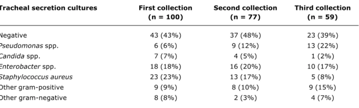
Pseudomonas spp. cultures, increasing from 6% at the first collection to 22% at the third (p = 0.002) with a relative risk (rr) of 2.08 (95% CI: 1.41 - 3.06). In contrast there was a significant decrease in cultures positive for Staphylococcus aureus from the first to the third collections, with percentages of 23 and 8% respectively (p = 0.009) while rr = 0.43 (95% CI: 0.19 - 0.98).
Of the 100 patients studied, 68 were admitted with no diagnosis of pneumonia. Sixteen (23.5%) of these developed VAP. The relation between these patients and tracheal secretion cultures and their antimicrobial resistance profiles is as follows: there were 10 positive cultures being six of S. aureus with three concomitant A. baumanni cultures, two of Klebsiella spp. with one concomitant Enterobacter spp. culture, one Candida spp. and one P.aeruginosa with four cultures of methicillin-resistant Staphylococcus aureus (MRSA) identified, two of ß-lactamase producing Klebsiella spp. and the P. aeroginosa culture was resistant to just carbenicillin.
Results of cultures collected concomitantly from other sites (blood cultures, urine cultures and catheter tip cultures) were as follows: 31 patients had positive cultures (18 blood cultures, five catheter tip cultures, and 15 urine cultures) with six blood cultures being the same as the findings from the tracheal secretion - four Staphylococcus aureus and two Candida spp. - and two urine cultures and one catheter tip culture were positive for Candida spp., also concomitant with the tracheal secretion and being from the same patients who had blood cultures positive for this microorganism.
Figure 1 demonstrates the microbiological profile of the first tracheal secretion culture for the groups of patients who had used antimicrobials for more than 48 hours before admission and for those who had not previously used antimicrobials. Applying the Mann-Whitney test to identify differences between the two groups revealed a significant increase in the presence of Candida spp. in the subset that had used antimicrobials previously (p < 0.05).
Discussion
For all three sequential samples there was a greater than 40% level of negative cultures irrespective of previous length of hospital stay or previous antimicrobial use. Earlier studies had indicated the possibility of negative results due to previous antimicrobial use and failures in the culturing technique.17,18 Notwithstanding, our study revealed higher levels of negative cultures for the three sequential samples than are described in the literature.12,17,19
No significant differences were observed with respect of negative cultures when the two groups of patients - those who had and those who hadnt received antimicrobials -were compared. The only pathogen for which a difference
Figure 1 - Comparison of the microbiological profile of the first tracheal secretion culture for the groups of patients who had used antimicrobials for more than 48 hours before admission to the PICU of Santa Casa de São Paulo and for those who had not previously used antimicrobials
Tracheal secretion cultures First collection Second collection Third collection
(n = 100) (n = 77) (n = 59)
Negative 43 (43%) 37 (48%) 23 (39%)
Pseudomonas spp. 6 (6%) 9 (12%) 13 (22%)
Candida spp. 7 (7%) 4 (5%) 1 (2%)
Enterobacter spp. 18 (18%) 16 (20%) 10 (17%)
Staphylococcus aureus 23 (23%) 13 (17%) 5 (8%)
Other gram-positive 9 (9%) 8 (10%) 9 (15%)
Other gram-negative 8 (8%) 2 (3%) 4 (7%)
25
21
13
10
5
2 1
2
6 6
22
20
55
10
5
Negative cultures
S. aureus Entero-bacter
Gram + Gram Candida
spp.
Pseudo-monas spp.
4 4
9 9
No antimicrobial therapy Antimicrobial therapy
was observed in terms of isolation between these two groups was Candida spp. with the group that had previously used antimicrobials presenting a greater number of isolations. This fact is due to the microorganisms predisposition towards colonization of mucosa because of the use of antimicrobials.17
When the results of the tracheal secretion cultures were associated with cultures collected concomitantly from other sites, six blood cultures grew the same pathogen as the tracheal secretion. Of these four were positive for Staphylococcus aureus and two for Candida spp. Two urine cultures positive for Candida spp. were also concomitant with the tracheal secretion, being from the same patients who had blood cultures positive for this microorganism. A finding of Candida spp. in tracheal secretions is more often associated with colonization, however, the cases described here are compatible with generalized fungal infection.20
Among the bacteria isolated during the first days after starting mechanical ventilation, there was a predominance
of Staphylococcus aureus, which, in the literature,
corresponds with around 20 to 30% of infections respiratory in PICUs and particularly so among those that manifest earlier.8
There was a significant increase in cultures positive for
Pseudomonas spp. which indicates that endotracheal
intubation breaks the barriers between the environment and the tracheal mucosa of patients allowing progressive colonization by Pseudomonas spp., which is a common pathogen in the ICU environment, as the length of intubation period increases.20,21 Pseudomonas spp. is a bacterium that is capable of surviving in nature with minimum nutritional requirements, being able to survive for long periods in humidifiers and solutions.11 The fact that colonization by Pseudomonas spp. increases as the patient remains longer in the ICU is an important finding since this bacteria is the largest cause of VAP out of the gram-negative bacteria and is the most associated with increased mortality.8,21
In this study the incidence of VAP was 23% (16 out of 68 patients), which is close to the 1610 and 29.1% levels found in literature.11 One of the agents isolated from the VAP patients was Staphylococcus aureus, in six out of 16 cases (37.5%), with four of these being strains resistant to oxacillin.
Gram-positive bacteria are becoming important pathogens in these situations, particularly Staphylococcus aureus, frequently resistant to oxacillin.8,12,13
Four (25%) of the 16 patients with VAP in our study had two different microorganisms isolated from the same sample of tracheal secretion which is in agreement with the literature.17,18
In our study there were difficulties with the diagnosis of VAP due to the difficulty of diagnostic standardization, specifically because the patients were pediatric. Two Brazilian studies have assessed the utility of tracheal secretions for etiologic VAP diagnosis.
The study undertaken by Camargo et al.15 compared the evaluation of tracheal secretions with qualitative and quantitative microbiological evaluation, concluding that
qualitative methods had greater sensitivity and quantitative methods greater specificity. Other studies, including one domestic study, have shown associations between the results of broncho-alveolar lavage and cultures from tracheal secretions.13,14
The colonization by pathogenic bacteria that occurs among patients during long hospital stays, particularly in PICU, represents a high risk for the patient and those who come into contact with them. We must remember that when these patients are discharged from the PICU they go to the general pediatric wards where they could become sources of contamination for other patients.22,23
We conclude that sequential monitoring of tracheal secretions could be of use in the evaluation of microbial flora as an indicator of the changes that take place in the intensive care unit environment.
References
1. Gilio AE, Stape A, Pereira CR, Cardoso MF, Silva CV, Troster EJ. Risk factors for nosocomial infections in a critically ill pediatric population: a 25-month prospective cohort study. Infect Control Hosp Epidemiol.2000;21:340-2.
2. Arantes A, da Silva Carvalho E, Medeiros EA, Farhat CK, Mantese OC. Pediatric risk of mortality and hospital infection. Infect Control Hosp Epidemiol. 2004;25:783-6.
3. Urrea M, Pons M, Serra M, Latorre C, Palomeque A. A prospective incidence study of nosocomial infections in a pediatric intensive care unit. Pediatr Infect Dis. 2003;22:490-3.
4. Milliken J, Tait GA, Ford-Jones EL, Mindorff CM, Gold R, Mullins G. Nosocomial infections in a pediatric intensive care unit. Crit Care Med. 1988;16:233-7.
5. Richards MJ, EdwardsJR, Culver DH, Gaynes RP. Nosocomial infections in pediatric intensive care in the United States. Pediatrics. 1999;103:39-45.
6. Singh-Naz N, Sprangue BM, Patel KM, Pollack MM. Risk factors for nosocomial infection in critically ill children: a prospective cohort study. Crit Care Med. 1996;24:875-8.
7. Niederman MS, Mantovani R, Schoch P, Papas J, Fein AM. Patterns and routes of tracheobronchial colonization in mechanically ventilated patients: the role of nutritional status in colonization of the lower airway by Pseudomonas species. Chest. 1989;95:155-61.
8. Elward AM, Warren DK, Fraser VJ. Ventilator-associated pneumonia in pediatric intensive care unit patients: risk factors and outcomes. Pediatrics. 2002;109:758-64.
9. Koleff MH. The prevention of ventilator associated pneumonia. NEJM 1999:340:627-41.
10. Johanson WG, PierceAK, Sanford JP, Thomas GD. Nosocomial respiratory infections with gram negative bacilli: the significance of the colonization of the respiratory tract. Ann Intern Med. 1972;77:701-6.
11. Rello J, Gallego M, Mariscal D, Sonora R, Valles J. The value of routine microbial investigation in ventilator-associated pneumonia. Am J Respir Crit Care Med. 1997;156:196-200. 12. Sanjai N, Paul B, Stephen B, Michael D, Mark L, Gary D.
Sampling variability in the microbiological evaluation of expectorated sputa and endotracheal aspirates. J Clin Microbiol. 2001;39:2344-7.
13. Fagon J, Chastre J, Hance AJ, Gujet M, Trouillet JL, Domart Y, et al. Detection of nosocomial pneumonia in ventilated patients. Am Rev Respir Dis. 1988;138:110-6.
14. Carvalho CF, Winkeler GF, Costa FA, Bandeira TJ, Pereira ED, Holanda MA. Concordância entre o aspirado traqueal e o lavado broncoalveolar no diagnóstico das pneumonias associadas à ventilação mecânica. J Bras Pneumol. 2004;30:26-38. 15. Camargo LF, De Marco FV, Hoelz C, Bueno MA, Rodrigues Jr M,
Corresponding author: Cid E. Carvalho
Av. do Guacá, 445/94, Bloco B CEP 02435-000 São Paulo, SP Brazil
Phone: +55 (11) 81262400 E-mail: [email protected] 16. Garner JS, Jarvis WR, Emori TG, Horan TC, Hughes JM. CDC
definitions for nosocomial infections. In: Olmsted RN, editor. APIC Infection Control and Applied Epidemiology: Principles and Practice. St Louis, MO: Mosby; 1996. p. A1-A2.
17. Olivier L, Timsit JF, Garrouste M, Misset B, Benali A, Carlet J. The Significance of distal bronchial samples with commensals in ventilator-associated pneumonia: colonizer or pathogen? Chest. 2002;122:1389-99.
18. Carrel TP, Eisinger E, Vogt M. Pneumonia after cardiac surgery is predictable by tracheal aspirates but cannot be prevented by prolonged antibiotic prophylaxis. Ann Thorac Surg. 2001;72: 143-8.
19. Mahul P, Auboyer C, Jospe R, Ros A, Guerin C, el Khouri Z, et al. Prevention of nosocomial pneumonia in intubated patients: respective role of mechanical subglottic secretions drainage and stress ulcer prophylaxis. Intensive Care Med.1992;18:20-5. 20. Fagon J, Lavarde V, Novara A. Nosocomial candida infections of
the lower respiratory tract in icu patients. Am J Resp Crit Care Med.1994;1(Suppl):A650.
21. Cordero L, Sananes M, Coley B, Hogan M, Gelman M, Ayers LW. Ventilator-associated pneumonia in very low-birth-weight infants at the time of nosocomial bloodstream infection and during airway colonization with Pseudomonas aeruginosa.AJIC. 2000:28:333-9.
22. George DL. Epidemiology of nosocomial ventilator-associated pneumonia. Infect Control Hosp Epidemiol.1993;14:163-9. 23. Klein BS, Perloff WH, Maki DG. Reduction of nosocomial infection