Abstract
Submitted: February 15, 2016 0RGL¿FDWLRQ-XO\ Accepted: July 5, 2016
Molecular and morphological surface
cleaning agents on root dentin
The quality of the dentin root is the most important factor for restoration resin sealing and drives the outcome of endodontic treatment. Objective: This
root dentin of primary teeth using Fourier-transformed Raman spectroscopy (FT-μ
electron microscopic (SEM) analysis. Material and Methods: Eighty roots of primary teeth were endodontically prepared and distributed into 4 groups and ®+zinc
oxide (CZ), Calcipex II® (CII), Vitapex®
were distributed to 4 subgroups according to cleaning agents (n=5): Control-no cleaning (C), Ethanol (E), Tergenform® (T), 35% Phosphoric acid (PA). Then, the roots were sectioned and the dentin root sections were internally evaluated by FT-Raman, μ-EDXRF and SEM. Data was submitted to two-way ANOVA and
D
difference in organic content. CP provided the lowest calcium values and, calcium/ phosphoric ratio (Ca/P), and the highest phosphoric values. For cleaning agents there was no difference in organic content when compared to the C; however,
similar results for phosphorus. The dentin smear layer was present after use
inorganic content, however they did not change the organic content. Cleaning agents did not alter the inorganic and organic content. PA cleaned and opened dentin tubules.
Keyw ords:
X-Ray Emission Spectrometry. Scanning electron microscopy.
Vanessa Benetello DAINEZI1
Alexsandra Shizue IWAMOTO1
Airton Abrahão MARTIN4
Luís Eduardo Silva SOARES2
Yumiko HOSOYA3
Fernanda Miori PASCON1
Regina Maria PUPPIN-RONTANI1
http://dx.doi.org/10.1590/1678-77572016-0053
1Universidade Estadual de Campinas, Faculdade de Odontologia de Piracicaba,
Departamento de Odontopediatria, Piracicaba, SP, Brasil.
2Universidade do Vale do Paraíba, Univap, Faculdade de Ciências da Saúde, Odontologia,
São José dos Campos, SP, Brasil, São José dos Campos, SP, Brasil.
3
Department of Oral Health and Development Sciences, Sendai, Japan.
4Universidade Federal do Piauí, Campus Ministro Petrônio Portella, Departamento de
Física , CCN Bairro Ininga, Teresina, PI, Brasil.
Corresponding address: Regina Maria Puppin-Rontani Avenida Limeira 901 - Vila Rezende Piracicaba - SP - Brazil - 13414-903 Phone: +55 19 2106 5286 - Fax: +55 19 2106 5218
Introduction
After root canal treatment of primary teeth, the tooth can be restored with an adhesive material, which 22. However, the
The composition of the materials, such as oily excipient and calcium hydroxide, would act as contaminants and decrease bonding strength in dentin9,12. In addition,
polymerized in clean dentin surface, for a successful retention of adhesive restoration15. Thus, in order to obtain satisfactory restoration longevity, the dentin surface has to be free of debris and with ideal features to allow for an adequate hybridization of the resin monomers and dentin5
and the restoration sealing are the most important factors for the endodontic treatment’s outcome6.
on severely damaged primary anterior teeth need to
Calcium hydroxide’s effect on the physical and sealing properties of canal sealers was investigated10. It was observed that Calcipex®
paste, composed of calcium hydroxide, barium sulfate and distilled water was easy to handle, and also easily removed10. In addition, Vitapex®, a silicon oil-based, composed of calcium hydroxide, iodoform and silicone
paste after cleaning the root surface. Therefore, it is supposed that Vitapex® and Calcipex® when used as
removed depending on the excipient contained. In order to clean and prepare the root canal surface for dentin bonding, cleaning agents can be used to
anionic detergent solution (sodium diethylene glycol lauryl ether sulfate) can be used to clean the dentin
liquids and suspension of the debris that results from root preparation. In addition, it could also 14. Ethanol is another
cleaning agent that can be used to remove some oil
is an organic solvent26. The use of ethanol on dentin promotes drying, inducing a hydrophobic dentin
of resin monomers to wet dentin, increasing resin retention23.
Phosphoric acid used as an etching agent is used in
the substrate surface during adhesive procedures, it can also Benetello considered as a surface cleaning agent that can clean dentin, since it removes the
peritubular dentin and exposes the collagen matrix without changing the intrinsic characteristics of the inorganic dentin content2,3.
of the canal preparation and cleaning procedures. It
modify the dentin surface18 and can jeopardize the bonding strength of adhesive restorative materials, compromising the endodontic treatment’s success.
agents, the evaluation of organic and inorganic content of dentin is an important factor for observing alterations in the dentin and could be helpful for selecting the best cleaning agent to be used in filled roots, without reducing adhesion. The calcium hydroxide-based pastes can alter root dentin properties such as hardness and modulus of elasticity29. In addition, depending on size and shape, the calcium hydroxide particles can penetrate into the dentin tubules11, occluding them and be detrimental to adhesion. On the other hand, cleaning agents, such
of dentin20. Therefore, anionic detergent solution removed the smear layer and opened the dentin tubule
17; its use combined with
phosphoric acid demineralizes the dentin, which could modify both the organic and the inorganic content16.
Therefore, the dentin structure and composition can be evaluated through sensitive analysis of the dentin’s chemical aspects to determine inorganic
transformed Raman spectroscopy (FT-Raman) and micro energy-dispersive X-ray fluorescence (μ-EDXRF ) are established methods for simple and nondestructive analyses for obtaining information with respect to molecular composition and substrate
μ
2940-2942 cm-1 band (C-H stretching) of the organic content1-3,13. Also, the scanning electron microscopic (SEM) can be used to observe details, post-treatment, of the root canal dentin surface of primary teeth.
Thus, the aim of this in vit ro study was to verify
agents on primary teeth root dentin. The hypothesis
with different cleaning agents, affect the molecular and surface features of the root canal dentin surface of primary teeth.
Material and methods
This study was conducted after receiving approval from The Ethics Committee in Research of the Dental School.
Sample selection
Eighty extracted, human anterior primary teeth were cleaned with saline solution to remove any remaining debris and soft tissue. The samples were frozen and stored for no more than 6 months until analysis. The selection criteria included those with at least two thirds of the root length and with no prior exposure to endodontic therapy.
Specimen preparation
The crowns were sectioned at the cementoenamel junction (CEJ) using a high-speed diamond saw with water-cooling and were subsequently discarded. Then, the roots were sectioned longitudinally, and only one slice from each root was selected, 80 specimens were obtained. The specimens were selected under a stereo
structural alterations were excluded. Thereafter, the
320-grit SiC paper, and sonicated for 60 seconds with deionized water to remove the residues from the lumen of the root canal of primary teeth.
Maillefer, Baillagues, Switzerland; batch number 8313220), simulating the clinical preparation of a
irrigated with 2 mL of 0.5% NaOCl solution (RioLab, Brazil; batch number CH95436). During preparation, EndoPTC (Biodinâmica Química e Farmacêutica Ltda.,
Ibiporã, PR, Brazil; batch number 83510) + 0.5% NaOCl solution was used20. The canals were dried with paper points.
The specimens were distributed into 4 groups
®
(1.0 g:1.0 g); Calcipex II® (CII); Vitapex® (V). The CZ paste was placed onto the lumen of the primary
directly on the lumen of the root canal with a
pre-stored at a relative humidity of 100% at 37°C for seven days so as to simulate a clinical procedure.
root canal were removed with the aid of curettes and
into 4 groups (n=5) according to cleaning agents: control group (C) (no cleaning agent); Ethanol (E):
10 seconds and rubbed onto the lumen of the root canal; Tergenform®
of the root canal; Phosphoric acid (PA): the lumen of the root canal was etched with 35% phosphoric acid for 15 seconds and later rinsed under water for 30 seconds. All specimens were dried with paper points. After that, they were stored at a relative humidity of 100% at 37°C until the molecular and morphological analyses were conducted (Figure 1B). The application times of E and T were established in a pilot study. All materials used (including manufacturer details) are described in Figure 2.
Molecular analysis
The cervical third of each lumen of the root canal was analyzed by Fourier-transformed Raman spectroscopy (FT-Raman) and μ-EDXRF spectroscopy in order to evaluate changes in the dentin’s organic and inorganic components, respectively. Both analytical methods were nondestructive and the data collection was based on laser or X-ray interactions with the sample, without contact.
FT-Raman spectroscopic analysis
Spectra of the samples were obtained using an
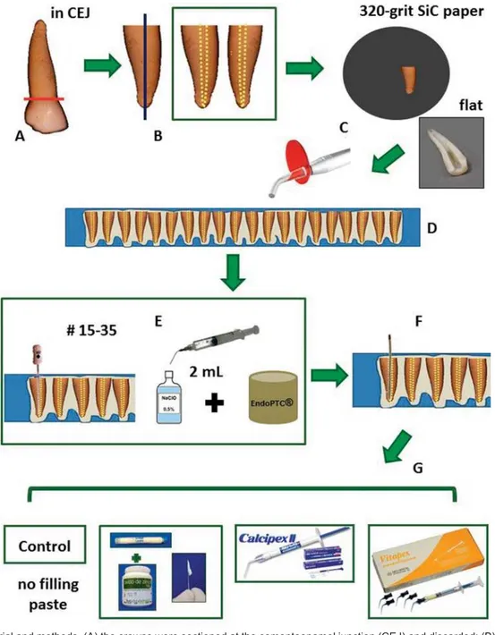
Figure 1A-0DWHULDODQGPHWKRGV$WKHFURZQVZHUHVHFWLRQHGDWWKHFHPHQWRHQDPHOMXQFWLRQ&(-DQGGLVFDUGHG%WKHURRWVZHUH
VHFWLRQHGORQJLWXGLQDOO\&WKHODWHUDOZDOOVZHUHJURXQGÀDWDQGVRQLFDWHGWRUHPRYHWKHUHVLGXHVIURPWKHOXPHQRIWKHSULPDU\WHHWK URRWFDQDO'WKHVSHFLPHQVZHUH¿[HGLQFRPSRVLWHUHVLQWKHOXPHQZDVZLWKRXWFRQWDFWZLWKUHVLQ(WKHOXPHQRIWKHURRWFDQDOZDV SUHSDUHGWRDZRUNLQJOHQJWKXQWLOWKHDSH[XVLQJ.¿OHVVL]HV%HWZHHQHDFK¿OHWKHOXPHQRIWKHFDQDOVZDVLUULJDWHGZLWK P/RI1D2&OVROXWLRQ'XULQJSUHSDUDWLRQ(QGR37&1D2&OVROXWLRQZDVXVHG)WKHFDQDOVZHUHGULHGZLWKSDSHUSRLQWV *WKHVSHFLPHQVZHUHGLVWULEXWHGLQWRJURXSVQ DFFRUGLQJWRWKH¿OOLQJSDVWH&RQWURO&3QR¿OOLQJ&DOHQ® paste thickened zinc
R[LGH&=JJ&DOFLSH[,,®&,,9LWDSH[®-V
sample surface was approximately 150 mW and the spectrum resolution was 4 cm-1.
The samples were positioned in the sample holder compartment and an IR352 lens collected radiation scattered through 90° on the dentin surface. For each sample, one spectrum was collected at a central point on the cervical dentin root. In order to obtain a good signal to noise ratio, 100 scans were co-added for each spectra. Five spectra were obtained in each group.
The changes in the organic dentin components were analyzed by comparing the integrated areas –1. The
software Microcal Origin 6.0 (Microcal Software, Inc., Northampton, MA, USA).
analysis
Semi-quantitative elemental analyses of calcium (Ca) and phosphorus (P) were carried out with
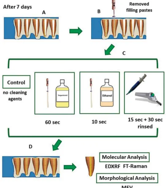
Figure 1B-0DWHULDODQGPHWKRGV$VSHFLPHQVZHUHVWRUHGDWDUHODWLYHKXPLGLW\RIDW&IRUVHYHQGD\V%DOOWKH¿OOLQJ
SDVWHVZKLFKZHUHYLVLEOHRQWKHOXPHQRIWKHURRWFDQDOZHUHUHPRYHGZLWKWKHDLGRIFXUHWWHVDQG.¿OHV&HDFKJURXSZDVGLVWULEXWHG LQWRJURXSVQ DFFRUGLQJWRFOHDQLQJDJHQWVFRQWUROJURXS&QRFOHDQLQJDJHQW(WKDQRO(IRUVHFRQGV7HUJHQIRUP®-T: for 60
VHFRQGV3KRVSKRULFDFLG3$SKRVSKRULFDFLGIRUVHFRQGVDQGODWHUULQVHGXQGHUZDWHUIRUVHFRQGV'WKHVSHFLPHQVZHUH VWRUHGDWDUHODWLYHKXPLGLW\RIDW&XQWLOWKHPROHFXODUDQGPRUSKRORJLFDODQDO\VHVZHUHFRQGXFWHG
computer system for data processing. The voltage in
of the current. Three spectra from each specimen were collected after the treatments. The measurements were performed with a count rate of 100 sec. per point. The equipment calibration and the chemical balance were performed as previously reported25.
μ
analyzed by μ-EDXRF using three points.
Morphological analysis
Scanning elect ron m icroscopic ( SEM) analysis
The specimens were dried and mounted on a holder using double-sided adhesive carbon tape. The
samples were sputter-coated with gold (Balzers-SCD 050 Sputter Coater, Liechtenstein) and examined with a scanning electron microscope (JEOL JSM 5600 LV,
morphological analysis was described according to the images obtained by SEM.
Statistical analysis
All data obtained is expressed in average and standard deviations. Data was submitted to the Lilliefors test for normality and only for the Ca/P data log transformation was used. The study factors were
tatistical analysis
Pastes EDXRF FT- Raman
Ca P Ca/P 2940 cm-1
&RQWUROQR¿OOLQJ 21.57±0.82b 12.15±0.73a 1.77±0.04b 1.08±0.14a
Calen® + Zinc Oxide -CZ 23.71±1.08a 10.77±0.76b 2.22±0.12a 1.06±0.13a
Calcipex II®-CII 23.51±1.04a 9.98±0.81b 2.41±0.27a 1.04±0.10a
Vitapex® 24.15±1.63a 10.80±0.44b 2.25±0.16a 1.04±0.09a
$ORZHUFDVHOHWWHUIROORZLQJDYDOXHLQGLFDWHVVLJQL¿FDQWGLIIHUHQFHVEHWZHHQJURXSV
Table 1-0HDQDQGVWDQGDUGGHYLDWLRQVRIFDOFLXPZWSKRVSKRUXVZWDQG&D3UDWLRFRQFHUQLQJ;UD\ÀXRUHVFHQFHDQDO\VLVDQG
FT-Raman spectroscopic analysis for peak area (2940 cm-1LQSULPDU\URRWGHQWLQIRUWKH¿OOLQJSDVWHV
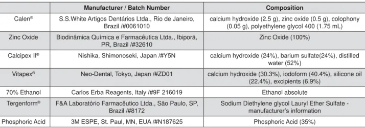
Manufacturer / Batch Number Composition
Calen® S.S.White Artigos Dentários Ltda., Rio de Janeiro,
%UD]LO calcium hydroxide (2.5 g), zinc oxide (0.5 g), colophony (0.05 g), polyethylene glycol 400 (1.75 mL) Zinc Oxide Biodinâmica Química e Farmacêutica Ltda., Ibiporã,
35%UD]LO Zinc Oxide (100%)
Calcipex II® 1LVKLND6KLPRQRVHNL-DSDQ<1 calcium hydroxide (24%), barium sulfate(24%), distilled
ZDWHU
Vitapex® 1HR'HQWDO7RN\R-DSDQ=' calcium hydroxide (30.3%), iodoform (40.4%), silicone oil
(22.4%), excipients (6.9%)
70% Ethanol &DUORV(UED5HDJHQWV,WDO\) Ethanol absolute
Tergenform® F&A Laboratório Farmacêutico Ltda., São Paulo, SP,
%UD]LO Sodium Diethylene glycol Lauryl Ether Sulfate - PDQXIDFWXUHU¶VLQIRUPDWLRQ
Phosphoric Acid 0(63(6W3DXO01(8$1 Phosphoric Acid (35%)
Figure 2- Details of the materials
Results
According to the two-way ANOVA statistical analysis applied to the FT-Raman and μ-EDXRF , there was no
pastes in the FT-Raman analysis (2940 cm-1 band), no
change in the organic content of the dentin. (Table 1)
paste (Table 1), μ-EDXRF analysis showed that the Ca content and Ca/P ratio of the control group (no
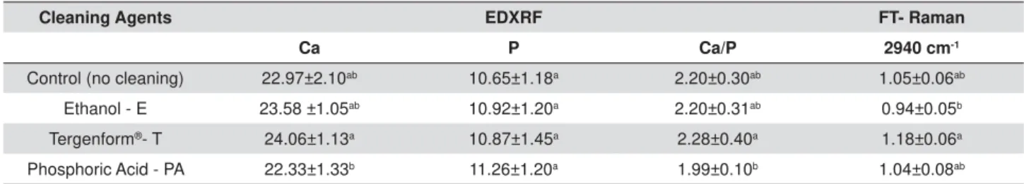
Regarding cleaning agents (Table 2), concerning
organic content than the ethanol group (p<0.05). In μ-EDXRF, T showed higher values for Ca content and Ca/P ratio than the phosphoric acid group. All groups were similar for P content with regards to the μ-EDXRF
analysis.
μ
pastes contained calcium in their composition. The CZ
titanium and sulfur, but had a higher percentage of
in higher proportions than calcium.
Morphological analysis of the root dentin of primary
paste amount and smear layers upon the dentin surface for all cleaning agent groups, except when
pastes on the dentin surface (Figure 4). For the control
or T, similar characteristics of the dentin surface were found: dentin tubules and dentin surface covered by dense smear layer, whereas cleaned up with PA
Cleaning Agents EDXRF FT- Raman
Ca P Ca/P 2940 cm-1
Control (no cleaning) 22.97±2.10ab 10.65±1.18a 2.20±0.30ab 1.05±0.06ab
Ethanol - E 23.58 ±1.05ab 10.92±1.20a 2.20±0.31ab 0.94±0.05b
Tergenform®- T 24.06±1.13a 10.87±1.45a 2.28±0.40a 1.18±0.06a
Phosphoric Acid - PA 22.33±1.33b 11.26±1.20a 1.99±0.10b 1.04±0.08ab
$ORZHUFDVHOHWWHUIROORZLQJDYDOXHLQGLFDWHVVLJQL¿FDQWGLIIHUHQFHVEHWZHHQJURXSV
Table 2-0HDQDQGVWDQGDUGGHYLDWLRQVRIFDOFLXPZWSKRVSKRUXVZWDQG&D3UDWLRZLWKUHJDUGVWR;UD\ÀXRUHVFHQFHDQDO\VLV
and FT-Raman spectroscopic analysis for peak area (2940 cm-1) in primary root dentin for the cleaning agents
$ORZHUFDVHOHWWHUIROORZLQJDYDOXHLQGLFDWHVVLJQL¿FDQWGLIIHUHQFHVEHWZHHQJURXSV
Elements Calen® + Zinc Oxide Calcipex II® Vitapex®
(%) (%) (%)
Si - - 42.29
S - 8.14
-Ca 26.79 40.48 27.91
Ti - 12.46
-Zn 73.21 -
-I - - 29.80
Ba - 38.92
-Table 3-5HVXOWVREWDLQHGE\;UD\ÀXRUHVFHQFHDQDO\VLVVKRZWKHFKHPLFDOHOHPHQWVVLOLFRQ6LVXOIXU6FDOFLXP&DWLWDQLXP7L
]LQF=QLRGLQH,EDULXP%DLGHQWL¿HGLQ¿OOLQJSDVWHVDVSHUFHQWDJHV
study, did not show any ability to remove the smear
debris remaining on the dentin surface.
Discussion
pastes associated with different cleaning agents affect the molecular and surface features of the root canal dentin surface of primary teeth was not accepted. Regarding the FT-Raman and μ-EDXRF, there was no
and cleaning agents).
content regarding Ca, P and Ca/P for the μ-EDXRF
1). For Ca and Ca/P, the values (%w) were higher for
organic content of dentin when FT-Raman analysis was conducted (Table 1). The increased Ca values could
calcium-based (Table 3) and they remained on dentin surface associated with the smear layer. However, based on the microanalysis of chemical containing pastes, it was noticed that the amount of Ca in the
dentin, since CII presented the highest Ca (Table 3) value and it did not provide a higher Ca %w (Table 1) available on the dentin when compared with the
phosphate analysis. Similar results were described by Rythén, et al.21 (2010), in which exfoliated primary teeth showed higher Ca than P on the dentin, the study was performed using X-ray microanalyses.
Figure 3-&KHPLFDOHOHPHQWVVSHFWUDVLOLFRQ6LVXOIXU6FDOFLXP&DWLWDQLXP7L]LQF=QLRGLQH,DQGEDULXP%DVKRZHGE\
;UD\ÀXRUHVFHQFHDQDO\VLVOLQHVFDQLQWKH¿OOLQJSDVWHVJURXSV
used as cleaning solutions did not provide changes in collagen related to control groups, it seems that
the organic content. This is clinically important, because dentin is composed of, 20% in weight8 or 30% in volume8, hydrated organic matrix, most of which consists of type I collagen. It constitutes ~90% of the organic matrix8. This can be good for bonding
when using those cleaning agents, dentin collagen was
integrated resin-collagen biopolymer hybrid that
30. The result of
this study corroborates with Borges, et al.1 (2007) and Borges, et al.2 (2008) showing that no alterations were found in the organic content of primary teeth dentin when an acid etch procedure was used.
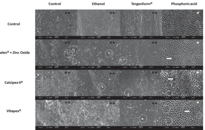
Figure 4- Representative scanning electron microscopy (SEM) images of root dentin surfaces of primary teeth, regarding the pastes and
FOHDQLQJDJHQWVRULJLQDOPDJQL¿FDWLRQV[6(0REVHUYDWLRQVUHYHDOHGWKHSUHVHQFHRIDKHDY\VPHDUOD\HULQDOOJURXSV)RU WKHSKRVSKRULFDFLGJURXSQRVPHDUOD\HUZDVVKRZQDQGDOOWXEXOHVZHUHFOHDQHGDQGRSHQHG:KLWHFLUFOHVUHSUHVHQWSDUWLFOHVRI ¿OOLQJSDVWHVUHPDLQLQJRQWKHGHQWLQVXUIDFH:KLWHDUURZVSRLQWRXWQRQGHPLQHUDOL]HGLQWHUWXEXODUGHQWLQDUHDV:KLWHFLUFOHVVLJQLI\ ¿OOLQJSDVWHUHVLGXHV
which did not show dentin demineralization nor remove
was a difference between them, they were similar to the control group (no cleaning agent). PA was similar to the control group (no cleaning agent), probably due
would buffer phosphoric acid, an effect associated with increasing pH, and the demineralization rate decreased in those groups28,with no time interference4. Another explanation or additional effect would be that the high carbonate content in primary teeth dentin is inversely proportional to the calcium content27, which probably contributed or added to the buffering effect. In addition, regarding the buffering effect, while acid etching removes up to 30% of the calcium phosphates, it dissolves 75% of the carbonates7. Therefore, the calcium phosphate was less affected, while the carbonate was removed, showing that the Ca% was similar to the etched PA and the control groups (no cleaning agent).
Representative SEM images (Figure 4) of the root dentin surfaces of primary teeth showed clean and
and were etched by PA. Borges, et al.2 (2008)also showed that primary teeth dentin etched by phosphoric acid enhanced the tubules ability to open and enlarge.
pastes and phosphoric acid showed some areas of non-demineralized intertubular dentin. Studies showed that after use of calcium hydroxide dressings on the root canal and cleaning there is residual of calcium hydroxide that causes decreases to the bond strength9,12 and even after the use of phosphoric acid there was no increase in the bond strength12. However, other cleaning agents studied showed
pastes and cleaning agents were used.
The results from this study provided important
pastes used in Pediatric Dentistry and the possible
an adequate surface for bonding, as it is commonly
dentin15. The results obtained from this study are promising, since it does not use up any cleansing agent, but uses two-step etching and adhesive rinsing, the adhesion of restorative materials is feasible, since it is less time consuming. In addition, the use of a self-etching system can be used with E or T or PA, which should be the best choice to cleaning the root
paste, since, they did not change the inorganic or organic content and also allowed for a smear layer on the dentin surface.
Conclusion
Based on our findings, regardless of filling pastes, all cleaning agents, in spite of providing some disturbance on the dentin surface, cannot be considered as harmful for cleaning the root dentin.
alter the organic content of primary teeth root dentin. The inorganic content (Ca, P and Ca/P) on the root
showing a residual mineral amount; however, cleaning agents did not change this. Morphological features of the root dentin of primary teeth were affected by phosphoric acid group treatment, showing cleaned and open dentin tubules.
The authors are grateful to the Department of Pediatric Dentistry (University of Campinas) and the Laboratory of Biomedical Vibrational Spectroscopy (Vale do Paraíba University).
This study was supported by FAPESP – São Paulo Research Support Foundation (grant numbers 09/10837-0, 10/12757-0, 01/14384-8, 05/50811-9) and CNPq- National Council for Scientific and Technological Development (grant number 307809/2013-7).
The authors are grateful to Mr. Marcos Blanco Cangiani, Mr. Marcelo Correa Maistro, Mrs. Eliene Orsini N. Romani and Mr. Adriano Luis Martins for their support of this research.
References
1- Borges AF, Bitar RA, Kantovitz KR, Correr AB, Martin AA, Puppin-Rontani RM. New perspectives about molecular arrangement of primary and permanent dentin. Appl Surf Sci. 2007;254:1498-505.
2- Borges AF, Bittar RA, Pascon FM, Sobrinho LC, Martin AA, Puppin Rontani RM. NaOCl effects on primary and permanent pulp chamber dentin. J Dent. 2008;36(9):745-53.
3- Borges AF, Puppin-Rontani RM, Bittar RA, Kantowitz KR, Pascon FM, Martin AA. Effects of acidic primer/adhesives on primary and permanent dentin. Am J Dent. 2009;22(1):30-6.
4- Camps J, Pashley DH. Buffering action of human dentin in vitro. J Adhes Dent. 2000;2(1):39-50.
5- Carvalho RM, Manso AP, Geraldeli S, Tay FR, Pashley DH. Durability of bonds and clinical success of adhesive restorations. Dent Mater. 2012;28(1):72-86.
6- Chala S, Abouqal R, Abdallaoui F. Prevalence of apical periodontitis and factors associated with the periradicular status. Acta Odontol Scand. 2011;69(6):355-9.
dentin particles of sub-millimeter size. Dent Mater. 2002;18(7):529-34.
composition and mineralization. Front Biosci (Elite Ed). 2011;3:711-35. 9- Guiotti FA, Kuga MC, Duarte MA, Sant'Anna AJ, Faria G. Effect of calcium hydroxide dressing on push-out bond strength of endodontic sealers to root canal dentin. Braz Oral Res. 2014;28. doi: 10.1590/ S1806-83242014.50000002.
10- Hosoya N, Kurayama H, Iino F, Arai T. Effects of calcium hydroxide on physical and sealing properties of canal sealers. Int Endod J. 2004;37(3):178-84.
11- Komabayashi T, D'souza RN, Dechow PC, Safavi KE, Spångberg LS. Particle size and shape of calcium hydroxide. J Endod. 2009;35(2):284-7.
calcium hydroxide dressing and acid etching on the push-out bond strengths of three luting resins to root canal dentin. Clin Oral Investig. 2014;18(2):489-98.
13- Liu Y, Hsu CY. Laser-induced compositional changes on enamel: a FT-Raman study. J Dent. 2007;35(3):226-30.
of oil contamination on in vitro bond strength of bonding agents to dental substrates. Am J Dent. 2008;21(2):101-4.
Res. 1982;16(3):265-73.
collapse and expansion of phosphoric acid-demineralized dentin. J Biomed Mater Res A. 2004;68(3):558-65.
surfaces after different chemical treatment modalities: an in vitro scanning electron microscopic study. J Oral Sci. 2000;42(3):139-46. 18- Pascon FM, Kantovitz KR, Borges AF, Puppin-Rontani RM. Effect of cleansers and irrigation methods on primary root dentin permeability. J Dent Child (Chic). 2007;74(1):30-5.
permeability index of primary tooth root dentin. J Clin Pediatr Dent. 2006;31(2):93-7.
20- Pashley DH, Tay FR, Carvalho RM, Rueggeberg FA, Agee KA, Carrilho M, et al. From dry bonding to water-wet bonding to ethanol-wet bonding. A review of the interactions between dentin matrix and solvated resins using a macromodel of the hybrid layer. Am J Dent. 2007;20(1):7-20.
22- Santos GL, Beltrame AP, Triches TC, Ximenes-Filho M, Baptista D,
in primary teeth. J Indian Soc Pedod Prev Dent. 2014;32(2):130-4. 23- Sauro S, Di Renzo S, Castagnola R, Grande NM, Plotino G, Foschi F, et al. Comparison between water and ethanol wet bonding of resin composite to root canal dentin. Am J Dent. 2011;24(1):25-30. 24- Soares LE, Brugnera A Jr, Zanin FA, Santo AM, Martin AA. Effects of heating by steam autoclaving and Er:YAG laser etching on dentin components. Lasers Med Sci. 2011;26(5):605-13.
25- Soares LE, Espírito Santo AM, Brugnera A, Zanin FA, Martin AA. Effects of Er:YAG laser irradiation and manipulation treatments on
spectrometry study. J Biomed Opt. 2009;14(2):024002.
26- Taylor TI, Larson L, Johnson W. Miscibility of alcohol and oils. Ind Eng Chem. 1936;28:616-8.
27- Tesch W, Eidelman N, Roschger P, Goldenberg F, Klaushofer K, Fratzl P. Graded microstructure and mechanical properties of human crown dentin. Calcif Tissue Int. 2001;69(3):147-57.
time, degree of saturation, pH and acid concentration of buffer solutions on the rate of in-vitro demineralization of human enamel. Arch Oral Biol. 1985;30(1):37-42.
![Figure 3-&KHPLFDOHOHPHQWVVSHFWUDVLOLFRQ6LVXOIXU6FDOFLXP&DWLWDQLXP7L]LQF=QLRGLQH,DQGEDULXP%DVKRZHGE\](https://thumb-eu.123doks.com/thumbv2/123dok_br/14984699.511597/8.892.217.679.93.857/figure-khplfdohohphqwvvshfwudvlolfrq-lvxoixu-fdoflxp-dwlwdqlxp-qlrglqh-dqgedulxp-dvkrzhge.webp)