C
AS
E
S
TUD
Correspondence to: Maísa Soares Gui – Avenida Limeira, 901 – Caixa Postal 52 – CEP: 13414-903 – Piracicaba (SP), Brazil – E-mail: [email protected]
Presentation: dec. 2012 – Accepted for publication: aug. 2013 – Financing source: none – Conflict of interests: nothing to declare – Presentation at a scientific event: II Congresso
ABSTRACT | The treatment of cutaneous ulcers by electrical stimulation in clinical practice has grown, however there are few studies investigating the effectiveness of these individual resources in monitoring and complete healing of the lesions. Thus, High Voltage Pulsed Stimulation (HVPS) was used in chronic skin ulcers with the aim of reduce the area of the lesion. Four male subjects with chronic cutaneous ulcers participated in the study. The treatment of the injury consisted on HVPS application (15 microseconds, 100/150 V, 100 Hz) for 30 min-utes, 2 times weekly. The electrode with negative polarity was placed on the lesion and positive on vascular path. The ulcers were assessed pre and post-intervention by photogrammetry, and it was calculated the area of the lesion. As a result, we ob-served the complete healing in the subjects I and II (respec-tively, area of 4.66 cm2 to 0 after 21 sessions and 1.74 cm2 to 0
after 16 sessions). The area of subject III right ulcer obtained reduction of 93% after 100° session (2.02 to 0.14 cm²) and left ulcer obtained reduction of 80.40% (2.50 to 0.49 cm²). In sub-ject IV there was a complete healing of the sacral lesion after 75 sessions (10.74 cm² to 0) and decrease sciatic lesion of 11.01 to 2.43 cm². Thus we conclude that HVPS facilitated the heal-ing process of stimulated ulcers because the areas of all ulcers had decreased more than 78%, and in three of them there was complete healing.
Keywords | electric stimulation; wound healing; photogrammetry; wound closure techniques.
High voltage pulsed stimulation
increases cicatrization of chronic
cutaneous ulcers: analysis of six cases
Estimulação elétrica de alta voltagem incrementa a
cicatrização de lesões cutâneas crônicas: análise de seis casos
Estimulación eléctrica de alto voltaje incrementa la
cicatrización de úlceras cutáneas crónicas: análisis de seis casos
Maísa Soares Gui1, Rinaldo Roberto de Jesus Guirro2, Daniel Iwai Sakabe3, Fabiana Forti Sakabe3
Study conducted at the Physical Therapy Department of Faculdades Integradas Einstein de Limeira – Limeira (SP), Brazil.
1Piracicaba Dentistry School at Universidade Estadual de Campinas (UNICAMP) – Piracicaba (SP), Brazil.
2Department of Biomechanics, Medicine and Rehabilitation of the Locomotor Apparatus in Ribeirão Preto Medicine School at
Universidade de São Paulo (USP) – Ribeirão Preto (SP), Brazil.
3Physical Therapy Course at Faculdades Integradas Einstein de Limeira – Limeira (SP), Brazil.
RESUMO | O tratamento de úlceras cutâneas por
estimula-ção elétrica tem crescido na prática clínica, no entanto, faltam estudos que investiguem a efetividade desse recurso em acompanhamento prolongado ou até que ocorra a cicatriza-ção completa das lesões. Assim, a estimulacicatriza-ção elétrica de alta voltagem (EEAV) foi aplicada em úlceras cutâneas crônicas com o objetivo de reduzir a área da lesão. Para tanto, partici-param do estudo quatro homens que apresentavam seis úl-ceras cutâneas que receberam a EEAV (fase=15ms; F=100 Hz; T: 100 a 150 V; fases gêmeas), 2 vezes por semana, durante 30 minutos. O eletrodo com polaridade negativa foi colocado sobre a lesão e o positivo no trajeto vascular. As úlceras foram avaliadas pré e pós-intervenção por meio da fotogrametria, sendo calculada a área da lesão. Como resultado, observa-mos o fechamento completo da lesão nos sujeitos I e II (área de 4,66 cm2 para 0 após 21 sessões e de 1,74 cm2 para 0 após
16 sessões, respectivamente). O sujeito III obteve redução de 93% na área da lesão direita (de 2,02 para 0,14 cm2) e na
es-querda de 80,40% (de 2,50 para 0,49 cm²), após 100 sessões. No sujeito IV ocorreu o fechamento completo da lesão sacral (de 10,74 cm2 para 0) e a redução da lesão isquiática de 11,01
INTRODUCTION
Skin injuries present several etiologies and can become chronic when the tissue formation is interrupted or de-stroyed by repeated damage or if one or two chemical or cellular elements of the healing process are deicient1.
In Brazil, the prevention work in general does not happen or it is not properly carried out, therefore the prevalence of pressure ulcers in hospitals is extremely high2,3. Diiculty in treatment success is increased due to problems like deiciencies in the patient’s nutritional status and in mobility4.
In 211 assessed risk patients, a 39.8% incidence of pressure ulcers was seen5. Also, higher rates of morbid-ity and mortalmorbid-ity were found in these patients1,6. hese results demonstrate the urgent need to create a program for the prevention and treatment of these wounds.
Not only the wounds of this etiology, but also the chronic injuries, present a relevant repercussion for the health of individuals at risk7. he slow healing in vascular ulcers has serious consequences for people, in-cluding pain, loss of job, and quality of life decrease8.
Depending on the injury level and depth in the tis-sues, the ulcers may bring complications like osteomyelitis, septicemia, or death. Besides the inancial losses caused to patients and their relatives, the problem also has psycho-logical disorders and hinders or makes it diicult for the participation of the subject in rehabilitation programs9,10.
Skin ulcer treatment using electrical stimulation has been growing in the clinical practice11, because it is a low-cost option and it may accelerate the heal-ing process, reducheal-ing treatment expenses12. In studies
using animals, it has been suggested that electrical stim-ulation improves wound healing, increasing growth fac-tors in the epidermis and dermis13.
Regan et al.14, in a systematic review, presented evi-dence supporting the use of electrical stimulation to accelerate tissue repair rate in pressure ulcers. Among these resources, the high-voltage pulsed stimulation (HVPS) promotes the acceleration of the healing pro-cess in chronic ulcers of several etiologies7,15-18, due to its signiicant efects to improve circulation15.
However, the majority of studies7,16-19 followed-up the wounds for a short period (4–6 weeks of stimula-tion)14, and they did not try to investigate if this re-source would lead to the complete lesion healing. hus, we observed the evolution of the areas of chronic skin ulcers during the treatment with HVPS for 12 months and/or until its complete cicatrization.
METHODOLOGY
Study outline
One case series describing the treatment results of six skin ulcers by HVPS, followed-up for 12 months, is presented in this study.
Participants
Four male subjects (aged 54.75±20.71 years) with chronic skin ulcers were invited to take part in this case
Descritores | estimulação elétrica; cicatrização; fotogrametria; técnicas de fechamento de ferimentos.
RESUMEN | El tratamiento de úlceras cutáneas con el uso de
es-timulación eléctrica tiene crecido en la práctica clínica, pero no hay muchos estudios que investigaron la efectividad de eso recurso en el acompañamiento prolongado o hasta la ocurrencia de la cica-trización completa de las lesiones. Así, la estimulación eléctrica de alto voltaje (EEAV) fue aplicada en úlceras cutáneas crónicas con el objetivo de reducir la área de la lesión. Para eso, cuatro hombres con seis úlceras cutáneas crónicas participaron del estudio, los cua-les habían recibido la EEAV (fase=15ms; F=100 Hz; T: el 100 al 150 V; fases), dos veces por semana, por 30 minutos. Lo electrodo con polaridad negativa fue posicionado sobre la lesión y lo positivo en el trayecto vascular. Las úlceras fueron evaluadas antes y después
de la intervención por medio de la fotogrametría, y la área de la le-sión fue calculada. Se observó, como resultado, el cierre completo de la lesión en los sujetos I y II (área de 4,66 cm2 para 0 después de
21 sesiones y de 1,74 cm2 para 0 después de 16 sesiones,
respectiva-mente). El sujeto III obtuvo reducción del 93% en el local de la lesión derecha (de 2,02 para 0,14 cm2) y en la izquierda del 80,40% (del 2,50
para 0,49 cm2) después de 100 sesiones. El cierre completo de la
lesión del sacro (del 10,74 cm2 para 0) y la reducción de la isquiática
del 11,01 para 2,43 cm2 ocurrieron en el sujeto IV después de 75
sesio-nes. Por lo tanto, se concluyo que la EEAV ha facilitado el proceso de cicatrización de las úlceras estimuladas, pues las áreas de todas las úlceras presentaron disminución superior al 78% con cicatrización completa en tres de ellas.
series. he inclusion criterion consisted of skin lesion appearance of any etiology, and infection in the le-sion to be treated was the exclusion criterion.
Ethics procedures
his study was carried out according to the resolution 196/96 of the Brazilian National Health Board, ap-proved by the Research Ethics Committee, under pro-tocol 08-3/019. he evaluation and intervention using HVPS was done in the Physical herapy School Clinic. All subjects were informed about the experimental pro-cedures and signed the free informed consent to take part in this study.
Intervention
he suggested treatment was performed with the Neurodyn High Volt® high-voltage electrical stimulation equipment (IBRAMED®, registration in M.S. 5122).
Intervention consisted of HVPS application (phase=15 ms; F=100 Hz; T: 100–150 V; twin phases) with a similar protocol to that described by Houghton et al.7, the treatment was carried out twice a week, and each ses-sion lasted 30 minutes.
he negative polarity active electrodes were wrapped up in sterilized gauze moistened by saline, being later placed and ixed with an adhesive tape inside the wound. he 10×18 cm self-adhesive dispersive electrode (VALUTRODE® Axelgaard Manufacturing Co., Ltd.) was ixed with positive polarity in the vascular path of the area. After every 5 minutes of treatment, the gauze under the electrodes was humidiied with saline using a sterilized syringe as support.
At the end of each session, asepsis of the electrodes was carried out for later use. he polarity of electrodes re-mained the same throughout the treatment.
It is worth mentioning that each subject had his/her own electrode kit. It was not used with any other medi-cine treatment in the treated ulcer, it was only necessary to apply, on a daily basis, sunlower oil — essential fatty acids, which are derived from linoleic acid — for skin hydration, and gauze for protection. his procedure was adopted because all patients were already making use of such oil for years.
Evaluation method
he treatment evolution was analyzed by means of standardized photographic records for the wound area
analysis, in squared centimeters. Hence, a digital camera was used (Panasonic®Lumix, model FX12; 7.2 mega-pixels), being placed at 40 cm from the ulcer, perpen-dicularly, including in the picture a millimeter-scaled ruler touching the skin.
After the recordings, the images were processed in the software used by Davini et al.19, which calculates the area. A 1-cm distance is marked in the ruler and re-ported to the software that automatically calculates it in pixels (DPixels).
Data analysis
Data presentation was performed in a descriptive manner to each subject individually. his procedure was adopted because the ulcers have diferent etiolo-gies and the number of HVPS sessions may vary be-tween the subjects.
RESULTS
Subject I
A 73-year-old patient presenting a supericial venous ulcer, plain crater, in the anterior region of the right tibia, with partial loss of the skin continuity involving epidermis and dermis, since a year. In the initial evalua-tion, the wound had a 4.66 cm2 area. After 10 sessions, it reduced to 0.65 cm², and the complete healing was obtained after 21 sessions (Figure 1).
Subject II
Twenty-six years old, paraplegic, with pressure ulcer in the sacral area, for 1 year, classiied as degree II (plain crater). In the initial photogrammetry, it registered an area of 1.74 cm2. After 10 sessions, there was a reduc-tion of the injury lesion to 0.75 cm2, and the complete closure happened after 4 months using HVPS, totaling 26 sessions (Figure 2).
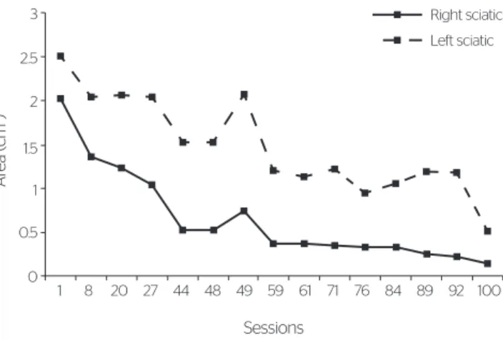
Subject III
as seen in Figure 3. Also, it should be mentioned that the lesion was classiied as degree IV (deep crater with extensive destruction).
he left sciatic pressure ulcer was also a degree IV lesion, which sufered three surgical intervention at-tempts (graft), with no success. It presented an initial area of 2.50 cm2 and after the 44th session, this sur-face reduced to 1.53 cm2 and in the 100th session to 0.49 cm2 (80.40% decrease).
It was also seen that there was an increase of the lesion area between the 48th and 50th sessions from 1.53 to 2.07 cm2. In such a period, the treatment was suspended for 2 months due to an infection in the lesion (Figure 3).
Subject IV
Sixty-six years old, triplegic due to cerebrovascular ac-cident, presents two pressure ulcers being one sacral and the other sciatic to the right, with 4 years of ex-istence. he sacral lesion, degree III (deep crater), had an initial area of 10.74 cm2, and after 37 sessions it decreased to 3.79 cm2, reaching complete healing after 75 sessions (Figure 4).
he sciatic ulcer, degree III, presented in the initial evaluation an 11.01 cm2 area, which, after 34 sessions, decreased to 2.94 cm2 and by the end of the 75th ses-sion, to 2.82 cm2 (74.38% decrease).
DISCUSSION
In this study, we could notice complete closure of the three wounds and an important reduction of other three lesions treated with HVPS. he ulcers had diferent eti-ologies and classiications; therefore, there were distinct responses as to cicatrization speed/time and percentage. Furthermore, the follow-up of these patients for a short period enabled a better comprehension of the lesion re-action to stimulation.
Wound classiication comprises stage I–IV with regard to depth of tissue compromising instead of le-sion severity20,21. Cicatrization happens with the sup-port of granulation tissue, by second intention, and the ulcer improvement or worsening evaluation is carried out by measuring its dimension22. hus, we veriied
5.00
2 4.50
1 3 6 9 12 15 16 17
4.00 3.50 3.00 2.50 2.00 1.50 1.00 0.50 0.00
Á
re
a (cm
2)
Sessions
Figure 1. Evolution of the ulcer area (cm2) from subject I’s leg during the
high-voltage pulsed stimulation treatment
Sessions
1 11 21 26
2.00
1.50
1.00
0.50
0.00
Ar
ea (cm
2)
Figure 2. Evolution of the ulcer area (cm2) of sacral pressure from subject
II in the high-voltage pulsed stimulation treatment
Sessions
3
2.5
2
1.5
1
0.5
0
Ar
ea (cm
2)
Right sciatic Left sciatic
8
1 20 27 44 48 49 59 61 71 76 84 89 92 100
Figure 3. Evolution of the area (cm2) of right and left sciatic pressure ulcers
from subject III in the high-voltage pulsed stimulation treatment
11
Sessions
1 23 34 43 46 51 75
12
10
8
6
4
2
0
Ár
ea (cm
2)
Sciatic Sacral
Figure 4. Evolution of the area (cm2) of right sacral and sciatic pressure
that supericial ulcers presented an accelerated healing speed, because in subjects I and II (degree II) there was complete closure of the lesion after 21 and 26 sessions, respectively, while the sacral lesion healing of subject IV (degree III) was achieved after 75 sessions.
In subjects with degree IV ulcers, there was a de-crease from 74 to 93% of the lesions areas; however, it happened more slowly. his reaction is somehow ex-pected, since cicatrization by second intention occurs from inside to outside the ulcer and, due to the fact that these lesions were deep, changes would be seen in depth and not in the lesion area20. his fact could be seen by the visualization of the photographed pictures, but it was not measured in this study and, therefore, it is a limitation of this work.
All lesions that did not completely heal were in the gluteal fold, that is, an area with much humidity and con-tamination and also a constant pressured place, which are caused by shearing and compression strengths.
Studies have indicated an improvement preferen-tially in pressure ulcers19. Griin et al.17 observed, after 20 consecutive stimulation days, an 80% decrease of pressure ulcers places and of 52% in the control group. he protocol consisted of negative polarity use in the lesion (60 minutes). Unger et al.16, however, performed polarity inversion of the electrodes, initiating by nega-tive and after 6 days of treatment, it changed to posinega-tive. Eight of the nine stimulated patients had an 88.9% heal-ing, while in the control group three of the eight subjects had a 37.5% improvement. In another study23, stage II ulcers presented increment of the cicatrization with sig-niicant increase in the granulation tissue concerning the control group, after 6 weeks of treatment.
However, HVPS in vascular chronic ulcers in the leg produced a reduction in the size of wounds when com-pared to those that were considered treated7. A similar result was seen in this study, as in one of the subjects who had his/her lesion healed, the etiology was ve-nous. On the other side, in another study24, nine pa-tients had their venous ulcers healed or reduced, while four had them increased, and the authors reported the reason for this as the incidence time of the lesions. Concerning time of the lesion, it is worth mentioning that the unhealed ulcers were aged between four and eight years old, which is a higher time than those pre-senting complete cicatrization (one year).
We also saw a fast lowering of the lesion areas in the beginning of treatment, which may be stabilized. his decrease in healing speed may be explained due to the better reaction to the polarity inversion according
to the phases in which the tissue repair is found than in the maintenance of the same polarity until the treatment ends25,26.
Recio et al.27 started the HVPS treatment with neg-ative polarity and then changed it weekly, thus achiev-ing complete healachiev-ing of chronic pressure ulcers (exis-tent from 11 to 14 months).
he tissue repair is divided into four phases: hemo-stasia, inlammation, proliferative, and remodeling1; therefore, Sussman and Byl26 preconized the begin-ning of the treatment with negative polarity, changing it every 3 days in the proliferation phase, and in the remodeling stage, it was altered every day. It is quite probable that the polarity inversion be necessary in these chronic and deep lesions, in order to not slow the cicatrization process.
he mechanisms for which the HVPS reaches posi-tive healing results have not been well established yet. he current, despite its polarity (single-phase wave)28,29, passes through the skin with despicable thermal and electrochemical efects and the highest current density is available for target tissues, afecting the cellular level directly30. In addition, it can be eicient to contain and absorb acute edemas, to fasten the dermal and sub-der-mal tissues repair, and to control pain31. Other aspect that should be considered is galvanotaxis, which is the migration of electric-charged cells with regard to an electrical ield of opposed polarity. As to skin cicatriza-tion, the electrical ields exogenously applied with the same size as those found in lesions, promote migration of human keratinocytes for the cathode32, which is the polarity used on the lesion in this study.
Besides its circulatory and regenerative actions, the HVPS presents bactericide action18, because it leads to local changes in the pH, electrochemical changes in the injured tissue, and recruitment of anti-microbial factors of the organism.
he increase in microcirculation around the infra-malleolar ischemic wounds would explain the quick decay in the area of 11 ulcers stimulated by HVPS37. Furthermore, it was veriied that there is an increase of the oxygen transcutaneous pressure and capillary perfu-sion in the edge of venous ulcers, which indicates that HVPS can result in oxygenation and cicatrization due to the increase of tissue perfusion38,39.
Finally, the eicacy of HVPS treatment may be as-sociated with lesion etiology, occurrence time, wound location, and polarity of electrodes. herefore, clinical perspectives of our results reinforce the extensive need for multidisciplinary treatment when handling this pa-thology. Considering the limitations of this outline as to the generalization possibility, the next step should be HVPS intervention use in a randomized and represen-tative sample in patients with skin ulcers, determining the polarity based on cicatrization phase and associat-ing it with conventional treatments.
CONCLUSION
From the experimental conditions that were performed, it could be veriied that the areas of all ulcers stimulated by HVPS decreased and there was complete healing in three of them.
REFERENCES
1. Gonçalves G, Parizotto NA. Fisiopatologia da reparação cutânea:
atuação da fisioterapia. Rev Bras Fisioter. 1998;3:5-13.
2. Bryant RA, Bar BW, Beshara M, Broussard CI, Cooper DM, Doughty DB, et al. Acute and chronic wounds: nursing management. 2. ed. Missouri: Mosby; 2000.
3. Petrolino HMBS. Úlcera de pressão em pacientes de unidade de terapia intensiva: incidência, avaliação de risco e medidas de prevenção. São Paulo. [Dissertação] – Escola de Enfermagem, Universidade de São Paulo (USP);2002.
4. Serpa LF, Santos V LCG. Malnutrition as a risk factor for the development of pressure. Ulcers Acta Paul Enferm. 2008;21(2):367-9. 5. Rogenski NMB, Santos VLCG. Estudo sobre a incidência de úlceras
de pressão em um Hospital Universitário. Rev Latino-am Enferm. 2005;13(4):474-80.
6. Roach R. The practicing physicians guide to pressure ulcers in 2008. Med Health. R I.2008;91(12):382-3.
7. Houghton PE, Kincaid CB, Lovell M, Campbell KE, Keast DH, Gail WM. Efect of electrical stimulation on chronic leg ulcer size and appearance. Phys Ther. 2003;83(1):17-28.
8. Gogia PP. Clinical Wound Management: Ulcers of the lower extremities. Thorofare: Alpine;1995.
9. Faro ACM.Fatores de risco para úlcera de pressão: subsídios para a prevenção. Rev Esc Enferm USP. 1999;33(3):279-83.
10. Langemo DK, Anderson J, Voldem CM. Nursing quality outcome indicators. North Dakota Study. J Nurs Adm. 2002;32(2):98-105. 11. Dissemond J. Physical treatment modalities for chronic leg ulcers.
Hautarzt. 2010;61(5):387-96.
12. Mittmann N, Chan BC, Craven BC, Isogai PK, Houghton P. Evaluation of the cost-efectiveness of electrical stimulation therapy for pressure ulcers in spinal cord injury. Arch Phys Med Rehabil. 2011;92:866-72. 13. Kutlu AK, Çeçen D, Gürgen SG, Sayin O, Çetin F. A Comparison Study of
Growth Factor Expression following Treatment with Transcutaneous Electrical Nerve Stimulation, Saline Solution, Povidone-Iodine, and Lavender Oil in Wounds Healing. Evid Based Complement Alternat Med. Epub 2013;2013:361832. doi:10.1155/2013/361832. Epub 2013 jun 3. 14. Regan MA, Teasell RW, Wolfe DL, Keast D, Mortenson WB, Aubut JL.
A systematic review of therapeutic interventions for pressure ulcers after spinal cord injury. Arch Phys Med Rehabil. 2009;90:213-31. 15. Davini R, Nunes CV, Guirro ECO, Guirro RRJ. Estimulação Elétrica
de Alta Voltagem uma opção de tratamento. Rev Bras Fisioter. 2005;9(3):249-56.
16. Unger P, Eddy J, Raimastry S. A controlled study of de efect of high voltage pulsed current (HVPC) on would healing. Phys Ther. 1991;71:S119. 17. Grifin JW, Tooms RE, Mendius RA. Eficacy of high voltage pulsed
current for healing of pressure ulcers in patients with spinal cord injury. Phys Ther. 1991;71(6):433-44.
18. Szuminsky NJ, Albers AC, Unger P, John GE. Efect of narrow, pulsed high voltages on bacterial viability. Phys Ther. 1994;74(7):660-7. 19. Davini R, Nunes CV, Guirro ECO, Guirro RRJ. Tratamento de úlceras
cutâneas crônicas por meio da estimulação elétrica de alta voltagem. Rev Cienc Med. 2005;14(3):249-58.
20. Costa MP, Sturtz G, Costa FP. Epidemiologia e tratamento das úlceras de pressão: experiência de 77 casos. Acta Ortop Bras. 2005;13(3):124-33. 21. Nogueira PC, Caliri MHL, Santos CB. Fatores de risco e medidas
preventivas para úlcera de pressão no lesado medular. Experiência da Equipe de Enfermagem do HCFMRP-USP. Rev Bras Med. 2002;35. 22. Bergstrom N, Allman RM, Alvarez OM, Carlson CE, Eaglesetein W,
Frantz RA, et al; U.S. Departament of Health and Human Services - Public Health Service. Pressure Ulcers Treatment. Clinical Practice: Guideline Number 15. Agency for Health Care Policy and Research (AHCPR) Publication. 1994; 95(0652):40.
23. Franek A, Kostur R, Polak A, Taradaj J, Szlachta Z, Blaszczak E, et al. Using high-voltage electrical stimulation in the treatment of recalcitrant pressure ulcers: results of a randomized, controlled clinical study. Ostomy Wound Manage. 2012;58(3):30-44.
24. Pires EJ. Fisioterapia na cicatrização e recuperação funcional nos portadores de úlcera de hipertensão venosa crônica: uso da estimulação elétrica com corrente de alta voltagem. São Paulo. [Dissertação] – Faculdade de Medicina, Universidade de São Paulo (USP); 2006. 25. Reich JD, Tarjan PP. Electrical stimulation of skin. Int J Dermatol.
1990;29:395-400.
26. Sussman C, Byl N. Electrical Stimulation for Wound Healing. In: Sussman C, Bates-Jensen BM editors. Wound Care Collaborative Practice Manual for Physical Therapists and Nurses. Aspen Publishers. 1998;16. 27. Recio AC, Felter CE, Schneider AC, McDonald JW. High-voltage
utility for recalcitrant wounds below the level of injury. J Spinal Cord Med. 2012;35(1):58-63.
28. Rodrigues-Bigaton D, Almeida AFN, Berni, KCS, Pedroni CR; Gonçalves RN, Bérzin F. Utilização de diferentes estimulações elétricas para o tratamento da dor em mulheres com disfunção temporomandibular. Rev Bras Fisioter. 2008;12(6):476-81.
29. Sandoval MC, Ramirez C, Camargo DM, Salvini TF. Efect of high-voltage pulsed current plus conventional treatment on acute ankle sprain. Rev Bras Fisioter. 2010;14(3):193-9.
30. Low J, Reed A. Eletroterapia Explicada: princípios e prática. São Paulo: Manole; 2001.
31. Garcia LB, Guirro ECO. Efeitos da estimulação de alta voltagem no linfedema pós-mastectomia. Rev Bras Fisioter. 2005;9(2):243-8. 32. Nishimura K, Isserof R, Nuccitelli R. Human keratinocytes migrate to
the negative pole in direct current electric fields comparable to those measured in mammalian wounds. J Cell Sci. 1996;109:199-207. 33. Li L, Gu W, Du J, Reid B, Deng X, Liu Z, et al. Electric fields guide
migration of epidermal stem cells and promote skin wound healing. Wound Repair Regen. 2012;20:840-51.
34. Weiss DS, Kirsner R, Eagistein WH. Electrical stimulation and wound healing. Arch Dermatol. 1991;126:222-5.
35. Gentzkow GD, Miller KH. Electrical stimulation for dermal wound healing. Clin Podiatr Med Surg. 1991;8(4):827-41.
36. Sebastian A, Syed F, Perry D, Balamurugan V, Colthurst J, Chaudhry IH, et al. Acceleration of cutaneous healing by electrical stimulation: Degenerate electrical waveform down-regulates inflammation, up-regulates angiogenesis and advances remodeling in temporal punch biopsies in a human volunteer study. Wound Repair Regen. 2011;19:693-708.
37. Goldman RJ, Brewley BI, Golden MA. Electrotherapy reoxygenates inframalleolar ischemic wounds on diabetic patients: a case series. Adv Skin Wound Care. 2003;15:112-20.
38. Goldman RJ, Rosen M, Brewley, BI, Golden M. Electrotherapy Promotes Healing and Microcirculation of Infrapopliteal Ischemic Wounds: A Prospective Pilot Study. Adv Skin Wound Care. 2004;17(6):284-94. 39. Ud-Din S, Perry D, Giddings P, Colthurst J, Zaman K, Cotton S, et al.