Authors
Virna Nowotny Carpio1 Carolina Rech2
Evlyn Isabel Eickhoff2 Karla Laís Pegas3 Maria Isabel Albano Edelweiss3
Luiz Felipe Santos Gonçalves1,2
Roberto Ceratti Manfro1,2 Francisco Veríssimo Veronese1,2
1Post Graduate Medical
Sciences Program of Uni-versidade Federal do Rio Grande do Sul – UFRGS.
2Division of Nephrology
of Hospital de Clínicas de Porto Alegre.
3Division of Pathology of
Hospital de Clínicas de Porto Alegre.
Submitted on: 05/04/2011 Approved on: 01/06/2011
Correspondence to: Francisco José Veríssimo Veronese
Hospital de Clínicas de Porto Alegre
Ramiro Barcelos, 2.350 Porto Alegre (RS) – Brazil Zip code: 90035-003 E-mail: fveronese@hcpa. ufrgs.br
This study was undertaken at UFRGS.
Financial support: Fundo de Incentivo à Pesquisa – FIPE – of Hospital de Clínicas de Porto Alegre and Coordenação de Aperfeiçoamento de Pessoal de Nível Superior – CAPES.
The authors report no conl ict of interest.
R
ESUMOIntrodução: A fração do complemento C4d é um marcador de rejeição mediada por anticorpos (RMA) em aloenxertos re-nais, embora na rejeição celular também se observem depósitos de C4d. Objetivos:
Correlacionar a expressão de C4d com pa-râmetros clínicopatológicos e a evolução do enxerto renal em três anos. Métodos:
Foram incluídos 146 receptores de trans-plante renal com biópsias por indicação. A marcação de C4d foi feita por imuno-histo-química em parafina. Foram medidas a fun-ção e a sobrevida do enxerto e determina-das as variáveis preditivas de sua evolução por meio de modelo de regressão de Cox.
Resultados: A marcação positiva para C4d foi detectada em 48 (31%) biópsias, das quais 23 (14,7%) tinham marcação difusa e 25 (16%), focal. A reatividade contra pai-nel (%PRA) de classe I e II pré-transplante foi significativamente maior nos pacientes C4d+ quando comparada aos C4d-. Tanto
glomerulite quanto pericapilarite foram as-sociadas com C4d (p = 0,002 e p < 0,001, respectivamente). A presença de C4d em biópsias sem rejeição (SR), rejeição celular aguda (RCA) ou fibrose intersticial/atrofia tubular (FI/AT) não teve impacto na função ou na sobrevida do enxerto. Comparados a indivíduos com SR, RCA e FI/AT C4d-,
pa-cientes com RMA C4d+ tiveram pior
sobre-vida do enxerto em 3 anos (p = 0,034), mas não houve diferença entre RMA versus SR, RCA e FI/AT C4d+ (p = 0,10). Na regressão
de Cox, função do enxerto no momento da biópsia e %PRA alto foram preditores de perda do enxerto. Conclusões: A pesquisa de C4d em biópsias do enxerto renal é útil para identificar RMA, com correlações clí-nicopatológicas bem definidas. O impacto do C4d em outros diagnósticos histológicos necessita de investigação adicional.
A
BSTRACTIntroduction: C4d is a marker of antibo-dy-mediated rejection (ABMR) in kidney allografts, although cellular rejection also have C4d deposits. Objective: To correlate C4d expression with clinico-pathological parameters and graft outcomes at three years. Methods: One hundred forty six renal transplantation recipients with graft biopsies by indication were included. C4d staining was performed by paraffin-im-munohistochemistry. Graft function and survival were measured, and predictive variables of the outcome were determined by multivariate Cox regression. Results:
C4d staining was detected in 48 (31%) biopsies, of which 23 (14.7%) had diffu-se and 25 (16%) focal distribution. Pre-transplantation panel reactive antibodies (%PRA) class I and II were significantly higher in C4d positive patients as compa-red to those C4d negative. Both glomeru-litis and pericapillaritis were associated to C4d (p = 0.002 and p < 0.001, respective-ly). The presence of C4d in biopsies diag-nosed as no rejection (NR), acute cellular rejection (ACR) or interstitial fibrosis/ tubular atrophy (IF/TA) did not impact graft function or survival. Compared to NR, ACR and IF/TA C4d-, patients wi-th ABMR C4d+ had the worst graft sur-vival over 3 years (p = 0.034), but there was no difference between ABMR versus NR, ACR and IF/TA that were C4d po-sitive (p = 0.10). In Cox regression, graft function at biopsy and high %PRA levels were predictors of graft loss. Conclusions:
This study confirmed that C4d staining in kidney graft biopsies is a clinically useful marker of ABMR, with well defined clini-cal and pathologiclini-cal correlations. The im-pact of C4d deposition in other histologic diagnoses deserves further investigation.
Correlações clinico-patológicas da marcação de C4d e sua
inl uência na evolução de receptores de transplante renal
I
NTRODUCTIONThe complement cleavage product C4d is a specific marker of antibody mediated rejection (ABMR) in kidney transplantation, and many studies have sho-wn C4d as an independent predictor of renal graft outcome.1-5 C4d represents one criterion to the diag-nosis of acute and chronic ABMR according to Banff classification.6 Staining for C4d in peritubular ca-pillaries is present in other histological diagnosis of kidney graft biopsies, such as acute cellular rejection and/or acute graft injury,3 interstitial fibrosis and tu-bular atrophy, calcineurin inhibitor toxicity, and even in grafts without dysfunction and with no morpho-logical signs of rejection.3,4,7 The influence of C4d on graft function and outcome over these different Banff categories is still unclear.
Another feature to consider about C4d staining is the accuracy and reproducibility of current techniques, which could affect its prevalence, clinical diagnosis and therapeutic decisions. In western countries, the prevalence of C4d positivity in renal allograft biop-sies using different techniques varies from 17 to 60% among biopsies by indication.3,8-13 In Brazil, Ludovico-Martins et al.14 showed a C4d prevalence of 45% by immunofluorescence (IF). In this study, frozen-immu-nohistochemistry (IHC) and paraffin-IF had a good concordance rate to frozen-IF, but paraffin-IHC had a much lower accuracy. Few information is available about the reproducibility of C4d interpretation when this marker is detected by paraffin-IHC.15
The aims of this study were to determine the prev-alence of C4d in graft biopsies according to Banff histopathology, and to correlate C4d expression with clinical and pathological parameters and graft func-tion and survival over time. The reproducibility of C4d interpretation, performed by immunohistochem-istry technique, was also measured.
M
ATERIALS AND METHODSThis study included prospectively 113 renal transplant recipients who had allograft biopsies by indication for acute or chronic graft dysfunction at Hospital de Clínicas de Porto Alegre (HCPA) from January 2007 to July 2009. Other 33 patients who also had biop-sies by indication at our centre from January 1991 to December 2006 were also included. In total, 156
biopsies of 146 patients were included. The study protocol was approved by the HCPA Research Ethics Committee.
CLINICAL DATA
The following variables were evaluated: age, gender, type of donor, re-transplants, pre- transplant serum panel reactive antibodies (PRA) against HLA class I and II, HLA mismatches in loci A, B, and Dr, se-rum creatinine at biopsy, occurrence of delayed graft function (need for dialysis in the first week), and ti-me between transplant and graft outcoti-me (months). Time interval between transplant and biopsy was defined as early (≤ 6 months) or late (> 6 months). Renal graft function was determined by estimated glomerular filtration rate (eGFR) using the re-expres-sed Modification of Diet in Renal Disease (MDRD) equation.16 Baseline immunosuppression consisted of prednisone, mycophenolate mofetil or sodium, and a calcineurin inhibitor (cyclosporine or tacroli-mus). As induction therapy, Basiliximab, OKT3 or Thymoglobulin were used. Acute cellular rejection (ACR) was treated with intravenous methylpredniso-lone, and resistant cases received Thymoglobulin or OKT3. Patients with ABMR were treated with plas-mapheresis and intravenous immunoglobulin.
HISTOPATHOLOGICALDIAGNOSIS
Histopathologic diagnosis was performed by a renal pathologist who was blinded to clinical data, accor-ding to the Banff’2005 Meeting Report.6 Patients were classified as: no rejection (NR), ACR, ABMR or interstitial fibrosis/tubular atrophy (IF/TA). No rejection included biopsies with normal tissue, acu-te tubular necrosis, calcineurin inhibitor toxicity, or borderline changes.
As we did not measure circulating anti-HLA an-tibodies, a “presumptive” diagnosis of ABMR was defined by C4d positivity and specific morphologi-cal features: margination of neutrophils in peritubu-lar capilperitubu-laries (PTC), fibrinoid necrosis, glomerulitis, thrombi in glomeruli and arteries, severe endarteritis (V3), and/or acute tubular injury. Patients who had a biopsy positive for C4d and morphological features suggestive of chronic ABMR – such as transplant glomerulopathy (glomerular basement membrane duplication or double contours, Banff score cg 1-3),
Keywords: Kidney transplantation. Complement Cd4. Graft survival. Graft rejection. Humoral rejection.
and/or fibrous intimal thickening in arteries without duplication of the internal elastic – were also included as ABMR. In ABMR group we included pure acute ABMR, ABMR with features of ACR, and chronic ABMR.
Graft function and survival were analyzed accord-ing to Banff category (SR, ACR, ABMR or IF/TA) and presence or absence of C4d, at a minimum follow-up of one year.
C4d IMMUNOSTAINING
C4d was detected by immunoperoxidase in formalin-fixed, paraffin-embedded sections. Slides were incu-bated overnight with a polyclonal anti-rabbit antibo-dy (Abcam, American Research Products®, Palo Alto, CA, US), followed by incubation with Universal Link Biotinylated Secondary Antibody (Biocare Medical®, Concord, CA, US) and after streptavidine-horseradish peroxidase (Biocare Medical®, Concord, CA, US). Reaction was developed with Romulin AEC chromo-gen (Biocare Medical®, Concord, CA, US). Criteria for C4d positivity were a linear and circumferential staining in at least 25% of PTCs in cortex or medulla, excluding fibrotic and necrotic areas. Distribution of C4d was established as: 1) diffuse when > 50% of PTC were stained; 2) focal when 25 to 50% of PTC were stained; and 3) negative if less than 25% of PTC were stained.11
Two observers blinded to clinical data scored C4d staining independently, when inter-observer variabil-ity was calculated. Intra-observer variabilvariabil-ity was cal-culated based on two separated interpretations of the same observer, at least six months apart.
STATISTICAL ANALYSIS
Data was described as means and standard deviation or median and interquartile ranges. Comparisons be-tween groups were performed by t independent test, Mann-Whitney test, one-way ANOVA, and χ2 test when appropriate. Intra-observer and inter-observer agreement rate was measured by Kappa statistic, in which values greater than 0.75 were considered as excellent concordance, between 0.40 and 0.75 fair to good, and less than 0.40 were considered as poor concordance.17 Graft survival at three years was mea-sured by Kaplan Meier. Multivariate Cox regression analysis was used to identify variables predicting graft loss at last follow-up, including variables with statis-tical significance in the univariate analysis. Data were processed and analyzed using SPSS for Windows, ver-sion 16.0 (Chicago, IL, US); p level < 0.05 was consi-dered significant.
R
ESULTSRENAL ALLOGRAFT BIOPSY FINDINGS AND C4d STAINING
Median time (and interquartile ranges) between trans-plant and graft biopsy were 11 (8-22) days and 23 (11-68) months for patients biopsed in early and late periods, respectively. Histopathological diagnosis ac-cording to Banff classification are shown in Table 1. Median time to biopsy of IF/TA group was 20 (8-66) months and for the other categories together was 13 (8-29) days (p < 0.001). Median times from trans-plantation to last follow-up were 56 (30-99) versus 27 (16-34) months for these two groups, respectively (p < 0.001).
Peritubular capillary staining for C4d was detect-ed in 48 (31%) biopsies, of which 23 (14.7%) had a diffuse staining and 25 (16%), a focal distribution. Positivity for C4d was found in 40 (83%) biopsies done early (< 6 months) post transplant as compared to 8(17%) in late (> 6 months) biopsies (p = 0.039).
CLINICALAND DEMOGRAPHICDATAACCORDINGTO
C4d STAINING
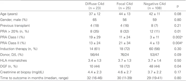
Baseline clinical and demographic characteristics are presented in Table 2. Patients whose graft biopsies stained diffusely for C4d had significantly higher
pre-Histological diagnosis n (%)
Normal tissue 5 (3)
Borderline changes 14 (9)
Acute cellular rejection 62 (40)
Type IA 37 (60)
Type IB 3 (5)
Type IIA 12 (19)
Type IIB 8 (13)
Type III 2 (3)
Humoral rejection 17 (11)
Pure acute humoral rejection 10 (59)
Humoral + cellular rejection 3 (17)
Chronic humoral rejection 4 (24)
Interstitial ibrosis/tubular atrophy 34 (22)
Grade I 11 (32)
Grade II 20 (59)
Grade III 3 (9)
Acute tubular necrosis 19 (12)
Calcineurin inhibitor toxicity 5 (3) Table 1 HISTOPATHOLOGICALDIAGNOSIS
ACCORDINGTO BANFF 2005
transplant %PRA. No difference was found in %PRA levels comparing patients with diffuse and focal C4d. Comparing patients with ABMR rejection against the other Banff categories together, %PRA class I was 27 ± 34 and 4 ± 12 (p < 0.001) and class II was 37 ± 37 and 3 ± 12 (p < 0.001), respectively.
Delayed graft function occurred more times in pa-tients who had focal C4d in biopsy than in diffuse and negative C4d groups (p = 0.04), and also in patients with ABMR as compared to the other Banff catego-ries (87 versus 46%; p = 0.003). No statistical dif-ference between the three groups was found for the other parameters (Table 2).
Positive staining for C4d was found in 83 and 17% of biopsies taken ≤ 6 months and > 6 months post transplantation, respectively (p = 0.04). Comparing
type of maintenance immunosuppression and type of induction therapy, no difference was observed in C4d positivity in these graft biopsies.
ASSOCIATIONOFC4dDEPOSITIONWITH BANFF
DIAGNOSIS AND MORPHOLOGIC FEATURES
The distribution of C4d in biopsies according to histo-pathology and morphologic Banff features are shown in Table 3. As the presence of C4d was a criterion to iden-tify ABMR, this group was taken as the reference group. C4d deposits were detected in 9 (21%), 18 (29%), and 4 (12%) of the biopsies with NR, ACR, and IF/TA, res-pectively (χ2 = 46.05; p = 0.001). The majority of biop-sies with histological criteria for acute cellular rejection (71%), histology other than rejection (79%) and chro-nic damage (88%) did not stain for C4d. Deposits of
Diffuse C4d (n = 23)
Focal C4d (n = 25)
Negative C4d
(n = 108) p
Age (years) 37 ± 12 44 ± 13 42 ± 11 0.08
Gender, male (%) 65 56 59 0.80
Previous transplant 4 (18) 4 (16) 8 (7) 0.21
PRA > 20% (n, %) 8 (35) 8 (32) 12 (11) 0.01
PRA Class I (%) 19 ± 29 11 ± 24 3 ± 11 0.002a
PRA Class II (%) 13 ± 24 21 ± 34 4 ± 13 0.004b
Induction therapy (n, %) 14 (61) 18 (72) 60 (56) 0.30
Donor, D/L (%) 56/44 76/24 53/47 0.09
HLA mismatches 3.4 ± 1.3 3.7 ± 1.3 3.7 ± 1.4 0.50
DGF (n, %) 10 (44) 18 (72) 48 (44) 0.04
Creatinine at biopsy (mg/dL) 4.4 ± 2.3 4.6 ± 2.7 3.7 ± 2.2 0.17
Time to outcome in months (median, range) 32 (16-46) 30 (11-39) 29 (19-41) 0.80 Table 2 PATIENTDEMOGRAPHICSANDCLINICALCHARACTERISTICSACCORDINGTO C4d STAININGPATTERNS
PRA: panel reactive antibodies; D/L: deceased/living; DGF: delayed graft function; MM: mismatches loci A, B, Dr; aDiffuse C4d versus Negative C4d; bFocal C4d versus Negative C4d; one-way ANOVA and Tukey Post Hoc test, Kruskal-Wallis.
Table 3 C4d STAINING, BANFFHISTOPATHOLOGYANDMORPHOLOGICFEATURESOFTRANSPLANTBIOPSIES
Diffuse C4d (n = 23)
Focal C4d (n = 25)
Negative C4d
(n = 108) p
Time to biopsy in days (median, range) 15 (10-36) 19 (11-64) 19 (9-227) 0.95
No rejection (n, %) 5 (22) 4 (16) 34 (32)
Acute cellular rejection (n, %) 8 (35) 10 (40) 44 (41) < 0.001*
Humoral rejection (n, %) 9 (39) 8 (32) 0
IF/TA (n, %) 1 (4) 3 (12) 30 (28)
Tubulitis (n, %) 13 (15) 14 (16) 61 (69) 0.99
Glomerulitis (n, %) 8 (35) 4 (17) 11 (48) 0.02
Arteritis (n, %) 7 (26) 7 (26) 13 (48) 0.04
Neutrophils in PTC (n, %) 7 (44) 8 (50) 1 (6) < 0.001
C4d in ACR types I, II and III were found in 25, 29 and 50% of the biopsies, respectively (p = 0.14).
Association between C4d positive staining and morphologic features of the Banff scheme was ana-lyzed individually. Twenty-seven (56%) biopsies with positive C4d staining showed tubulitis as compared with 11 (57%) biopsies with negative C4d (p = 0.56). Glomerulitis was present in 12 (25%) of C4d+ and in 11 (10%) of C4d- biopsies (p = 0.02), arteritis in 14 (29%) versus 13 (12%); p = 0.01, and margination of neutro-phils in PTC in 15 (32%) versus 1 (0.9%) (p < 0.001), respectively. Grading C4d staining as diffuse, focal or negative yielded similar results (Table 3).
C4d STAINING AND GRAFT FUNCTION
Follow-up graft function was determined at 12 mon-ths, 2 years and at last measured serum creatinine. Graft function (mL/min/1.73m2) at those time points comparing C4d+ and C4d- patients, in a median follow-up of 31 (17-42) months, were 47 ± 21 versus 54 ± 19 (p = 0.15), 50 ± 23 versus 53 ± 22 (p = 0.77) and 40 ± 26 versus 46 ± 28 (p = 0.30), respectively.
Analyzing graft function according to C4d grad-ing, patients with focal C4d had the lowest eGFR at 12 months: 44 ± 21, 51 ± 21 (diffuse C4d) and 54 ± 19 (negative C4d) mL/min/1.73 m2 (p = 0.19). At last follow-up, eGFR was also lower in patients with focal C4d in biopsy: 36 ± 28, 44 ± 24 and 46 ± 28 mL/min/1.73 m2,respectively (p = 0.32). However, these differences did not reach statistical significance, which is probably related to the small number of patients in each C4d positive group. Overall, there was no difference in graft function be-tween the three groups at the maximum follow-up period.
When we analyzed graft function according to Banff histology and C4d positivity, eGFR at last follow-up in patients with ACR was lower in the C4d posi-tive group (43 ± 23 versus 50 ± 227 mL/min/1.73 m2 in ACR C4d negative), but without statistical differ-ence (p = 0.43). In patients with NR, eGFR was similar in all time periods comparing C4d+ and C4d- groups. Patients with IF/TA and C4d+ had the worst graft func-tion at 2 years as compared to IF/TA C4d- (29 ± 7 versus 45 ± 23 mL/min/1.73 m2; p = 0.046), but not at last follow-up (21 ± 12 versus 30 ± 24 mL/min/1.73 m2, re-spectively; p = 0.49). However, the number of patients in the former subgroup was small (n = 4).
C4d STAINING AND GRAFT SURVIVAL
Analysis of graft loss according to each Banff category showed that significantly more patients with ABMR
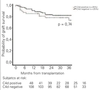
(7/17, 41%) and IF/TA (15/34, 44%) lost their grafts (χ2 = 13.32; p = 0.004) as compared to patients with NR (5/43, 12%) and ACR (13/62, 21%). Comparing C4d+ and C4d- groups, graft survival at nearly three years of follow-up was lower for patients with C4d+ but without statistical significance (Figure 1). Graft survival according to C4d grading was not statisti-cally different either, but patients with focal C4d had a lower survival rate than the group with diffuse C4d (67 versus 78%, log-rank = 1.398; p = 0.23) or nega-tive C4d (67 versus 83%, log-rank = 1.113; p = 0.29). Comparing graft survival at three years by type of do-nor and positivity for C4d in biopsy, we found that grafts from deceased donors with C4d survived less than those from deceased donors without C4d (63 versus 79%, respectively; p = 0.17) but this differen-ce did not reach statistical significandifferen-ce due to sample size. Irrespective of C4d, grafts from living donors survived the same (87 versus 85% for C4d+ and C4d-, respectively; p = 0.51).
The effect of C4d deposits in PTC at the time of biopsy on graft survival was evaluated for each Banff category. No difference in graft sur-vival rate was detected for ACR C4d+ as com-pared to ACR C4d- cases, 77 versus 83% (log-rank = 0.005; p = 0.94), as well as for NR C4d+ versus C4d- (log-rank = 1.519; p = 0.21) and IF/ TA C4d+versus C4d- (log-rank = 0.411; p = 0.41) groups. Additionally, we grouped patients with NR, ACR and FI/AT as one category and strati-fied them according to C4d positivity, and those
Figure 1. Graft survival rates according to C4d positivity in biopsy. No difference was found between C4d+ and C4d- patients at last follow-up (74 versus 83%, log-rank=0.11; p=0.74).
0 0,0 0,2 0,4
P
robabilit
y of graf
t sur
viv
al
0,6 0,8 1,0
p = 0,74
C4d positive (≥=25%) C4d negative (<=25%)
6 12 18 24 30 36
108 103 95 82 68 51 33 48
C4d negative C4d positive
Months from transplantation Subjetcs at risk:
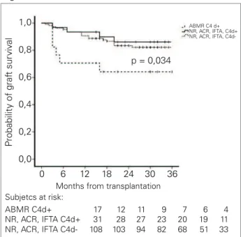
patients were compared to patients with ABMR. Graft survival was significantly lower in ABMR as compared to grouped categories C4d-, but the difference between ABMR and grouped categories C4d+ was not statistically significant (Figure 2).
Multivariate Cox regression showed eGFR at the time of graft biopsy and PRA > 20% as significant predictors of graft failure (Table 4). C4d deposition did not predict graft loss, but ABMR rejection was the Banff category associated with a trend towards graft failure. After adjustment, ABMR did not remain a significant predictor of graft loss.
INTERANDINTRA-OBSERVERREPRODUCIBILITYOFC4d
INTERPRETATION
Intra-observer concordance rate for the presence or absence of C4d was excellent, with a Kappa value (k-value) of 0.77 (p < 0.001). Agreement rate were 91.7% for the presence and 89.1% for the absence of C4d. A high k-value was also found for grading C4d as negative, focal or diffuse staining (k -va-lue = 0.73; p < 0.001). The agreement rates were 89.1, 72 and 91.3% for scoring C4d as negative, focal and diffuse, respectively. The lowest concordance rate was observed for interpreting C4d staining as focal, when ten biopsies were scored as C4d negative and two as C4d diffuse in the first interpretation changing to C4d focal in the second analysis.
Inter-observer reproducibility for the presence or absence of C4d was worse, with a k-value was 0.60 (p < 0.001). Agreement rates were 93% for presence and 75% for absence of C4d. For grading, k-value was even worse (0.57; p < 0.001), with an agreement rate of 75% for absence of C4d, 64% for grading C4d as focal and 95% as a diffuse staining.
D
ISCUSSIONSeveral studies have been showing that C4d accumu-lation in PTCs occurs not only in ABMR but also in cellular rejection and even in biopsies without rejec-tion (i.e., acute tubular necrosis, drug toxicity).2,3,8-11,18 So far, the pathogenic and clinical significance of tho-se findings is incompletely understood. Humoral and cellular alloresponses might affect the kidney graft either independently or concurrently, with endothe-lium antigens of capillaries and arteries acting as tar-gets for both humoral and cellular immune responses, respectively.9,19,20
This study showed a 31% prevalence of C4d de-position in biopsies of kidney grafts similar to other studies, in which C4d has varied from 30 to 45% in average.2,3,9,10,13,14 Lower prevalence of C4d positiv-ity has been reported in indication biopsies, as in the
Figure 2. Kaplan-Meier curves of graft survival rates showing a signiicantly lower survival for patients with antibody-mediated rejection (ABMR) as compared to patients grouped as no rejection (NR), acute cellular rejection (ACR) or interstitial ibrosis/tubular atrophy (IF/TA) negative for C4d (64 versus 82%; p = 0.034, log-rank = 4.48). When grouped NR, ACR or IF/TA positive for C4d was compared to ABMR, no difference in graft survival was found (78 and 64%, respectively; p = 0.10, log-rank = 2.76).
0 0,0 0,2 0,4
P
robabilit
y of graf
t sur
viv
al
0,6 0,8 1,0
p = 0,034
ABMR C4 d+ NR, ACR, IFTA, C4d+ NR, ACR, IFTA,
C4d-6 12 18 24 30 36
31 28 27 23 20 19 11
17 NR, ACR, IFTA C4d+
108 103 94 82 68 51 33
NR, ACR, IFTA C4d-ABMR C4d+
Months from transplantation Subjetcs at risk:
12 11 9 7 6 4
Variable Crude Hazard Ratio (95%IC) p Adjusted Hazard Ratio (95%IC) p
eGFR (MDRD) 0.94 (0.91-0.97) < 0.001 0.95 (0.91-0.98) 0.005
PRA > 20% 1.03 (1.01-1.04) < 0.001 1.02 (1.00-1.04) 0.038
C4d deposition in PTC 0.89 (0.44-1.78) 0.74 Non included
Banff category 2.81 (0.86-9.20)* 0.08 0.47 (0.11-2.17)* 0.33
Table 4 CLINICALANDPATHOLOGICDETERMINANTSOFKIDNEYGRAFTOUTCOMEIN MULTIVARIATE COX REGRESSION
study of Mengel et al.11 (20.7%) and Cheunsuchon et al.12 (16.4%). This variability can be explained by several factors, such as different staining techniques and percentage of PTC positivity, different biopsy set-tings, time since transplant, and case selection.
Acute pathologic changes other than rejection and normal biopsies were grouped as NR category. Twenty one percent of these biopsies stained posi-tively for C4d, a prevalence that did not differ from other series,9,10,13,18 independently of the technique employed. However, it is not possible to exclude that in some of these first biopsies with acute graft injury C4d deposits preceded the development of cellular or ABMR, that would be later diagnosed in subsequent biopsies,21 which were not done in our study.
Mauiyyedi et al.3 reported IF C4d deposition in 15% of the biopsies with ACR type I and II. Using the same pathological criteria, Nickeleit et al.9 described the prevalence of C4d based on morphologic features present in biopsy: 43% in tubulo-interstitial rejection and 45% in transplant endarteritis. Using IHC, preva-lence rates of C4d positive staining were similar: 20% of indication biopsies with ACR,11,13 21.4% in ACR Banff I, II or III10, 24% in a series of protocol and di-agnostic biopsies,22 and 29% in the present study.
The correlation of Banff morphologic features and C4d in acute rejection has been emphasized in some studies.3,9-11,13 Our results are in agreement with pre-vious reports, since both tubulitis and arteritis were significantly more frequent in C4d negative biopsies (71% of ACR biopsies), whereas glomerulitis and neutrophils in PTC predominated in biopsies with C4d deposits, the majority of them with ABMR.
Mauiyyedi et al.8 found C4d in 61% of biopsies diagnosed as chronic rejection. Ranjan et al.13 re-ported C4d in 30% of patients with chronic allograft nephropathy, rate that increased to 83% if associated to ABMR. In the study of Nickeleit et al.,9 C4d was present in 23% of biopsies with striped fibrosis and in 14% of diffuse interstitial fibrosis. In our sample, C4d was present in 12% of biopsies with IF/TA, and we cannot exclude an antibody-mediated chronic re-jection in these cases. It is important to note that irre-spective of C4d, the presence of IF/TA correlates with late graft dysfunction and failure.
While the majority of the studies showed C4d positivity as predictive of a worse graft function and survival,2-4,10,21,23 some did not confirm this associa-tion.9 Moreover, in specific settings, such as protocol biopsies11 or late acute renal allograft rejection,24,25 there is controversy if C4d deposition is associated or not with poor function and higher rates of graft loss.
Overall, we could not demonstrate this association, but graft survival of patients without ABMR but posi-tive for C4d did not differ from those with ABMR, a finding to be further explored. We believe that our results indicate that ABMR was correctly identified in this cohort as it was associated with an adverse outcome. However, a question remains if C4d present in other histologic categories affects graft outcome. Certainly, numerous other mechanisms must be taken into account, as many different molecules have altered expression in rejection and in other types of injury po-tentially affecting graft outcome.
It has been discussed if the percentage of C4d dis-tribution has distinct clinical and morphologic correla-tions, and how it affects graft function and survival in the short and long term. Magil et al.2,26 showed a simi-lar clinical course and biopsy findings in patients with focal or diffuse C4d. Haririan et al.4 also demonstrated that graft survival was adversely influenced by C4d in-dependent of the staining pattern, and focal C4d was a significant independent predictor of graft failure. In our study, patients with focal C4d had the lowest eGFR and graft survival at last follow-up, but this group had either an increased prevalence of DGF and a trend to more deceased donors as graft source, both potentially related to this worse outcome (Table 2). The percent-age of C4d staining that accurately predicts kidney transplantation outcome is still debated, and even a cutoff of 10% for C4d positivity was reported to be a strong predictor of renal graft loss.27
Several studies had compared IF and IHC on fro-zen and paraffin sections for C4d identification and grading in kidney graft biopsies. Some of them showed higher accuracy for frozen-IF with monoclonal anti-body in relation to paraffin-IHC,14,15,28 but other au-thors reported an acceptable sensitivity (87.5%) and specificity (98%) of paraffin-IHC with anti-C4d poly-clonal antibody.22 Nadasdy et al.29 compared frozen-IF and paraffin-IHC, suggesting a loss of sensitivity of 31% for IHC. Technical factors can contribute to a lower sensitivity of IHC, such as tissue fixation, paraf-fin-embeddeding process, the quality of paraffin, and a more variable intensity of staining.15,21,28 Moreover, the staining of plasma proteins in PTCs that results from soluble C4 fixation sometimes makes interpre-tation of C4d less reliable.24 Nonetheless, potential advantages of paraffin-IHC are better preservation of morphologic features, and most important, IHC can be done retrospectively in stored paraffin sections when diagnosis of ABMR is doubtful.3,22
reproducibility. Agreement rate were good for inter-observer and excellent for intra-inter-observer interpreta-tions, suggesting that our C4d analysis has been con-sistent. In only one study the agreement rates of C4d in paraffin-IHC was evaluated.15 This study showed similar results: a good inter-observer concordance (k value = 0.63) and an excellent agreement for intra-observer interpretation (k-value = 0.83).
A limitation of our study is the lack of measure-ment of donor specific antibodies (DSA). There are consistent data showing DSA as predictive of worse graft function and outcome,3,22,30 irrespective of C4d staining at least in the first year of transplant.4 DSA were not measured in our centre when these biopsies were carried out, so we should call ABMR as “pre-sumptive antibody-mediated rejection” based only in C4d positivity and pathologic features.
In conclusion, this study confirms that C4d stain-ing in kidney allograft biopsies is clinically useful as a marker of antibody-mediated rejection, with defined clinical and pathological correlations in renal trans-plant recipients. The impact of C4d deposition in other Banff histology was not defined by the present investigation. We can speculate that even with a good reproducibility, C4d staining by immunohistochemis-try in paraffin can render immunopathology less sen-sitive to detect antibody-mediated graft injury, which might be present in some cases of acute rejection and therefore affect graft outcome.
A
CKNOWLEDGMENTSWe are grateful to Robert Colvin, Patricia Della Pelle and Nicole Brousaides for their expert technical advi-ce. This study was supported by Fundo de Incentivo à Pesquisa (FIPE) of Hospital de Clínicas de Porto Alegre, Porto Alegre, RS, Brazil. Virna N. Carpio was supported by a scholarship of Coordenação de Aperfeiçoamento de Pessoal de Nível Superior (CAPES) of Brazilian Ministry of Education and Culture, Brazil.
R
EFERENCES1. Lederer SR, Kluth-Pepper B, Schneeberger H, Albert E, Land W, Feucht HE. Impact of humoral alloreactivity early after transplantation on the long-term survival of renal allografts. Kidney Int 2001;59:334-41.
2. Herzenberg AM, Gill JS, Djurdjev O, Magil AB. C4d deposition in acute rejection: an independent long-term prognostic factor. J Am Soc Nephrol 2002;13:234-41. 3. Mauiyyedi S, Crespo M, Collins AB, Schneeberger
EE, Pascual MA, Saidman SL, et al. Acute humoral rejection in kidney transplantation: II. Morphology,
immunopathology, and pathologic classification. J Am Soc Nephrol 2002;13:779-87.
4. Haririan A, Kiangkitiwan B, Kukuruga D, Cooper M, Hurley H, Drachenberg C, et al. The impact of c4d pattern and donor-specific antibody on graft survival in recipients requiring indication renal allograft biopsy. Am J Transplant 2009;9:2758-67.
5. Feucht HE, Schneeberger H, Hillebrand G, Burkhardt K, Weiss M, Riethmüller G, et al. Capillary deposition of C4d complement fragment and early renal graft loss. Kidney Int 1993;43:1333-8.
6. Solez K, Colvin RB, Racusen LC, Sis B, Halloran PF, Birk PE, et al. Banff’05 Meeting Report: differential diagnosis of chronic allograft injury and elimination of chronic allograft nephropathy (‘CAN’). Am J Transplant 2007;7:518-26.
7. Nickeleit V, Mihatsch MJ. Kidney transplants, antibodies and rejection: is C4d a magic marker? Nephrol Dial Transplant. 2003;18:2232-9.
8. Mauiyyedi S, Pelle PD, Saidman S, Collins AB, Pascual M, Tolkoff-Rubin NE, et al. Chronic humoral rejection: identification of antibody-mediated chronic renal allograft rejection by C4d deposits in peritubular capillaries. J Am Soc Nephrol 2001;12:574-82. 9. Nickeleit V, Zeiler M, Gudat F, Thiel G, Mihatsch
MJ. Detection of the complement degradation product C4d in renal allografts: diagnostic and therapeutic implications. J Am Soc Nephrol 2002;13:242-51. 10. Regele H, Exner M, Watschinger B, Wenter C,
Wahrmann M, Osterreicher C, et al. Endothelial C4d deposition is associated with inferior kidney allograft outcome independently of cellular rejection. Nephrol Dial Transplant 2001;16:2058-66.
11. Mengel M, Bogers J, Bosmans JL, Serón D, Moreso F, Carrera M, et al. Incidence of C4d stain in protocol biopsies from renal allografts: results from a multicenter trial. Am J Transplant 2005;5:1050-6.
12. Cheunsuchon B, Vongwiwatana A, Premasathian N, Shayakul C, Parichatikanond P. The prevalence of C4d-positive renal allografts in 134 consecutive biopsies in Thai patients. Transplant Proc 2009;41:3697-700. 13. Ranjan P, Nada R, Jha V, Sakhuja V, Joshi K. The role
of C4d immunostaining in the evaluation of the causes of renal allograft dysfunction. Nephrol Dial Transplant 2008;23:1735-41.
14. Ludovico-Martins H, Silva C, Teodoro WR, Martini Filho D, Noronha IL. Analysis of different staining techniques for c4d detection in renal allograft biopsies. Transplant Proc 2009;41:862-5.
15. Seemayer CA, Gaspert A, Nickeleit V, Mihatsch MJ. C4d staining of renal allograft biopsies: a comparative analysis of different staining techniques. Nephrol Dial Transplant 2007;22:568-76.
16. Levey AS, Bosch JP, Lewis JB, Greene T, Rogers N, Roth D. A more accurate method to estimate glomerular filtration rate from serum creatinine: a new prediction equation. Modification of Diet in Renal Disease Study Group. Ann Intern Med 1999;130:461-70.
18. Worthington JE, McEwen A, McWilliam LJ, Picton ML, Martin S. Association between C4d staining in renal transplant biopsies, production of donor-specific HLA antibodies, and graft outcome. Transplantation 2007;83:398-403.
19. Halloran PF, Schlaut J, Solez K, Srinivasa NS. The significance of the anti-class I response. II. Clinical and pathologic features of renal transplants with anti-class I-like antibody. Transplantation 1992;53:550-5. 20. Collins AB, Schneeberger EE, Pascual MA, Saidman SL,
Williams WW, Tolkoff-Rubin N, et al. Complement activation in acute humoral renal allograft rejection: diagnostic significance of C4d deposits in peritubular capillaries. J Am Soc Nephrol 1999;10:2208-14. 21. Feucht HE. Complement C4d in graft capillaries -- the
missing link in the recognition of humoral alloreactivity. Am J Transplant 2003;3:646-52.
22. Troxell ML, Weintraub LA, Higgins JP, Kambham N. Comparison of C4d immunostaining methods in renal allograft biopsies. Clin J Am Soc Nephrol 2006;1:583-91.
23. Böhmig GA, Exner M, Habicht A, Schillinger M, Lang U, Kletzmayr J, et al. Capillary C4d deposition in kidney allografts: a specific marker of alloantibody-dependent graft injury. J Am Soc Nephrol 2002;13:1091-9. 24. Poduval RD, Kadambi PV, Josephson MA, Cohn
RA, Harland RC, Javaid B, et al. Implications
of immunohistochemical detection of C4d along peritubular capillaries in late acute renal allograft rejection. Transplantation 2005;79:228-35.
25. Satoskar AA, Lehman AM, Nadasdy GM, Sedmak DD, Pesavento TE, Henry ML, et al. Peritubular capillary C4d staining in late acute renal allograft rejection -- is it relevant? Clin Transplant 2008;22:61-7.
26. Magil AB, Tinckam KJ. Focal peritubular capillary C4d deposition in acute rejection. Nephrol Dial Transplant 2006;21:1382-8.
27. Crary GS, Raissian Y, Gaston RC, Gourishankar SM, Leduc RE, Mannon RB, et al. Optimal cutoff point for immunoperoxidase detection of C4d in the renal allograft: results from a multicenter study. Transplantation 2010;90:1099-105.
28. Colvin RB. Antibody-mediated renal allograft rejection: diagnosis and pathogenesis. J Am Soc Nephrol 2007;18:1046-56.
29. Nadasdy GM, Bott C, Cowden D, Pelletier R, Ferguson R, Nadasdy T. Comparative study for the detection of peritubular capillary C4d deposition in human renal allografts using different methodologies. Hum Pathol 2005;36:1178-85.