Parallel Determination of NeuroD1, Chromogranin-A, KI67 and
Androgen Receptor Expression in Surgically Treated Prostate
Cancers
L. Cindolo, M. Cantile, R. Franco, P. Chiodini, G. Schiavo, I. Forte, I. Zlobec, L. Salzano,
G. Botti, S. Gidaro, L. Terracciano, C. Cillo
Department of Urology, S. Pio da Pietrelcina Hospital, Vasto, Italy (LC), Department of Clinical
and Experimental Medicine (MC, CC), Federico II University, Naples, Italy, Surgical Pathology (RF,
IF), G. Pascale National Cancer Institute, Naples, Italy, Department of Medicine and Public Health
(PC), Second University, Naples, Italy, Institute of Pathology (GS, IZ, LT, CC), University of Basel,
Basel, Switzerland, Department of Urology (LS), G. Rummo Hospital, Benevento, Italy, Department
of Surgical and Experimental Sciences (SG), Chieti-Pescara University, Chieti, Italy
ABSTRACT
Purpose: Neuroendocrine differentiation is a hallmark of prostate cancer. The aim of our study was the detection of the parallel expression of neuroendocrine related markers using a prostate tissue microarray (TMA).
Materials and Methods: Our study was aimed at detecting the parallel expression of NeuroD1, Chromogranin-A (ChrA), Androgen Receptor (AR) and Ki-67 by immunohistochemistry on prostate cancer tissue microarray. The data was ana-lyzed using SAS version 8.2 (SAS Inc, Cary, NC). The relationships between NeuroD1, ChrA and AR expressions and patients’ characteristics were investigated by multivariate logistic regression analysis. Progression and Overall Survival (OS) distributions were calculated using Kaplan-Meier method.
Results: Tissue reactivity for NeuroD1, ChrA and AR concerned 73%, 49% and 77% of the available cases, respectively. Regarding overall survival, there were 87 deaths and 295 patients alive/censored (6 years of median follow-up).
Seventy-seven disease progressions occurred at the median follow-up 5.4y. A signiicant correlation between NeuroD1, ChrA and
AR expression was observed (p < 0.001 and p < 0.03, respectively). Additionally, ChrA was strongly associated in multi-variate analysis to Gleason score and Ki67 expression (p < 0.009 and p < 0.0052, respectively). Survival analysis showed
no association between markers neither for overall nor for cancer-speciic survival.
Conclusions: The results highlight that NeuroD1, Chromogranin-A and Androgen Receptor are strongly associated, how-ever their expression does not correlate with overall survival or disease progression.
Key words: prostatic neoplasms; neuroendocrine cells; neuroD1 protein; ki-67 antigen; chromogranin A, receptors, androgen prognosis
Int Braz J Urol. 2011; 37: 57-66
INTRODUCTION
Prostate cancer (PCa) is the most frequent cancer in Western countries and the second leading
doi: 10.1590/S1677-55382011000700008
with a strong increase in biological aggressiveness and
a signiicant decrease in survival (3). Only a few stud -ies on docetaxel-based chemotherapy have reported results in terms of survival, pain control, quality of life and progression in patients with metastatic castra-tion-resistant prostate cancer (CRPC) (4,5), albeit the risk of cytotoxic chemotherapy should be individually weighted.
In recent years, the presence of neuroendo-crine differentiation (NED) features has been reported as a variable associated with the development of the CRPC (6,7) during the natural history of this PCa. In general, pure neuroendocrine (NE) tumor cells do not express androgen receptors (AR), are resistant to androgen deprivation therapy and do not proliferate in response to androgens (8). Autocrine-paracrine epithelial interactions and/or transdifferentiation are the mechanisms through which NE cells act in PCa homeostasis (9).
The early detection of NE activity in pros-tate adenocarcinoma could suggest or anticipate an early diagnosis of hormones refractoriness behavior and thus justify changes in therapeutic approaches.
Unfortunately, the diagnosis and the quantiication of
prostatic NE cell activity remains a problem. Chro-mogranin A (ChrA), consistently expressed during NE cell differentiation (8), is the most frequently used marker to detect NE differentiation in PCa patients, both at tissue and at serum level (10,11). Neverthe-less, differences between assays for serum ChrA
pro-vided a signiicant discordance rate, suggesting that
the commercial kits for serum detection might elicit different information (12). Moreover, tissue ChrA
lack prognostic signiicance in patients with bone
metastatic PCa (13). Other NE markers (such as tissue CD56, synaptophysin) add only little information on the acquisition of NE phenotype in human prostate
(14). Neuron-speciic enolase (NSE) could become a
valuable tumor progression marker and could serve as predictor of survival together with clinical parameters but only in advanced and hormone refractory prostate neoplasms (15,16).
These evidences highlight that the identii -cation of new diagnostic and prognostic markers is relevant for the clinical management of PCa patients, especially related to neuroendocrine differentiation.
Following the identiication of the neurogenic char
-acteristic of the 2q31-33 genome region (HOX D locus) which houses genes involved with epithelial-neuronal cell conversion (17), we investigated the role of NeuroD1 in normal and neoplastic human prostates. We have previously reported that Neu-roD1 tissue reactivity correlates with the indicators of malignancy in moderately to poorly differentiated PCa and it could be involved in the pathophysiology of PCa neuroendocrine differentiation (14). Here we report on an immunohistochemical analysis using a tissue micro array (TMA) containing a high number of different naive prostate cancer specimens, in order to verify the prognostic relevance of NeuroD1 together with ChrA, AR and Ki67 tissue reactivity and their correlations.
MATERIALS AND METHODS
A total of 732 patients (members of the Kaiser Foundation Health Plan) treated for clinically local-ized PCa by radical prostatectomy or transurethral resection (TURP) (incidental diagnosis) at one of two Kaiser Hospitals in Portland (OR, USA) between 1971 and 1996, were retrospectively evaluated. The full study protocol, including access to the slides and blocks, was reviewed and approved by the Commit-tee for the Protection of Human Subjects of Kaiser
Permanente, Portland, OR. All patient identiiers
were removed and replaced by unique study numbers,
linked to the original identiiers by a single ile kept
under high security. Medical records for the entire cohort were abstracted at one time, 1999-2001, to assure uniform criteria for diagnosis, progression, and staging.
Selection of the specimens, classiication,
as well as patient management and follow-up have extensively been described elsewhere (18). Before
1992 (pre-PSA era), progression was deined clini -cally based on the results of bone scans, chest x-rays, and/or digital rectal examination. After 1992,
Benign prostatic hyperplasia (BPH), as con-trol, was also evaluated in 89 specimens (not included in the analysis).
Tissue Microarray Design
The prostate TMA was constructed as
pre-viously described (18,19). Briely, one core
tissue-biopsy (diameter 0.6 mm) was taken from the least
differentiated region of individual parafin-embedded
prostate tumors (donor blocks) and precisely arrayed
into a new recipient parafin block (35-20 mm) with
a custom-built precision instrument (Beecher Instru-ments, Silver Spring, MD). The core-tissue biopsies were put into one of the two recipient blocks that
de-ined one replicate TMA. Six replicate TMAs contain -ing the identical set of tumors were constructed. After the block construction, 5 mm sections were cut using a microtome. Originally, 732 donor tissue blocks were available for the construction of this TMA. Specimens from 74 tumors could not be included in the study because of incomplete follow-up data, lack of tumor in the arrayed sample (sampling error), damaged tis-sue (heat or crush artifacts), or a total lack of tistis-sue at some array positions (‘empty spots’). The number of patients varies between the individual marker analyses because of variability in the number of interpretable specimens on consecutive sections.
The presence of tumor tissue on the arrayed
samples was veriied on a hematoxylin-eosin-stained
section. All data in this study are based upon the analysis of 658 PCa specimens.
Immunohistochemistry
Sections (4 µm) of TMA blocks were trans-ferred to an adhesive-coated slide system (Instrumed-ics Inc, Hackensack, NJ, USA). After incubation, immunodetection was performed following a stan-dard avidin-biotin complex method (LSAB-DAKO; Glostrup, Denmark, and DAB; Vector Laboratories, Burlingame, CA,). The slides were immunoassayed for neuroD1 (sc-20805, 1:150; Santa Cruz Biotechnol-ogy, Santa Cruz, CA.), Ki-67 (MIB1, 1:800; Dako, Glostrup, Denmark), chromogranin A (DAK-A3,
1:100; Dako, Milan, Italy) and androgen receptor (clone AR 441 1:300 DAKO, Glostrup, Denmark).
Stained TMA sections were evaluated by pa-thologists using uniform criteria. In particular, single markers expression was recorded as negative/positive, considering expression in normal versus neoplastic, being the discrepancies resolved in a reviewed joint analysis.
The fraction of immunohistochemically positive cells per punch was evaluated. NeuroD1 was
classiied as 0%, 1-50%, > 50%. Chromogranin A was classiied as 0-4%, 5-9%, ≥ 10%. For Ki67 and
androgen receptor, only nuclear staining was
consid-ered. AR was classiied as 0-10%, 11-50%, > 50%; whereas Ki67 was visually scored and stratiied into two groups (low ≤ 10%; high > 10%) (18). The cut-off
values used in the analyses have been selected on the bases of the best possible discriminatory effect.
Statistical Analysis
The data was analyzed using SAS version 8.2 (SAS Inc, Cary, NC). A two-tailed P value < 0.05
was considered signiicant. Continuous variables
were expressed as mean and Standard Deviation and compared with ANOVA. Categorical variables were expressed as a number or a percentage and compared by using Fisher’s exact test. The relationships between NeuroD1, ChrA and AR expressions and patients’ characteristics were investigated by multivariate logistic regression analysis. Progression and Overall Survival (OS) distributions were calculated using the Kaplan-Meier method.
RESULTS
The main clinical-pathological characteristics of the biopsies are listed in Table-1. Follow-up data for progression (median 5.4, range 0.5-20 years) were available in 631 cases. For the overall survival were useful data from 623 patients (median 6, range 2-20 years). Gleason score was assessed for all the PCa
specimens on TMA (658 punches) and classiied as
well, moderately, or poorly differentiated (Gleason
pathologic stage were highly predictive for progres-sion (p < 0.0001) and overall survival (p < 0.0001).
Immunohistochemistry
A total of 409 PCa punches were available to detect for NeuroD1 protein expression. Among these, 302 (73%) showed a NeuroD1 positive cytoplasmic staining (Table-1). Only few cases showed a faint nuclear stain. Results according to Gleason score were reported in Table-2. NeuroD1 expression has
shown signiicant association with ChrA (p < 0.001)
and AR expression (p < 0.004) (Table-3). Only 3/89 (3%) cases of BPH showed a weak positivity. Failure of analysis occurred in 249 cases mostly for unreli-ability of staining or missing/damaged tissue.
Of 628 PCa punches valuable for ChrA ex-pression, 270 (43%) showed a moderately-to-high positive staining (Table-1). For ChrA, 30 cases are invaluable or missing tissue, due to technical prob-lems. The immunohistochemical analysis revealed a cytoplasmic positivity, whereas 206 cases were completely negative. Twenty cases of BPH were focally positive. Results according to Gleason score are reported in Table-3. ChrA expression is associated with Gleason score, NeuroD1, AR and Ki67 index (p = 0.002, p < 0.001, p = 0.004 and p < 0.001, respec-tively) (Tables 2 and 3).
The staining for the AR was available for 373 punches of PCa (Table-1), displaying predominantly a nuclear localization. We detected a low, intermedi-ate and high AR tissue reactivity in 38%, 35% and 27%, respectively. AR expression is associated with NeuroD1 and ChrA (p = 0.004 and p = 0.004, respec-tively).
A high Ki67 Labelling Index (missing 121 cases) was found in 14.5% of the 537 evaluated
punches and it was signiicantly associated with a high
ChrA expression (p < 0.001) (Table-3). The univariate analysis associates ChrA and Ki67 with Gleason score (p = 0.002 and p < 0.001) (Table-2). The multivariate analysis (Table-4) further shows all markers but AR
in signiicant test trend association with the Gleason
score. Neither ChrA, nor AR and NeuroD1 positive staining were found to be associated with the presence of seminal vesicles, urethral or perineural invasion.
The Kaplan-Meier model curves showed that Gleason score (data not shown) and Ki67 level
had a signiicant inluence on survival parameters
(p < 0.001), whereas ChrA (p = 0.7), AR (p = 0.8)
Table 1 – Main clinical and pathological indings of 658
patients.
Characteristics N (%)*
Median age (range), years 65 (45-92) Surgery
RP 589 (89)
TURP 71 (11)
Gleason score
<7 378 (57.5)
7 224 (34.0)
>7 56 (8.5)
Stage
pT2 467 (71.0)
pT3 105 (16.0)
pT4 27 (4.1)
pTx 59 (9.0)
High Grade PIN 25 (3.8) Perineural invasion 270 (41.0) Seminal vesicles invasion 35 (5.3) Urethral invasion 42 (6.4) NeuroD1
Absent 107 (26.2)
Intermediate 130 (31.8)
High 172 (42.1)
ChrA
Low 358 (57.0)
Intermediate 165 (26.3)
High 105 (16.7)
AR
Low 142 (38.1)
Intermediate 131 (35.1)
High 100 (26.8)
Ki67
Low 459 (85.5)
High 78 (14.5)
and NeuroD1 (p = 0.7) did not show any signiicant inluence on progression-free (Figure-1) and overall
survival (data not shown).
COMMENTS
Although several immunohistochemical stud-ies revealed the presence of NE cells in almost all PCa
(20), their prognostic relevance remain controversial
(21). The NED (mainly identiied by tissue ChrA
positive staining) seems to be useful as predictor for biochemical failure after radical prostatectomy in clinically localized PCa (21-23) and in low Gleason score PCa (23). As far as NE activity is concerned it
will be dificult to detect as the knowledge of NED
pathophysiology remains obscure, prompting the search for new biomarkers (14). Therefore, we
previ-Table 2 – Pattern of markers expression distributed according to homogeneous pathological group of patients following Gleason score*.
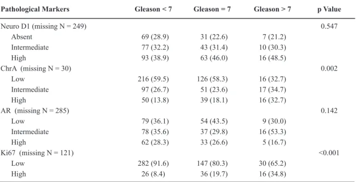
Pathological Markers Gleason < 7 Gleason = 7 Gleason > 7 p Value
Neuro D1 (missing N = 249) 0.547
Absent 69 (28.9) 31 (22.6) 7 (21.2)
Intermediate 77 (32.2) 43 (31.4) 10 (30.3)
High 93 (38.9) 63 (46.0) 16 (48.5)
ChrA (missing N = 30) 0.002
Low 216 (59.5) 126 (58.3) 16 (32.7)
Intermediate 97 (26.7) 51 (23.6) 17 (34.7)
High 50 (13.8) 39 (18.1) 16 (32.7)
AR (missing N = 285) 0.142
Low 79 (36.1) 54 (43.5) 9 (30.0)
Intermediate 78 (35.6) 37 (29.8) 16 (53.3)
High 62 (28.3) 33 (26.6) 5 (16.7)
Ki67 (missing N = 121) <0.001
Low 282 (91.6) 147 (80.3) 30 (65.2)
High 26 (8.4) 36 (19.7) 16 (34.8)
* tables entries are absolute numbers and percentages. ChrA = chromogranin-A; AR = androgen receptor; Ki67 = Ki67 label index.
Table 3 – Spearman’s correlation matrix of marker tissue reactivity*.
* tables entries are correlation coeficients and p Values. ChrA = chromogranin-A; AR = androgen receptor; Ki6 7= Ki67 label in -dex.
NeuroD1 ChrA AR Ki67
NeuroD1 - 0.187 (<0.001) 0.189 (0.004) 0.013 (0.806)
ChrA - - 0.151 (0.004) 0.164 (<0.001)
AR - - - 0.091 (0.115)
-ously investigated the effects of cAMP on epithelial
prostate cancer cell lines detecting a signiicant varia -tion of HOX-D gene expression and identifying the upstream area of the HOX-D locus on chromosome 2q31-33 as potentially involved in a neurogenic program connected to NED (17). Among the genes located in this genomic area, NeuroD1 expression has been related to PCa (14). New evidences have further stressed the use of pro-neural transcription factors, including NeuroD1, as cancer biomarkers (24), sug-gesting that the aberrant initiation of differentiation programs may confer a selective advantage. The ob-servation that in different PCa models (human derived neoplastic cell-lines, transgenic mouse tumors and patient samples) the hallmarks of neural transdiffer-entiation along the progression to metastatic disease were associated with changes in the expression of activator-type beta-Helix-Loop-Helix transcription factors including Hes6 and Ascl1 (24) strongly
cor-roborates our indings. The activation of pro-neural
transcription factors may well be a crucial step in PCa progression even in a naïve prostate cancer. Through the use of TMA methodology, we have compared dif-ferent NE markers in patients who underwent radical
prostatectomy for surgically treated naïve PCa. This immunohistochemical assay (IHC) showed a very low expression of NE markers in BPH (data not shown), as previously reported (14). On the other hand, in PCa we found a higher prevalence of NeuroD1 (73% of the cases), Ki-67 (85%) and AR (62%) over ChrA expression (42%), respectively. Herein, we showed that all the markers in our study are mutually and strongly associated (Tables 2 and 3).
T h e w e l l - d o c u m e n t e d c o r r e l a t i o n s (18,23,25,26) between the Ki-67 expression and the
aggressive features of PCa were conirmed here by the demonstration of its signiicant association with
Gleason score, ChrA expression and survival. On the other hand, the absence of correlation with the NeuroD1 and AR (Figure-2) could be explained by the fact that Ki-67 is only a marker of proliferation, whereas NeuroD1 and AR are implicated into the neu-roendocrine differentiation pathway (9,14,27,28).
The evidence of signiicant associations
between ChrA, NeuroD1 and AR probably sug-gests that their expression is not only correlated, but
also that the biological signiicance remains rather
obscure. We can speculate about the functional
rela-Table 4 – Multivariate analysis of association with markers distributed according to homogeneous pathological groups*.
NeuroD1 ChrA AR Ki67
Age, year 0.99 (0.96-1.01) 0.99 (0.97-1.01) 0.99 (0.97-1.02) 1.00 (0.96-1.03) Gleason score ^
<7 (reference)
7 1.44 (0.96-2.16) 1.06 (0.75-1.49) 0.77 (0.50-1.17) 2.53 (1.44-4.45)
>7 1.72 (0.82-3.61) 2.88 (1.59-5.22) 0.92 (0.45-1.91) 4.86 (2.21-10.7) Stage
pT2 (reference)
pT3 0.99 (0.57-1.73) 1.02 (0.67-1.56) 1.03 (0.62-1.73) 0.87 (0.43-1.73) pT4 0.70 (0.28-1.75) 1.14 (0.52-2.54) 0.93 (0.34-2.53) 1.76 (0.67-4.68) pTx 0.91 (0.47-1.76) 0.71 (0.38-1.32) 0.94 (0.48-1.87) 2.24 (0.95-5.27) Perineural invasion
Yes vs No 0.87 (0.59-1.27) 1.23 (0.89-1.69) 1.19 (0.80-1.77) 1.49 (0.89-2.51)
Figure 1 – Progression free survival according to the markers tissue reactivity. ChrA = chromogranin-A; AR = androgen receptor; Ki67 = Ki67 label index. In the irst panel, the blue line is referred to the group without NeuroD1 tissue reactivity indicated as “low”.
tionships in induction or sustain of a neuroendocrine activity or NED in PCa. In the low-grade (Gleason score < 7) group NeuroD1 and ChrA were detected in 71.1 and 40.5% of the cases, respectively. In our
opinion this inding is interesting and suggests that
NeuroD1 could be activated in prostate tumorigen-esis and that it probably is a more accurate marker of transdifferentiated cells or cells predisposed to an early NED.
Further experiments are needed to demon-strate that for the early detection of NE activity an integrated diagnostic panel (e.g. Dopa-Decarboxylase,
a-methylacyl-CoA racemase, IL-8 receptors) should be proposed (9).
A limitation of our study concerns the cut-off values used in the analyses, selected on the bases of the best possible discriminatory effect. This approach may predispose to detect false positive results. However, as Figure-1 indicates, only Ki67 robustly emerged as prognostic variable between the markers tested for
prognostic implication. A clear inding of the study is the easy identiication of high- and low-progression
intermediate group includes a signiicant fraction of
patients who experience progression of disease, urging for additional markers. Furthermore, we have used an historical (1971-1996) series of surgically treated patients (members of the Kaiser Foundation Health
Plan) for the evaluation of the prognostic signiicance
and the internal relationships of the markers. Thus, the likelihood of biases due to patient selection, surgical management, follow-up data and tissue quality is not negligible. On the other hand, the long median follow-up time (almost 6 years with the longest follow-follow-up time being over 12 years) is an interesting argument suggesting that PCa cells may remain dormant for long periods of time (PCa progression can also take place 10 years after prostatectomy). Moreover, data concerning the kind of progression detection (by the use of the preoperative and during the follow-up PSA values or traditional imaging test) are lacking, hinder-ing any possible inference relationship between kind of progression, PSA, NE markers and prognosis.
CONCLUSIONS
Our study highlights the utility of TMAs to
eficiently evaluate candidate prognostic markers in PCa. While some results conirm previous ind
-ings, for the irst time, to our knowledge, ChrA, AR
and NeuroD1 were evaluated together on a prostate TMA. The lack of association between the ChrA, AR and NeuroD1 tissue reactivity and survival suggest that these markers cannot be considered prognostic marker in patients surgically treated for
PCa. Nevertheless, a better identiication of such
neuroendocrine differentiation could advise about a better response rate after carboplatin-etoposide regimen chemotherapy (29).
Also, the highest reactivity of NeuroD1 over ChrA suggests its possible use, for example, as a target for antisense oligonucletide therapy (30).
CONFLICT OF INTEREST
None declared.
REFERENCES
1. Prostate Cancer Incidence and Mortality Worldwide in 2008: Globocan 2002. Available at http://globocan. iarc.fr/factsheets/cancers/prostate.asp Last access 11 Nov. 2010.
2. Altekruse SF, Kosary CL, Krapcho M, Neyman N, Aminou R, Waldron W, et al.: Edwards BK (eds). SEER Cancer Statistics Review, 1975-2007, National Cancer Institute. Bethesda, MD. Available at http:// seer.cancer.gov/csr/1975_2007/, based on November 2009 SEER data submission, posted to the SEER web site, 2010.
3. Schröder FH: Progress in understanding androgen-independent prostate cancer (AIPC): a review of potential endocrine-mediated mechanisms. Eur Urol. 2008; 53: 1129-37.
4. Tannock IF, de Wit R, Berry WR, Horti J, Pluzanska A, Chi KN, et al.: Docetaxel plus prednisone or mito-xantrone plus prednisone for advanced prostate cancer. N Engl J Med. 2004; 351: 1502-12.
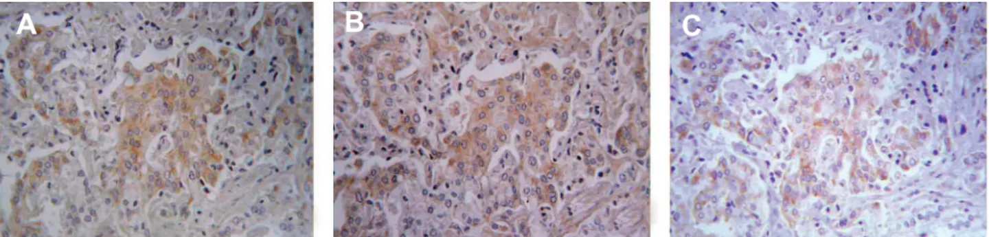
Figure 2 – Sequential slides of a same Gleason-4 prostate cancer, showing high chromogranin-A (A) and Neuro D1 (B) positivity with absent expression of androgen receptor (C). Immunohistochemical staining, X40.
5. Petrylak DP, Tangen CM, Hussain MH, Lara PN Jr, Jones JA, Taplin ME, et al.: Docetaxel and estramus-tine compared with mitoxantrone and prednisone for advanced refractory prostate cancer. N Engl J Med. 2004; 351: 1513-20.
6. Jiborn T, Bjartell A, Abrahamsson PA: Neuroendocrine differentiation in prostatic carcinoma during hormonal treatment. Urology. 1998; 51: 585-9.
7. Hirano D, Okada Y, Minei S, Takimoto Y, Nemoto N: Neuroendocrine differentiation in hormone refrac-tory prostate cancer following androgen deprivation therapy. Eur Urol. 2004; 45: 586-92; discussion 592.
8. Kokubo H, Yamada Y, Nishio Y, Fukatsu H, Honda N, Nakagawa A, et al.: Immunohistochemical study of chromogranin A in Stage D2 prostate cancer. Urology. 2005; 66: 135-40.
9. Cindolo L, Cantile M, Vacherot F, Terry S, de la Taille A: Neuroendocrine differentiation in prostate cancer: from lab to bedside. Urol Int. 2007; 79: 287-96. 10. Vashchenko N, Abrahamsson PA: Neuroendocrine
differentiation in prostate cancer: implications for new treatment modalities. Eur Urol. 2005; 47: 147-55. 11. Sciarra A, Gentile V, Monti S, Dattilo C, Gomez AA,
Salciccia S, et al.: Comparison of chromogranin A,
in-sulin-like growth factor 1 and prostate-speciic antigen
serum markers in prostate adenocarcinoma and benign prostatic hyperplasia. Urol Int. 2008; 80: 68-73. 12. Zitella A, Berruti A, Destefanis P, Mengozzi G, Torta
M, Ceruti C, et al.: Comparison between two com-mercially available chromogranin A assays in detecting neuroendocrine differentiation in prostate cancer and benign prostate hyperplasia. Clin Chim Acta. 2007; 377: 103-7.
13. Yamada Y, Nakamura K, Aoki S, Taki T, Matsubara H, Sai S, et al.: Is neuroendocrine cell differentiation detected using chromogranin A from patients with bone metastatic prostate cancer a prognostic factor for outcome? Oncol Rep. 2006; 15: 1309-13.
14. Cindolo L, Franco R, Cantile M, Schiavo G, Liguori G, Chiodini P, et al.: NeuroD1 expression in human prostate cancer: can it contribute to neuroendocrine differentiation comprehension? Eur Urol. 2007; 52: 1365-73.
15. Hvamstad T, Jordal A, Hekmat N, Paus E, Fosså SD: Neuroendocrine serum tumour markers in hormone-re-sistant prostate cancer. Eur Urol. 2003; 44: 215-21. 16. Berruti A, Dogliotti L, Mosca A, Bellina M, Mari M,
Torta M, et al.: Circulating neuroendocrine markers in patients with prostate carcinoma. Cancer. 2000; 88: 2590-7.
17. Cantile M, Kisslinger A, Cindolo L, Schiavo G, D’Antò
V, Franco R, et al.: cAMP induced modiications of
HOX D gene expression in prostate cells allow the identification of a chromosomal area involved in vivo with neuroendocrine differentiation of human advanced prostate cancers. J Cell Physiol. 2005; 205: 202-10.
18. Zellweger T, Ninck C, Mirlacher M, Annefeld M, Glass AG, Gasser TC, et al.: Tissue microarray analysis
re-veals prognostic signiicance of syndecan-1 expression
in prostate cancer. Prostate. 2003; 55: 20-9.
19. Kononen J, Bubendorf L, Kallioniemi A, Bärlund M, Schraml P, Leighton S, et al.: Tissue microarrays for
high-throughput molecular proiling of tumor speci -mens. Nat Med. 1998; 4: 844-7.
20. Abrahamsson PA, Wadström LB, Alumets J, Falkmer S, Grimelius L: Peptide-hormone- and serotonin-im-munoreactive cells in normal and hyperplastic prostate glands. Pathol Res Pract. 1986; 181: 675-83.
21. Komiya A, Suzuki H, Imamoto T, Kamiya N, Nihei N, Naya Y, et al.: Neuroendocrine differentiation in the progression of prostate cancer. Int J Urol. 2009; 16: 37-44.
22. Revelos K, Petraki C, Scorilas A, Stefanakis S, Malovrouvas D, Alevizopoulos N, et al.: Correlation of androgen receptor status, neuroendocrine differ-entiation and angiogenesis with time-to-biochemi-cal failure after raditime-to-biochemi-cal prostatectomy in clinitime-to-biochemi-cally localized prostate cancer. Anticancer Res. 2007; 27: 3651-60.
23. May M, Siegsmund M, Hammermann F, Loy V, Gunia
S: Prognostic signiicance of proliferation activity
and neuroendocrine differentiation to predict treat-ment failure after radical prostatectomy. Scand J Urol Nephrol. 2007; 41: 375-81.
24. Vias M, Massie CE, East P, Scott H, Warren A, Zhou Z, et al.: Pro-neural transcription factors as cancer markers. BMC Med Genomics. 2008; 1: 17.
25. Laitinen S, Martikainen PM, Tolonen T, Isola J, Tam-mela TL, Visakorpi T: EZH2, Ki-67 and MCM7 are prognostic markers in prostatectomy treated patients. Int J Cancer. 2008; 122: 595-602.
26. Rubio J, Ramos D, López-Guerrero JA, Iborra I, Collado A, Solsona E, et al.: Immunohistochemical expression of Ki-67 antigen, cox-2 and Bax/Bcl-2 in prostate cancer; prognostic value in biopsies and radical prostatectomy specimens. Eur Urol. 2005; 48: 745-51.
phenotype. Biochim Biophys Acta. 2001; 1539: 28-43.
28. Yuan TC, Veeramani S, Lin MF: Neuroendocrine-like prostate cancer cells: neuroendocrine transdifferentia-tion of prostate adenocarcinoma cells. Endocr Relat Cancer. 2007; 14: 531-47.
29. Loriot Y, Massard C, Gross-Goupil M, Di Palma M, Escudier B, Bossi A, et al.: Combining carboplatin
and etoposide in docetaxel-pretreated patients with castration-resistant prostate cancer: a prospective study evaluating also neuroendocrine features. Ann Oncol. 2009; 20: 703-8.
30. Hadaschik BA, Sowery RD, Gleave ME: Novel targets and approaches in advanced prostate cancer. Curr Opin Urol. 2007; 17: 182-7.
Accepted after revision:
August 28, 2010
Correspondence address:
Dr. Luca Cindolo Department of Urology
“S. Pio da Pietrelcina” Hospital Via C. De Lellis, 1
Vasto, 66054, Italy