Rate of Renal Cell Carcinoma Subtypes in Different Races
Alexander Sankin, Jacob Cohen, Hongbei Wang, Richard J. Macchia, Nicholas Karanikolas
State University of New York Downstate Medical School, Brooklyn, New York, USA
ABSTRACT
Purpose: We sought to identify racial differences among histological subtypes of renal cell carcinoma (RCC) between black and non-black patients in an equal-access health care system.
Materials and Methods: We established a multi-institutional, prospective database of patients undergoing partial or radi-cal nephrectomy between January 1, 2000 and Sept 31, 2009. For the purposes of this study, data captured included age at diagnosis, race, tumor size, presence of lymphovascular invasion, presence of capsular invasion, margin status, and tumor histology.
Results: 204 kidney tumors were identiied (Table-1). Of these, 117 (57.4%) were in black patients and 87 (42.6%) were
in non-black patients. Age at surgery ranged from 37 to 87 with a median of 62. Tumor size ranged from 1.0 to 22.0 cm with a median of 5.0 cm. Overall, tumors were composed of clear cell RCC in 97 cases (47.5%), papillary RCC in 65 cases (31.9%), chromophobe RCC in 13 cases (6.4%), collecting duct/medullary RCC in 2 cases (1.0%), RCC with multiple histological subtypes in 8 cases (3.9%), malignant tumors of other origin in 6 cases (2.9%), and benign histology in 13 cases (6.4%). Among black patients, papillary RCC was seen in 56 cases (47.9%), compared to 9 cases (10.3%) among non-black patients (p < 0.001) (Table-2). Clear cell RCC was present in 38 (32.5%) of black patients and in 59 (67.8%)
of non-blacks (p < 0.001).
Conclusions: In our study, papillary RCC had a much higher occurrence among black patients compared to non-black
patients. This is the irst study to document such a great racial disparity among RCC subtypes.
Key words: kidney neoplasm; renal cell; carcinoma Int Braz J Urol. 2011; 37: 29-34
INTRODUCTION
Renal cell carcinoma (RCC) is the seventh most common human malignancy, having accounted
for approximately 59,702 new cancer cases in the United States in 2009 (1). The histological classiica -tion of RCC has undergone several major revisions,
with the current 2004 World Health Organization (WHO) guidelines incorporating new cytogenetic
findings and molecular markers that help guide diagnosis, prognosis, and therapy (2). RCC is now recognized to be a complex neoplasm consisting of several different tumor subtypes, each with distinct
doi: 10.1590/S1677-55382011000700004
genetic and clinical features. Previous population-based studies on racial differences in incidence pat-terns and outcomes of RCC have failed to account for the different histological subtypes of RCC (3). We sought to identify racial differences in the patterns of histological subtypes of RCC in a racially diverse, equal-access health care system.
MATERIALS AND METHODS
2009. Our hospitals included a tertiary care academic
center, a veteran’s administration hospital, and an inner-city county hospital, ensuring a mixed ethnic population. All slides underwent centralized pathology
review using the WHO 2004 Classiication of renal cell
histology to classify tumors into clear cell, papillary,
chromophobe, or medullary/collecting duct varieties.
We did not differentiate between papillary RCC type 1 and papillary RCC type 2. Data was collected by reviewing patient charts and capturing the following parameters: age at diagnosis, race, tumor size, pres-ence of lymphovascular invasion, prespres-ence of capsular invasion, margin status, and tumor histology.
RESULTS
Two hundred and four kidney tumors were identiied (Table-1). Of these, 117 (57.4%) were in black patients and 87 (42.6%) were in non-black patients. Age at surgery ranged from 37 to 87 with a median age of 62. Among black patients the age range was 37 to 82 with a median of 63 and among non-black patients the age range was 40 to 87 with a median of 60.
Tumor size ranged from 1.0 to 22.0 cm with a median of 5.0 cm. The size of tumors among blacks
ranged from 1.4 to 20.0 cm and from 1.0 to 22.0 cm in
Table 1 – Baseline characteristics.
Black # (%) Non-Black # (%) Total
Patient characteristics
Number of patients 117 (57.4) 87 (42.6) 204 (100)
Age
Range 37 - 82 40 - 87 37 – 87
Mean 60.3 64.1 61.9
Median 63 60 62
Under 65 years old 74/117 (63.2) 52/87 (59.8) 126/204 (61.8)
65 years old or older 43/117 (36.8) 35/87 (40.2) 78/204 (38.2) p = 0.61 Tumor characteristics
Size (cm)
Range 1.4 – 20.0 1.0 – 22.0 1.0 – 22.0
Mean 5.9 5.1 5.6
Median 5.0 4.9 5.0
4 cm or less 41/113 (36.3) 35/82 (42.7) 76/195 (39.0) Greater than 4 cm and
less than or equal to 7 cm 38/113 (33.6) 31/82 (37.8) 69/195 (35.4)
Greater than 7 cm 34/113 (30.1) 16/82 (19.5) 50/195 (25.6) p = 0.25
Lymphovascular invasion
Yes 5/115 (4.3) 10/84 (11.9) 15/199 (7.5)
No 110/115 (95.7) 74/84 (88.1) 184/199 (92.5) p = 0.05
Capsular invasion
Yes 10/115 (8.7) 11/84 (13.1) 21/199 (10.6)
No 105/115 (91.3) 73/84 (86.9) 178/199 (89.4) p = 0.32
Margin status
Positive 6/115 (5.2) 5/84 (6.0) 11/199 (5.5)
non-blacks. There was no statistically signiicant dif -ference in tumor size between blacks and non-blacks when subdivided into groups of 4 cm or less, greater
than 4 cm and less than or equal to 7 cm, and greater than 7 cm ( p = 0.25).
The majority of tumors in blacks and non-blacks had absence of lymphovascular invasion (4.3% and 11.9%, p = 0.05), absence of capsular invasion (91.3% and 86.9%, p = 0.32), and negative surgical margins (94.8% and 94.0%, p = 0.82).
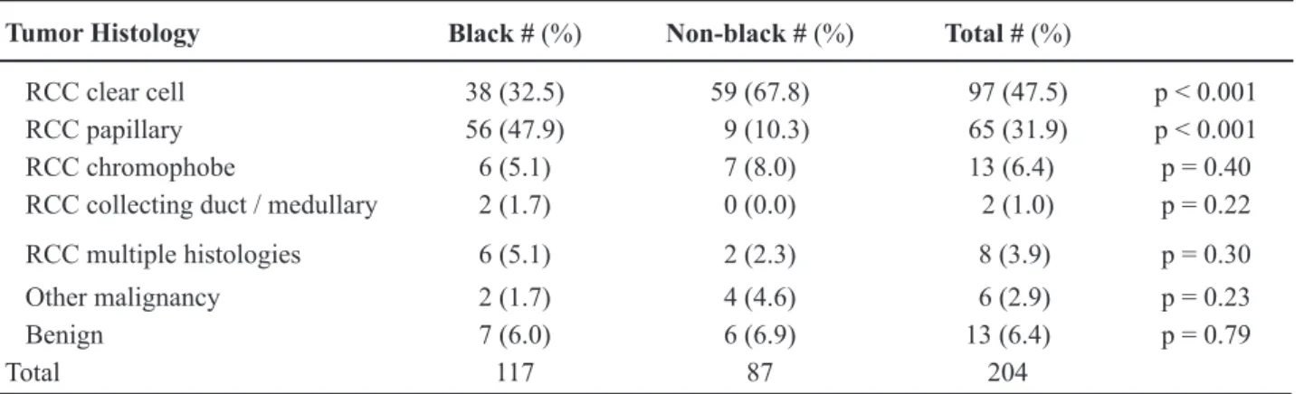
Overall, tumors were composed of clear cell RCC in 97 cases (47.5%), papillary RCC in 65 cases (31.9%), chromophobe RCC in 13 cases (6.4%), col
-lecting duct/medullary RCC in 2 cases (1.0%), RCC with multiple histological subtypes in 8 cases (3.9%), malignant tumors of other origin in 6 cases (2.9%), and benign histology in 13 cases (6.4%). Among black patients, papillary RCC was seen in 56 cases (47.9%), compared to 9 cases (10.3%) among non-black patients (p < 0.001) (Table-2). Clear cell RCC was present in 38 (32.5%) of black patients and in 59 (67.8%) of non-blacks (p < 0.001).
COMMENTS
Our study found a greater than four-fold
increase in occurrence of papillary RCC in blacks
when compared to non-blacks. This is the irst study
to document such a large ethnic disparity among RCC subtypes.
This phenomenon of neoplastic variability
among different races has been observed in other urologic cancers. Prostate cancer is a well-studied malignancy that tends to behave differently among different races. Compared with whites, blacks are more likely to be diagnosed with prostate cancer at a younger age (4). Black patients with prostate cancer are also less likely to be treated with surgical
interven-tion and have an overall lower survival (5).
There are many previously reported, large,
population-based studies elucidating the racial
dis-parities among patients with RCC. These reports,
however, have focused on topics such as overall survival and access to health care. A recent paper by Stafford et al. found that blacks had an overall increase in incidence of renal cell carcinoma and decrease in
survival rate when compared to other ethnicities (6).
Zini et al. concluded that black patients with RCC
were 50% less likely to undergo nephrectomy com
-pared to white patients (7). A study by Brendt et al.
also addresses the lower survival rate among blacks with renal cell carcinoma and proposes that this may be explained by both a higher number of comorbidities
and lower rate of surgical treatment (8). While these
studies have unearthed many interesting data trends among RCC cases, none have included information on histological subtypes of RCC.
Beyond the well-described association or renal medullary carcinoma with sickle-cell disease and trait in black patients, there has been very little re-ported regarding racial differences for the other RCC
Table 2 – Race and tumor histology.
Tumor Histology Black # (%) Non-black # (%) Total # (%)
RCC clear cell 38 (32.5) 59 (67.8) 97 (47.5) p < 0.001 RCC papillary 56 (47.9) 9 (10.3) 65 (31.9) p < 0.001
RCC chromophobe 6 (5.1) 7 (8.0) 13 (6.4) p = 0.40
RCC collecting duct / medullary 2 (1.7) 0 (0.0) 2 (1.0) p = 0.22 RCC multiple histologies 6 (5.1) 2 (2.3) 8 (3.9) p = 0.30
Other malignancy 2 (1.7) 4 (4.6) 6 (2.9) p = 0.23
Benign 7 (6.0) 6 (6.9) 13 (6.4) p = 0.79
Total 117 87 204
histologies (9). Historically, clear cell tumors have
accounted for 70-80% of RCC, followed by 10-15% for papillary, 3-5% for chromophobe, and 1-2% for medullary/collecting duct (10). Our reported 47.9%
incidence of papillary tumors among black patients is
the irst of its kind and is more than four-fold the inci -dence of papillary tumors among non-black patients in
our study. One explanation for this observation may be
a genomic predisposition for blacks to express genes unique for the development of papillary RCC.
Papillary RCC is commonly found in two classic genetic syndromes, hereditary papillary RCC and hereditary leiomyomatosis and renal cell cancer syndrome. Papillary tumors have also previously been reported to be associated with acquired renal cystic
disease. The chromosomal abnormalities for papillary
tumors are distinct from the chromosome 3 and Von-Hippel Lindau gene abnormalities of clear cell tumors. Papillary tumors typically demonstrate trisomy of
chromosomes 7 and 17 and loss of the Y chromo -some. Mutations in the met-oncogene, which encodes hepatocyte growth factor, is the usual mutation for papillary tumors, which usually display multicentric-ity and hypovascularmulticentric-ity. As expected with the new era of targeted therapies for RCC, unique molecular agents separate from tyrosine kinase inhibitors are being developed for papillary tumors. If black patients with RCC truly have a predisposition for expression
of papillary type tumors, they may beneit from early
counseling and treatment with novel agents targeting this unique tumor.
More recently, papillary RCC has been sub-divided into type 1 and type 2, with type 2 tumors ex-hibiting more aggressive clinicopathological features
and worse prognosis (11). Our retrospective analysis does not allow statements regarding the inluence
of tumor subtype on clinical course and prognosis following treatment. As such, we do not present any outcomes data and do not differentiate between
subtypes of papillary RCC. Of note, there have been
studies demonstrating no differences in disease-free survival between papillary and clear cell RCC (12).
One must also consider the possible inluence
of confounding factors on the variable expression of RCC subtypes. Previous studies have shown that there is a predisposition among males and patients with end stage renal disease to develop papillary RCC (13,14).
This potential bias should be taken into consideration
when designing future studies aimed to capture racial differences observed among RCC subtypes.
CONCLUSION
We report for the irst time, to our knowledge,
that black patients seem to have an increased risk of developing papillary RCC than the general population.
This result needs to be conirmed by large,
population-based studies that examine different RCC histologies against a wide range of demographic, geographic, and environmental factors. Given the complex genetic and molecular basis for RCC and its role in response to adjuvant treatments, race and ethnicity may be important factors when counseling patients regarding prognosis and treatment outcomes.
CONFLICT OF INTEREST
None declared.
REFERENCES
1. American Cancer Society: Cancer Facts & Figures 2009. Atlanta: American Cancer Society 2009.
Avail-able at. http://oralcancerfoundation.org/facts/pdf/
Us_Cancer_Facts.pdf
2. Lopez-Beltran A, Scarpelli M, Montironi R, Kirkali
Z: 2004 WHO classiication of the renal tumors of the adults. Eur Urol. 2006; 49: 798-805.
3. Vaishampayan UN, Do H, Hussain M, Schwartz K: Racial disparity in incidence patterns and outcome of
kidney cancer. Urology. 2003; 62: 1012-7.
4. Shao YH, Demissie K, Shih W, Mehta AR, Stein MN,
Roberts CB, et al.: Contemporary risk proile of pros -tate cancer in the United S-tates. J Natl Cancer Inst.
2009; 101: 1280-3.
5. Schwartz K, Powell IJ, Underwood W 3rd, George J,
Yee C, Banerjee M: Interplay of race, socioeconomic status, and treatment on survival of patients with
pros-tate cancer. Urology. 2009; 74: 1296-302.
6. Stafford HS, Saltzstein SL, Shimasaki S, Sanders C, Downs TM, Sadler GR: Racial/ethnic and gender dis -parities in renal cell carcinoma incidence and survival.
7. Zini L, Perrotte P, Capitanio U, Jeldres C, Duclos A,
Arjane P, et al.: Race affects access to nephrectomy
but not survival in renal cell carcinoma. BJU Int. 2009; 103: 889-93.
8. Berndt SI, Carter HB, Schoenberg MP, Newschaffer
CJ: Disparities in treatment and outcome for renal cell cancer among older black and white patients. J Clin
Oncol. 2007; 25: 3589-95.
9. Assad L, Resetkova E, Oliveira VL, Sun W, Stewart
JM, Katz RL, et al.: Cytologic features of renal
medul-lary carcinoma. Cancer. 2005; 105: 28-34.
10. Reuter VE, Presti JC Jr: Contemporary approach to the
classiication of renal epithelial tumors. Semin Oncol. 2000; 27: 124-37.
11. Yamashita S, Ioritani N, Oikawa K, Aizawa M, Endoh
M, Arai Y: Morphological subtyping of papillary renal
cell carcinoma: clinicopathological characteristics and
prognosis. Int J Urol. 2007; 14: 679-83.
12. Dall’Oglio MF, Antunes AA, Pompeo AC, Mosconi
A, Leite KR, Srougi M: Prognostic relevance of the histological subtype of renal cell carcinoma. Int Braz
J Urol. 2008; 34: 3-8.
13. Schrader AJ, Sevinc S, Olbert PJ, Hegele A, Varga Z, Hofmann R: Gender-speciic characteristics and survival of renal cell carcinoma. Urologe A. 2008; 47: 1182, 1184-6.
14. Ishikawa I, Kovacs G: High incidence of papillary re-nal cell tumours in patients on chronic haemodialysis.
Histopathology. 1993; 22: 135-9.
Accepted after revision: July 30, 2010
Correspondence address: Dr. Alexander Sankin Division of Urology
State Univ. of New York Downstate Medical School
450 Clarkson Ave,
Brooklyn, NY, 11203, USA
Fax: + 1 718 270-3848
E-mail: alexsankin@gmail.com
EDITORIAL COMMENT
We have known for a while that renal cell carcinomas (RCC) are more common in blacks compared to non-Blacks and Asians. Not only more common, RCC tend to be more aggressive in blacks,
with a higher disease-speciic mortality even when
tumor size and stage are independent variables. Black
patients have a signiicantly higher incidence rate and lower survival rate than all other races/ethnicities even
when having more localized cancer.
This study lists the rates of different types
of renal cell carcinoma in a population composed of many ethnicities. In blacks, almost half of all tumors
were papillary RCC, compared to only 10% in non-blacks. This disparity is not commonly addressed.
In addition, it found a much lower rate of clear cell
RCC in blacks, which is not a common inding in the literature. The readers could have beneited by data
on survival or progression, which unfortunately the study lacks. Nonetheless, given such racial disparities in renal cell carcinoma incidence in this one study,
one should consider expanding and conirming these indings that may help elucidate biological, behav -ioral and environmental factors that can potentially be addressed.
Dr. Fabio Tavora
EDITORIAL COMMENT
Renal cell carcinoma (RCC) tends to manifest variable prognoses and outcomes. Previous articles have already pointed toward prognostic parameters including pathological subtypes and ethnic origin. Yet the issue of race was not fully addressed. Epide-miologic literature showed shorter survival in Black
Americans with RCC. Tripathi et al. concluded that the overall survival for metastatic RCC was signii -cantly shorter for Black Americans. Vaishampayan et al. had a similar conclusion for local RCC, empha-sizing a higher incidence, poorer outcome in Black patients with a similar age and stage. However, the histology subtypes disparities were not mentioned, demonstrating the advantage of the recent manuscript based on data that was retrieved from an equal-access health care system. Stafford published a major study comparing ethnic groups (White, Black, Hispanic and
Asian -Paciic) concluding that Black patients tend to
have a higher incidence and a shorter survival while Asian patients demonstrate the opposite. Yet even this article did not point to the histology subdivision.
Herein we can appreciate an important study advancing our knowledge regarding the correlation between race and RCC histology subtypes. Further studies should investigate the papillary predominance
of the RCC in the black population while paradoxi-cally demonstrating a poorer prognosis than what was known previously for that histological subtype.
REFERENCES
1. Tripathi RT, Heilbrun LK, Jain V, Vaishampayan UN:
Racial disparity in outcomes of a clinical trial popula-tion with metastatic renal cell carcinoma. Urology.
2006; 68: 296-301.
2. Vaishampayan UN, Do H, Hussain M, Schwartz K: Racial disparity in incidence patterns and outcome of
kidney cancer. Urology. 2003; 62: 1012-7.
3. Berndt SI, Carter HB, Schoenberg MP, Newschaffer CJ: Disparities in treatment and outcome for renal cell cancer among older black and white patients. J Clin
Oncol. 2007; 25: 3589-95.
4. Zini L, Perrotte P, Capitanio U, Jeldres C, Duclos A, Arjane P, et al.: Race affects access to nephrectomy
but not survival in renal cell carcinoma. BJU Int. 2009; 103: 889-93.
5. Stafford HS, Saltzstein SL, Shimasaki S, Sanders C, Downs TM, Sadler GR: Racial/ethnic and gender dis -parities in renal cell carcinoma incidence and survival.
J Urol. 2008; 179: 1704-8.