113
Radiology Page
International Braz J Urol Vol. 37 (1): 113-114, January - February, 2011
Emphysematous Cystitis as Complication in Chronic Rejection of
Renal Transplant
Erich K. Lang, Karl Zhang, Daniel Thorner, Quan D. Nguyen
Department of Radiology (EKL, KZ, DT, QDN), SUNY, Downstate Medical School, Brooklyn, New
York, USA and Johns Hopkins Medical Institutions, Bayview Center, Baltimore, Maryland, USA
Radiology PageRadiology Page
doi: 10.1590/S1677-55382010000700015
This 43-year-old Caucasian female presented in a septic condition in the emergency room. At the time of admission her temperature was 38.5 Celsius, white blood cells (WBC) 12800, red blood cells 3.8 mill, hemoglobin 10.8, hematocrit 36, urea 28 mg/dl, serum creatinine 3.2 mg/dl, K 5.8 meq/l, Na 128 meq/ l, alk ptsa 142 U/l. Urine analysis 50 WBC/hpf, in-numerable bacteria/hpf, cellular debries, gas bubbles. Both urine and later blood cultures revealed E. coli.
The lower abdomen and pelvis were exqui-sitely tender to palpation. Patient had been hemodia-lyzed 2 days earlier. Patient had received a cadaver-transplant 3 years ago, which functioned well until 4 months ago. At this time chronic rejection was diagnosed. An antegrade pyelogram revealed a stric-ture at the implant site of the cadaver ureter, dilatation and possible ulcerations of the mid – and upper ureter. Bullous edema of the native bladder was seen, most prominent near the implant site. Immunosuppressive therapy was instituted and despite these efforts, func-tion of the transplant kidney continued to deteriorate
and the patient was inally put on hemodialysis 1
month ago. It was contemplated to perform a uretero-neo-calicostomy with the still present native ureter to hopefully salvage the kidney
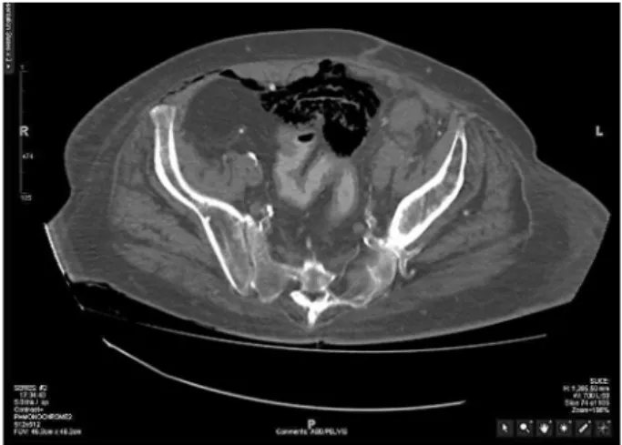
A non-contrast multi-detector computed tomography demonstrated gas in the submucosa and bladder, and also extravesical anterior to the bladder (Figure-1). Strands of debries and sloughed tissue sur-rounded by air are seen in the bladder lumen (arrow). Gas has dissected along the anterior abdominal wall
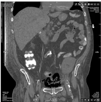
(Figure-2). A coronal reconstruction shows relatively little striation in the perirenal space. There is edema in the peripelvic area and around the upper ureter (Figure-3).
To control the fulminating gas forming in-fection, bladder, transplant kidney and ureter were removed, the space of Retzius drained. Depending on the severity of the infection and underlying conditions such as diabetes mere control of the diabetes and
ap-propriate antibiotic therapy may sufice while severe
forms may mandate surgical intervention to remove the necrotic debries (1-3). After prolonged antibiotic
114
Radiology Page
therapy and hemodialysis the patient recovered and received a second successful transplant kidney.
REFERENCES
1. Dinckan R, Tekin R, Turkyilmaz S, Kacak H, Gurkan A, Erdogen G, Tuncer M: Early and late urologic complications corrected surgically following renal transplantation. Transpl. Int. 2007; 20: 702-7. 2. Akalin E, Hyde C, Schmitt G, Kaufman J, Hamburger
RJ: Emphysematous cystitis and pyelitis in a diabetic
renal transplant recipient. Transplantation 1996; 62: 1024 -6.
3. Davavri HR, Yarmohammadi H, Malekhosseini SA, Salahi H, Bahador A, Salchipoun S: Urologic complications in 980 consecutive patients with renal transplantation. Int. J. Urol. 2006; 13: 1271-5.
Figure 2 – Gas is dissecting anteriorly into the space of Retzius and laterally toward the abdominal wall and inguinal fossa. Note tissue debries outlined by gas in the bladder and a soft tissue mass (ileal loop) indenting the bladder.
Figure 3 – A coronal reconstruction shows debries and an ad-herent ileal loop outlined by the gas-illed bladder. A transplant kidney is seen on the left side, with only minimal stranding in the perirenal space. However edema is seen peripelvic and peri-ureteral space.
Correspondence address: Dr. Erich K. Lang
Departments of Urology and Radiology SUNY, Downstate Health Science Center 455 Lenox Road