Risk of Catecholamine Crisis in Patients Undergoing Resection of
Unsuspected Pheochromocytoma
Gina Song, Bonnie N. Joe, Benjamin M. Yeh, Maxwell V. Meng, Antonio C. Westphalen, Fergus
V. Coakley
Departments of Radiology (GS, BNJ, BMY, ACW, FVC) and Urology (MVM), University of California
San Francisco, San Francisco, California, USA
ABSTRACT
Purpose: To report the risk of catecholamine crisis in patients undergoing resection of unsuspected pheochromocytoma. Materials and Methods: Over a four-year period, we retrospectively identiied four patients who underwent resection of adrenal pheochromocytoma in whom the diagnosis was unsuspected based on preoperative clinical, biochemical, and imaging evaluation.
Results: None of the patients exhibited preoperative clinical features of catecholamine excess. Preoperative biochemical
screening in two patients was normal. CT scan performed in all patients demonstrated a nonspeciic enhancing adrenal
mass. During surgical resection of the adrenal mass, hemodynamic instability was observed in two of four patients, and one of these two patients also suffered a myocardial infarct.
Conclusion: Both surgeons and radiologists should maintain a high index of suspicion for pheochromocytoma, as the
tu-mor can be asymptomatic, biochemically negative, and have nonspeciic imaging features. Resection of such unsuspected
pheochromocytomas carries a substantial risk of intraoperative hemodynamic instability.
Key words: adrenal gland neoplasms; imaging; surgery; pheochromocytoma; catecholamines
Int Braz J Urol. 2011; 37: 35-41
INTRODUCTION
While there is widespread awareness of the classical clinical and radiological features of pheochromocytoma, it is perhaps less well known that 10% of pheochromocytomas are not associated with symptoms of excess catecholamine production, and up to 35% of pheochromocytomas have atypical imaging indings (1-5). Accordingly, the diagnosis may be unsuspected, and an indeterminate adrenal or retroperitoneal mass that is actually a pheochromo-cytoma could undergo resection without preoperative commencement of biochemical blockade. Such a patient is then at risk for a potentially a life-threaten-ing intraoperative catecholamine crisis. However,
doi: 10.1590/S1677-55382011000700005
while the risks of iodinated contrast administration and percutaneous biopsy have been reported in these unsuspected pheochromocytomas (6-8), to our knowledge, the risk of surgery in this population has not been well described. We recently encountered several patients who underwent surgical resection of unsuspected pheochromocytomas. Therefore, we un-dertook this study to report the risk of catecholamine crisis in patients undergoing resection of unsuspected pheochromocytoma.
MATERIALS AND METHODS
the requirement for informed consent. Four cases of pathologically proven pheochromocytoma that underwent attempted surgical resection were identi-ied by the study authors between 2004 and 2007. All available imaging studies and medical records of these patients were reviewed by the principal investigator. Preoperative imaging consisted of CT scan examination obtained with multiple contiguous axial images performed in the venous phase following intravenous contrast agent in all four patients with
additional delayed phases obtained in two patients. One of the patients also underwent whole body PET scan after the administration of 16.4 milliCurie intravenous luoro-2-deoxy-D-glucose (FDG) and utilizing attenuation-corrected regional emission images. None of the patients underwent imaging with iodine-131-meta-iodobenzylguanidine. The diagnosis of pheochromocytoma was conirmed by pathology of the surgically resected specimen in all four patients.
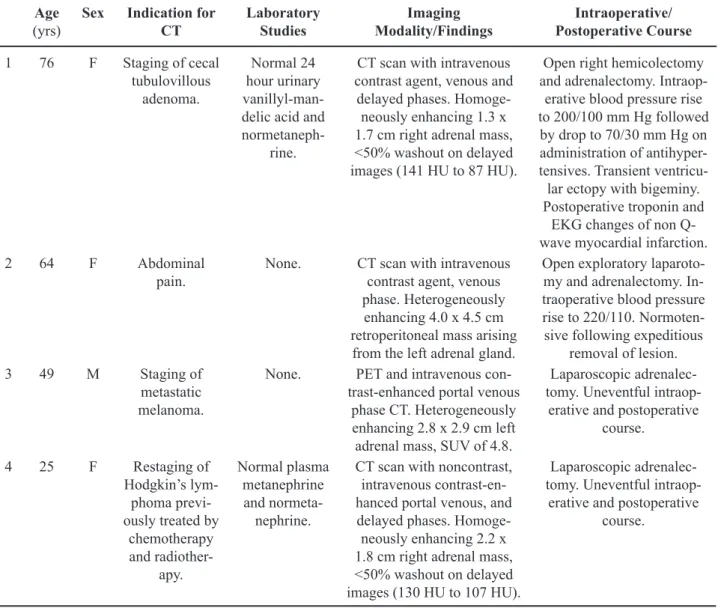
Table 1 – Clinical, imaging, and operative characteristics of four patients undergoing resection of an unsuspected pheo-chromocytoma.
Age
(yrs) Sex Indication for CT
Laboratory Studies Imaging Modality/Findings Intraoperative/ Postoperative Course
1 76 F Staging of cecal
tubulovillous adenoma.
Normal 24
hour urinary vanillyl-man-delic acid and
normetaneph-rine.
CT scan with intravenous contrast agent, venous and
delayed phases. Homoge-neously enhancing 1.3 x
1.7 cm right adrenal mass,
<50% washout on delayed
images (141 HU to 87 HU).
Open right hemicolectomy and adrenalectomy. Intraop-erative blood pressure rise
to 200/100 mm Hg followed by drop to 70/30 mm Hg on
administration of antihyper-tensives. Transient ventricu-lar ectopy with bigeminy. Postoperative troponin and
EKG changes of non
Q-wave myocardial infarction.
2 64 F Abdominal pain.
None. CT scan with intravenous contrast agent, venous phase. Heterogeneously
enhancing 4.0 x 4.5 cm
retroperitoneal mass arising from the left adrenal gland.
Open exploratory laparoto-my and adrenalectolaparoto-my. In-traoperative blood pressure
rise to 220/110. Normoten -sive following expeditious
removal of lesion.
3 49 M Staging of
metastatic melanoma.
None. PET and intravenous con-trast-enhanced portal venous
phase CT. Heterogeneously
enhancing 2.8 x 2.9 cm left adrenal mass, SUV of 4.8.
Laparoscopic
adrenalec-tomy. Uneventful intraop -erative and postop-erative
course.
4 25 F Restaging of Hodgkin’s
lym-phoma previ-ously treated by
chemotherapy and radiother-apy. Normal plasma metanephrine and normeta-nephrine.
CT scan with noncontrast, intravenous contrast-en-hanced portal venous, and delayed phases.
Homoge-neously enhancing 2.2 x 1.8 cm right adrenal mass,
<50% washout on delayed
images (130 HU to 107 HU).
Laparoscopic
adrenalec-tomy. Uneventful intraop -erative and postop-erative
RESULTS
The clinical and radiologic characteristics and intraoperative and postoperative course of the four patients in this study are summarized in Table-1. Three of the patients underwent imaging for purposes of staging a malignancy, and one patient underwent imaging for abdominal pain. None of the four patients exhibited preoperative clinical features of catecholamine excess. One patient underwent serologic analysis, which demonstrated normal lev-els of plasma metanephrine and normetanephrine. Another patient underwent urinary analysis, which demonstrated normal levels of urinary vanillyl-mandelic acid and normetanephrine. All patients
demonstrated an adrenal mass (mean diameter 2.7 cm, range 1.7 to 4.5 cm) with associated heteroge -neous (n = 2) or homoge-neous (n = 2) enhancement on CT scan. Delayed images obtained in two cases demonstrated less than ifty percent washout. One patient had additional imaging with PET, which demonstrated increased FDG uptake within the le -sion. The radiological indings of the four patients are highlighted in Figures-1 to 4. All 4 patients proceeded to adrenalectomy, either open (n = 2) or laparoscopic (n = 2). The intraoperative course of two patients was notable for blood pressure lability peaking at 200/100 to 220/110 mmHg systolic/dia -stolic. The postoperative course of one of these two patients was complicated by an elevated troponin level to 9.7 and an electrocardiogram consistent with a non Q-wave myocardial infarction.
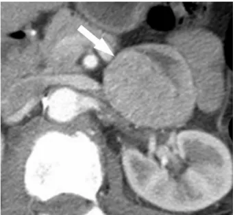
Figure 1 –Axial contrast-enhanced portal venous phase CT scan in a 76 year old woman undergoing preoperative staging of a cecal mass (Case 1 in Table-1). A homogeneously enhancing right adrenal mass (arrow) is seen, and showed less than 50% washout on delayed images (not shown). The patient proceeded to right hemicolectomy and adrenalectomy, because the working diagnosis before surgery was of possible adrenal metastasis. Surgery was complicated by hemodynamic instability and a postoperative non-Q wave myocardial infarction. Final pathol-ogy revealed a cecal tubulovillous adenoma and an adrenal pheochromocytoma.
Figure 2 – Axial contrast-enhanced portal venous phase CT scan in a 64 year old woman with abdominal pain (Case 2 in Table-1). A heterogeneously enhancing mass (arrow) is seen anterior to the left renal vessels. The left adrenal gland (not shown) was
identi-ied just superior to this mass. Open exploratory laparotomy and
adrenalectomy was performed, based on a preoperative working diagnosis of retroperitoneal sarcoma. Surgery demonstrated a mass arising exophytically from the inferior aspect of the left adrenal gland, and was complicated by intraoperative
hemody-namic instability. The mass was removed expeditiously and inal
Figure 3 – A) Axial contrast-enhanced portal venous phase CT scan in a 49 year old man with melanoma (Case 3 in Table-1) shows a homogeneously enhancing left adrenal mass (arrow). B)
Coronal PET image shows increased luoro-2-deoxy-D-glucose
(FDG) uptake in the mass (arrow). The patient proceeded to laparoscopic adrenalectomy for presumed metastatic disease, with an uneventful intraoperative and postoperative course. Final pathology revealed a diagnosis of pheochromocytoma.
Figure 4 –Axial contrast-enhanced portal venous phase CT scan in a 25 year old woman undergoing re-staging of Hodgkin’s lymphoma previously treated by chemotherapy and radiotherapy (Case 4 in Table-1) shows a homogeneously enhancing right adrenal mass (arrow). The patient proceeded to laparoscopic adrenalectomy for suspected recurrent lymphoma, with an un-eventful intraoperative and postoperative course. Final pathology revealed a diagnosis of pheochromocytoma.
COMMENTS
Our study illustrates the importance of keep-ing a high index of suspicion for the possibility of
pheochromocytoma for any retroperitoneal mass. While hemodynamic instability associated with surgi-cal and laparoscopic resection of known or suspected pheochromocytoma has been reported (9,10), the risk of resection in the population of unsuspected pheochromocytoma has not been well described to our knowledge. Two out of four of our patients dem-onstrated intraoperative hemodynamic instability that may have been prevented by appropriate preoperative alpha-blockade. The postoperative morbidity in our population might also have been averted, as the myo-cardial infarction of one of our patients was presum-ably related to the sudden intraoperative hypertensive challenge.
Deining typical characteristics of a pheo -chromocytoma by CT is dificult as heterogeneous enhancement and poor washout as well as FDG uptake on PET may also be seen in a metastasis or adenoma (1,4,11). Further imaging by magnetic resonance (MR) is also problematic, as up to 35% of pheochro -mocytomas do not exhibit the “lightbulb bright” high T2 signal classically associated with pheochromocy -tomas (1). Current guidelines on preoperative diagno
-A
sis thus include additional studies after diagnosis of an adrenal mass with nonspeciic characteristics on cross-sectional imaging. Metaiodobenzylguanidine (MIBG) reportedly has an excellent speciicity of up to 100% and may increase the sensitivity of pheochro-mocytoma detection to around 80% (10,12). Given the suboptimal sensitivity of biochemical markers alone, a combination of MIBG imaging supplemented with biochemical testing is currently recommended (12,13). Such an imaging approach may result in better preoperative identiication of pheochromocy -tomas, and facilitate commencement of pre-operative alpha blockade. Both appropriate pretreatment with alpha blockade and readily available intraoperative antihypertensive agents have been shown to decrease intraoperative lability (14). While pretreatment does not exclude the possibility of intraoperative luctua -tions in blood pressure (9,10), more favorable blood pressure control may be achieved with a combination of pretreatment and intraoperative medications (15). In a recent series of 24 patients who underwent laparo -scopic adrenalectomy for adrenal pheochromocytoma (most were treated pre-operatively with prazosin), no cases of intra-operative hemodynamic instability were reported (16). While such pharmacological blockade may prevent clinically significant hemodynamic changes, it may not prevent biochemical changes. For example, analysis of serial catecholamine levels in 11 patients undergoing 12 laparoscopic adrenalectomies while being maintained on an intravenous alpha 1 blocker showed signiicant elevations related to the induction of pneumoperitoneum and manipulation of the adrenal gland (17).
Our report has several limitations. The study is a small retrospective case series, and cases were not identiied systematically. Patients with enhanc -ing adrenal masses with poor washout or increased FDG uptake at PET imaging undergoing resection were not studied prospectively, and as such, the frequency of pheochromocytoma in adrenal masses with nonspeciic imaging characteristics is unknown. Only two out of four patients received screening for catecholamines, and systematic evaluation of the rate of false negatives either by urinary or plasma analysis was therefore not made. Further imaging with MIBG was not obtained in any of our patients, and the rate of false negatives by MIBG was thus not obtained.
In conclusion, we report that two of four pa-tients who underwent resection of unsuspected pheo-chromocytoma sustained intraoperative hemodynamic instability. This study emphasizes the asymptomatic presentation, nonspeciic imaging characteristics, potential for false negative preoperative laboratory analysis, and resultant risk of catecholamine crisis in patients with adrenal masses. Accordingly, both sur -geons and radiologists should maintain a high index of suspicion for pheochromocytoma before resection of nonspeciic adrenal masses even in asymptomatic patients. Further studies to better delineate the imag-ing and biochemical preoperative evaluation of these patients are required.
CONFLICT OF INTEREST
None declared.
REFERENCES
1. Blake MA, Kalra MK, Maher MM, Sahani DV, Swee
-ney AT, Mueller PR, et al.: Pheochromocytoma: an imaging chameleon. Radiographics. 2004; 24 (Suppl 1): S87-99.
2. Mayo-Smith WW, Boland GW, Noto RB, Lee MJ: State-of-the-art adrenal imaging. Radiographics. 2001; 21: 995-1012.
3. Sutton MG, Sheps SG, Lie JT: Prevalence of clinically unsuspected pheochromocytoma. Review of a 50-year autopsy series. Mayo Clin Proc. 1981; 56: 354-60. 4. Dunnick NR, Korobkin M: Imaging of adrenal inciden
-talomas: current status. AJR Am J Roentgenol. 2002; 179: 559-68.
5. Mansmann G, Lau J, Balk E, Rothberg M, Miyachi Y, Bornstein SR: The clinically inapparent adrenal mass: update in diagnosis and management. Endocr Rev. 2004; 25: 309-40.
6. Mukherjee JJ, Peppercorn PD, Reznek RH, Patel V, Kaltsas G, Besser M, et al.: Pheochromocytoma:
effect of nonionic contrast medium in CT on
circu-lating catecholamine levels. Radiology. 1997; 202: 227-31.
7. Bessell-Browne R, O’Malley ME: CT of pheochro
-mocytoma and paraganglioma: risk of adverse events
with i.v. administration of nonionic contrast material.
8. Casola G, Nicolet V, vanSonnenberg E, Withers C, Bretagnolle M, Saba RM, et al.: Unsuspected pheo
-chromocytoma: risk of blood-pressure alterations during percutaneous adrenal biopsy. Radiology. 1986; 159: 733-5.
9. Kinney MA, Warner ME, vanHeerden JA, Horlocker TT, Young WF Jr, Schroeder DR, et al.: Perianesthetic
risks and outcomes of pheochromocytoma and
para-ganglioma resection. Anesth Analg. 2000; 91: 1118-23.
10. Bravo EL, Tagle R: Pheochromocytoma: state-of-the-art and future prospects. Endocr Rev. 2003; 24: 539-53.
11. Yoon JK, Remer EM, Herts BR: Incidental pheochro -mocytoma mimicking adrenal adenoma because of
rapid contrast enhancement loss. AJR Am J Roent
-genol. 2006; 187: 1309-11.
12. Guller U, Turek J, Eubanks S, Delong ER, Oertli D, Feldman JM: Detecting pheochromocytoma: deining the most sensitive test. Ann Surg. 2006; 243: 102-7. 13. Kudva YC, Sawka AM, Young WF Jr: Clinical review
164: The laboratory diagnosis of adrenal pheochromo
-cytoma: the Mayo Clinic experience. J Clin Endocrinol Metab. 2003; 88: 4533-9.
14. Weismann D, Fassnacht M, Schubert B, Bonig R, Tschammler A, Timm S, et al.: A dangerous
liaison--pheochromocytoma in patients with malignant disease.
Ann Surg Oncol. 2006; 13: 1696-701.
15. Chung PC, Ng YT, Hsieh JR, Yang MW, Li AH: La -betalol pretreatment reduces blood pressure instability
during surgical resection of pheochromocytoma. J Formos Med Assoc. 2006; 105: 189-93.
16. Castilho LN, Simoes FA, Santos AM, Rodrigues TM, dos Santos Junior CA: Pheochromocytoma: a long-term follow-up of 24 patients undergoing laparoscopic adrenalectomy. Int Braz J Urol. 2009; 35: 24-31; dis
-cussion 32-5.
17. Rocha MF, Tauzin-Fin P, Vasconcelos PL, Ballanger P: Assessment of serum catecholamine concentrations
in patients with pheochromocytoma undergoing
vide-olaparoscopic adrenalectomy. Int Braz J Urol. 2005; 31: 299-307; discussion 307-8.
EDITORIAL COMMENT
The authors of this paper deserve to be com-plimented because, despite the modest data, that was analyzed retrospectively, they raise some issues that are very relevant for those who study and treat adrenal diseases.
First of all, they point out that adrenal masses, and I would add retroperitoneal masses in general, can be pheochromocytomas or paragangliomas without clinical signs or with very subtle symptoms, which do
not lead the physician to consider lesions that produce adrenergic substances. In my personal experience (1-3), I had the opportunity to ind some pheochromo -cytomas that had not been diagnosed preoperatively, much like the authors of this paper. Even worse, I found pheochromocytomas that had been diagnosed by endocrinologists as non-functioning, which pro-duced adrenergic discharges in the operating room, causing all of the risks described by the authors.
Accepted after revision: August 3, 2010
Correspondence address: Dr. Fergus Coakley
Chief, Abdominal Imaging
Secondly, the authors show that any surgeon is likely to encounter a patient that has not been properly diagnosed and reacts to what he believes to be a pheochromocytoma in the beginning of the procedure, causing hemodynamic instability. This creates a dilemma to the surgical team: move forward or abort the procedure? In my personal opinion, the safest measure is to stop the procedure and adequately prepare the patient for another surgery 30 or 45 days later. However, I acknowledge that if the team is very experienced (both surgeons and anesthesiologists), in a hospital with all the necessary resources (medica -tions and support), the procedure can be carried out with good chances of success.
Finally, and this is the main point of my analy-sis before the facts that were presented by the authors, each case of adrenal mass or retroperitoneal mass suspected of being a pheochromocytoma or a para-ganglioma, regardless of the existence of symptoms, must be exhaustively analyzed by an endocrinologist
with expertise in adrenal diseases. Personally, I do not consider myself capable of making such evalu-ation and I believe that most adrenal surgeons are not. From my own personal experience, I believe that most endocrinologists are not capable of performing this task.
REFERENCES
1. Castilho LN: Laparoscopic adrenalectomy--experience of 10 years. Arq Bras Endocrinol Metabol. 2004; 48: 776-83.
2. Castilho LN, Simoes FA, Santos AM, Rodrigues TM, dos Santos Junior CA: Pheochromocytoma: a long-term follow-up of 24 patients undergoing laparoscopic adrenalectomy. Int Braz J Urol. 2009; 35: 24-31; discussion 32-5. 3. Castilho LN, Castillo OA, Dénes FT, Mitre AI, Arap S:
Laparoscopic adrenal surgery in children. J Urol. 2002; 168: 221-4.