Response of Scallop
Chlamys farreri
Zhi Zhou1,2, Jialong Yang1,2, Lingling Wang1*, Huan Zhang1, Yang Gao1,2, Xiaowei Shi1,2, Mengqiang Wang1, Pengfei Kong1,2, Limei Qiu1, Linsheng Song1*
1Key Laboratory of Experimental Marine Biology, Institute of Oceanology, Chinese Academy of Sciences, Qingdao, China,2Graduate School, Chinese Academy of Sciences, Beijing, China
Abstract
Background: Dopa decarboxylase (DDC) is a pyridoxal 5-phosphate (PLP)-dependent enzyme that catalyzes the decarboxylation of L-Dopa to dopamine, and involved in complex neuroendocrine-immune regulatory network. The function for DDC in the immunomodulation remains unclear in invertebrate.
Methodology:The full-length cDNA encoding DDC (designated CfDDC) was cloned from mollusc scallopChlamys farreri. It contained an open reading frame encoding a polypeptide of 560 amino acids. The CfDDC mRNA transcripts could be detected in all the tested tissues, including the immune tissues haemocytes and hepatopancreas. After scallops were treated with LPS stimulation, the mRNA expression level of CfDDC in haemocytes increased significantly (5.5-fold, P,0.05) at 3 h and reached the peak at 12 h (9.8-fold, P,0.05), and then recovered to the baseline level. The recombinant protein of
CfDDC (rCfDDC) was expressed inEscherichia coliBL21 (DE3)-Transetta, and 1 mg rCfDDC could catalyze the production of
1.65160.22 ng dopamine within 1 hin vitro. When the haemocytes were incubated with rCfDDC-coated agarose beads, the
haemocyte encapsulation to the beads was increased significantly from 70% at 6 h to 93% at 24 hin vitroin comparison
with that in the control (23% at 6 h to 25% at 24 h), and the increased haemocyte encapsulation was repressed by the addition of rCfDDC antibody (which is acquired via immunization 6-week old rats with rCfDDC). After the injection of DDC inhibitor methyldopa, the ROS level in haemocytes of scallops was decreased significantly to 0.41-fold (P,0.05) of blank group at 12 h and 0.47-fold (P,0.05) at 24 h, respectively.
Conclusions:These results collectively suggested that CfDDC, as a homologue of DDC in scallop, modulated the immune responses such as haemocytes encapsulation as well as the ROS level through its catalytic activity, functioning as an indispensable immunomodulating enzyme in the neuroendocrine-immune regulatory network of mollusc.
Citation:Zhou Z, Yang J, Wang L, Zhang H, Gao Y, et al. (2011) A Dopa Decarboxylase Modulating the Immune Response of ScallopChlamys farreri. PLoS ONE 6(4): e18596. doi:10.1371/journal.pone.0018596
Editor:Sharyn Jane Goldstien, University of Canterbury, New Zealand
ReceivedDecember 12, 2010;AcceptedMarch 5, 2011;PublishedApril 13, 2011
Copyright:ß2011 Zhou et al. This is an open-access article distributed under the terms of the Creative Commons Attribution License, which permits unrestricted use, distribution, and reproduction in any medium, provided the original author and source are credited.
Funding:This work was supported by National Basic Research Program of China (No. 2010CB126404), http://www.973.gov.cn/; and grants (No. 30925028, 30730070 to L.S., 31072192 to L.W.) from NSFC, http://www.nsfc.gov.cn/. The funders had no role in study design, data collection and analysis, decision to publish, or preparation of the manuscript.
Competing Interests:The authors have declared that no competing interests exist. * E-mail: [email protected] (LS); [email protected] (LW)
Introduction
L-DOPA decarboxylase (DDC) is a pyridoxal 5-phosphate (PLP)-dependent enzyme that catalyses the decarboxylation of L-Dopa to dopamine, one of the important catecholamines present in a wide variety of animals. DDC is also referred as aromatic L-amino acid decarboxylase (AADC) in mammalian to decarboxyl-ate aromatic L-amino acids [1,2,3]. By catalyzing the reaction to produce dopamine, DDC is involved in many important metabolic processes and plays a central role in the complex neuroendocrine-immune regulatory network [4]. In vertebrate, the structure and distribution of DDC is essential for functioning. DDC is found to be a homodimer in vivo, and each subunit can bind one PLP molecule [5,6]. PLP serves as the cofactor of DDC which binds PLP-binding motif in the conserved Pyridoxal_deC domain to mediate the positive reaction [7]. Human DDC distributes in all catecholaminergic neurons of the central and peripheral nervous system [8], and it has been also detected in some peripheral organs
such as the liver, adrenal gland and leukocytes of rat and human [9,10]. In invertebrate, the studies about DDC are mainly focused on the identification and characterization from insects, such as
Pseudaletia separate, Aedes aegyptiand Drosophila [11,12,13], and the mRNA expression of DDCs is also detected in both the central nervous system and the immunocytes [14]. In contrast with mammal and insect, relatively little is known about DDC from other animals, especially in those more primitive animals than insects.
the immune response against various pathogenic microorganisms [10,15]. Recent available evidences have demonstrated the possible immunomodulation of DDC in invertebrate. For instance, Surface-associated DDC modulates some cellular immune re-sponse such as phagocytosis, nodulation in medfly haemocytes by the melanization reaction with the involvement of phenol oxidase [21,22,23]. And its end production dopamine depresses the immunity of shrimppenaeus monodon and freshwater giant prawn
macrobrachium rosenbergiithrough modulating phenoloxidase activity, reactive oxygen species (ROS), and superoxide dismutase activities [24,25]. Although there have been some studies on the function of DDCs from mammals and insects, cognition of DDCs from mollusc in the immunomodulation is found to be quite meagre.
The scallopChlamys farreriis a dioecious bivalve native to the coast of China, Korea and Japan, and contributes weightily to the aquaculture industry of northern China. In recent years, the outbreak of disease has resulted in severe mortality of scallops. Neuroendocrine regulation of immune defense system makes a major contribution to the accomplishment of immune response and the maintenance of the homeostasis during infection [26]. Investigation of dopamine metabolism gene could help further knowledge of neuroendocrine modulation to the innate immune system, and provide new insights into the disease control of the scallop. The purposes of this study were (1) to clone the full-length cDNA of DDC fromC. farreri, (2) to detect its mRNA distribution in different tissues and the temporal expression in haemocytes after LPS stimulation, (3) to investigate the influence of surface-associated recombinant CfDDC to the haemocyte encapsulation, (4) to survey the effect of the inhibited CfDDC activity on the ROS level in haemocytes.
Materials and Methods
Ethics statement
The scallops used in the present study are marine cultured animals, and all the experiments are conducted according to the regulations of local and central government. Female Wistar rats were from Qingdao institute for the control of drug products (Qingdao, China), and the animal experiments were approved by the Animal Care and Use Committee at Qingdao institute for the control of drug products with a permit number of SCXK (Shandong) 20090007, which complied with the National Institute of Health Guide for the Care and Use of Laboratory Animals.
Scallops, tissue collection, LPS and methyldopa treatment
Healthy scallopsC. farreriwere collected from a local farm in Qingdao, Shandong Province, China, and maintained in the aerated seawater at 15–18uC for two week before processing.
For the tissue distribution analysis of CfDDC mRNA, six tissues including hepatopancreas, kidney, adductor muscle, gonad, gill and mantle from six healthy adult scallops were collected as parallel samples. Haemolymph from these six scallops was also collected from the adductor muscle and then immediately centrifuged at 8006g, 4uC for 10 min to harvest the haemocytes. All these samples were stored at280uC after addition of 1 mL TRIzol reagent (Invitrogen) for subsequent RNA extraction.
In the LPS stimulation experiment, totally one hundred and twenty scallops were employed and divided into three groups. Fifty scallops in the first group received an injection of 50mL phosphate buffered saline (PBS, 377 mmol L21 NaCl, 2.7 mmol L21 KCl, 8.09 mmol L21Na2HPO4, 1.47 mmol L2
1
KH2PO4, pH 7.4), and
were employed as control group, while another fifty scallops in the second group were employed as stimulation group which received an
injection of 50mL LPS fromEscherichia coli0111:B4 (Sigma-Aldrich, 0.5 mg ml21in PBS). These scallops were returned to water tanks after treatment, and 6 individuals were randomly sampled at 3, 6, 12, 24 and 48 h post-injection from the stimulation and control group. The rest twenty untreated scallops were employed as blank group, and 6 individuals were randomly sampled at 0 h. The haemolymph was collected and stored as described above.
Eighty scallops were employed for the DDC inhibitor stimulation experiment, and they were divided into three groups. Thirty scallops in the first group received an injection of 50mL
DDC inhibitor methyldopa (Sigma-Aldrich, 1.0 mmol L21 in PBS) and were employed as inhibitor stimulation group, while another thirty scallops in the second group received an injection of 50mL PBS, and were employed as control group. These scallops were returned to water tanks after treatment, and 6 individuals were randomly sampled at 6, 12 and 24 h post-injection in the inhibitor stimulation and control group. The rest twenty untreated scallops were employed as blank group, and 6 individuals were randomly sampled at 0 h. About 300mL haemolymph was
collected, and centrifuged at 8006g, 4uC for 10 min to harvest the haemocytes for the determination of ROS.
RNA isolation and cDNA synthesis
Total RNA was isolated from the tissues of scallops using Trizol reagent (Invitrogen) according to its protocol. The first-strand synthesis was carried out based on Promega M-MLV RT Usage information using the DNase I (Promega)-treated total RNA as template and oligo(dT)-adaptor as primer (Table 1). The reaction was performed at 42uC for 1 h, terminated by heating at 95uC for 5 min. The cDNA mix was diluted to 1:100 and stored at280uC for subsequent SYBR Green fluorescent quantitative real-time PCR (RT-PCR).
EST analysis and cloning of the full-length CfDDC cDNA
A cDNA library was constructed with the whole body of a scallop challenged byListonella anguillarumas described by Wang et al. [27]. Random sequencing of the library using T3 primer yielded 6935 successful sequencing reactions. BLAST analysis of all the EST sequences revealed that one EST (no. rscag0_006949, 557 bp) was homologous to previously identified DDCs.
Six specific primers (Table 1) were designed based on the sequence of EST to clone the full-length cDNA of CfDDC by rapid amplification of cDNA ends (RACE) approach. PCR amplification to clone the 39 end of CfDDC was carried out using sense primer P1, P2 or P3 and antisense primer Oligo(dT)-adaptor P7, while sense primer Oligo(dG)-Oligo(dT)-adaptor P8 and antisense primer P4, P5 or P6 were used to get the 59 end according to the Usage Information of 59 RACE system (Invitrogen). All PCR amplification was performed in a PCR Thermal Cycle (TAKARA, GRADIENT PCR).
The PCR products were gel-purified and cloned into pMD18-T simple vector (Takara, Japan). After being transformed into the competent cells ofEscherichia coliTop10F’, the positive recombinants were identified through anti-ampicillin selection and PCR screening with sense vector primer RV-M and antisense vector primer M13-47 (Table 1). Three of the positive clones were sequenced on an ABI 3730 XL Automated Sequence (Applied Biosystems). The sequenc-ing results were verified and subjected to cluster analysis.
Sequence analysis
the Expert Protein Analysis System (http://www.expasy.org). SignalP 3.0 program was utilized to predict the presence and location of signal peptide, and the cleavage sites in amino acid sequence (http://www. cbs.dtu.dk/services/SignalP). The protein domain was predicted with the simple modular architecture research tool (SMART) version 5.1 (http://www.smart.emblheidelberg.de/). Multiple alignment of the CfDDC and other DDCs was performed with the ClustalW multiple alignment program (http://www.ebi.ac.uk/clustalw/) and multiple alignment show program (http://www.biosoft.net/sms/index.html). An unrooted phylogenic tree was constructed based on the deduced amino acid sequence of CfDDC and other known DDCs by the neighbor-joining (NJ) algorithm using the MEGA4.1 software (http://www.megasoftware.net). To derive the confidence value for the phylogeny analysis, bootstrap trials were replicated 1000 times.
Real-time PCR analysis of CfDDC mRNA expression
The quantitative real-time RT-PCR was carried out in a total volume of 25mL, containing 12.5mL of 26SYBR Green Master Mix (Applied Biosystems), 2mL of the 100 times diluted
cDNA, 0.5mL of each primers (10 mmol L21) and 9.5mL of
DEPC-water. A 110 bp product was amplified with the sense primer P9 and the antisense primer P10 (Table 1, designed on the basis of the full cDNA sequence of CfDDC) from cDNA template, and then sequenced to verify the PCR specificity. Two b-actin primers, the sense primer P11 and the antisense primer P12 (Table 1), were used to amplify a 94 bp fragment as an internal control to verify the successful reverse transcription and to calibrate the cDNA template. The SYBR Green RT-PCR assay was carried out in an ABI PRISM 7300 Sequence Detection System (Applied Biosystems) as described by Zhang et al [28].
ROS assay
The determination of ROS was performed as described by Zhang [29]. The blank group was used as the reference sample,
called the calibrator, and the value stood for an n-fold difference relative to the calibrator.
Recombinant expression of CfDDC
The cDNA fragment encoding the mature peptide of CfDDC was amplified using Promega Taq polymerase with specific primers P13 and P14 (Table 1). The PCR products were gel-purified and cloned into pEASY-E1 expression vector with a His tag (Transgen, China). The recombinant plasmid (pEASY-E1-CfDDC) was transformed into Trans1-T1 phage resistant chemically competent cell (Transgen). The forward positive clones were screened by PCR using vector primer T7 and recombination primer P14 (Table 1), and further confirmed by nucleotide sequencing with vector primer T7 and T7t (Table 1). The valid recombinant plasmid (pEASY-E1-CfDDC) was ex-tracted and transformed into E. coli BL21 (DE3)-Transetta (Transgen). Positive transformants were incubated in LB medium (containing 50 mg ml21 ampicillin) at 37uC with shaking at 220 rpm. When the culture mediums reached OD600of 0.5–0.7,
the cells were incubated for 4 additional hours with the induction of IPTG at the final concentration of 1 mmol L21. The recombinant protein CfDDC (designated rCfDDC) were purified by a Ni2+
chelating Sepharose column (GE healthcare), pooled by elution with 400 mmol L21imidazole under denatured condition (8 mol L21urea). The purified protein was refolded in gradient urea-TBS glycerol buffer (50 mmol L21Tris-HCl, 50 mmol L21 NaCl, 10% glycerol, 2 mmol L21 reduced glutathione, 0.2 mmol L21 oxide glutathione, a gradient urea concentration of 6, 5, 4, 3, 2, 1, 0 mol L21urea in each gradient, pH 7.4; each gradient at 4uC for 12 h). Then the resultant protein were separated by reducing 15% SDS-polyacrylamide gel electropho-resis (SDS-PAGE), and visualized with Coomassie bright blue R250. The concentration of purified rCfDDC was quantified by BCA method [28]. Then the obtained protein was stored at
280uC for subsequent experiment.
Table 1.Sequences of the primers used in the experiment.
Primer Sequence (59-39) Sequence information
P1 (forward) AAGGAGGGCACATCAAACTGG CfDDC specific primer
P2 (forward) CTGTGTGCAGAGAATGCTAACGAG CfDDC specific primer
P3 (forward) AGTCCAGCACCAGGATGAGAAG CfDDC specific primer
P4 (reverse) CTGACTCAGATGCCCGATATTCAC CfDDC specific primer
P5 (reverse) TCGTTAGCATTCTCTGCACACAG CfDDC specific primer
P6 (reverse) CAGGTGCCGACACTTCAAATC CfDDC specific primer
P7 (forward) GGCCACGCGTCGACTAGTACT17 Oligo(dT)-adaptor
P8 (reverse) GGCCACGCGTCGACTAGTACG10 Oligo(dG)-adaptor
P9 (forward) TGTTAGCCAGACCGTCAG Real-time CfDDC primer
P10 (reverse) ATTATCTCCTTCTTCGTCCTCC Real-time CfDDC primer
P11 (forward) CAAACAGCAGCCTCCTCGTCAT Real-time actin primer P12 (reverse) CTGGGCACCTGAACCTTTCGTT Real-time actin primer
P13 (forward) ATGGAGAACATTCACAACCGTAGG CfDDC recombination primer
P14 (reverse) TTAAAAATTAAATATAGGTTCATCGAACGA CfDDC recombination primer
M13-47 CGCCAGGGTTTTCCCAGTCACGAC pMD18-T vector primer
RV-M GAGCGGATAACAATTTCACACAGG pMD18-T vector primer
T7 TAATACGACTCACTATAGGG pEASY-E1 vector primer
T7t GCTAGTTATTGCTCAGCGG pEASY-E1 vector primer
Enzyme assay of rCfDDC
The activity of rCfDDC was assayed by the method described by Fragoulis and Sekeris with slight modifications [30]. Briefly, 50mL of rCfDDC (,18.35mg) was added into the reaction
mixture constituted by 30 mmol L21L-Dopa and 0.4 mmol L21 PLP in 500mL PBS to start the catalytic reaction, while adding 50mL of the final dialyzate served as negative control reaction.
The reaction lasted out at 37uC for 1 h, and then was terminated by cooling the mixture to 0uC. The amount of dopamine produced was measured by dopamine ELISA kit (USCNLIFE, E0851h) according to the operation instruction. Each assay was performed at three times for statistic analysis. The enzymatic activity of rCfDDC is defined as the amount of dopamine (ng) produced by 1 mg rCfDDC in 1 h (ng h21mg21).
Antibodies preparation and Western blotting analysis
The renatured rCfDDC was dialyzed against ddH2O
contin-uously and then frozen concentrated. The purified rCfDDC was immuned to 6 weeks old rats to acquire polyclonal antibody (designated anti-rCfDDC) as the method described by Cheng et al. [31].
After SDS-PAGE, the protein was electrophoretically trans-ferred onto a 0.45 mm pore nitrocellulose membrane at 200 mA for 5 h. The membrane was blocked with PBS containing 3% BSA at 37uC for 1 h, and incubated with polyclonal antibodies anti-rCfDDC at 37uC for 1 h, following by washing three times with PBS containing 0.05% Tween-20 (PBS-T). Then the membrane was incubated with goat-anti-rat Ig-alkaline phosphatase conju-gate (Southern Biotech) diluted 1:4000 in PBS at 37uC for 1 h, and washed three times with PBS-T. Protein band was stained in freshly prepared substrate solution (100 mmol L21 NaCl, 100 mmol L21Tris and 5 mmol L21MgCl2, pH 9.5) containing
nitroblue tetrazolium (NBT, Sigma) and 5-bromo-4-chloro-3-indolyphosphate (BCIP, Sigma) for 5 min and stopped by washing with distilled water. Rats’ pre-immune serum was used as negative control.
Encapsulationin vitro
Encapsulationin vitro was performed as described by Ao [32]. Briefly, the renatured His-tagged rCfDDC or the same concen-tration of BSA (as a control protein) were incubated with Ni-NTA agarose beads (Qiagen) equilibrated in Tris-buffer saline (TBS, 20 mmol L21 Tris-HCl, 500 mmol L21 NaCl, 10 mmol L21 CaCl2, pH 7.5) in a 1.5 mL tube with shaking at 4uC overnight.
The protein-coated beads were washed with TBS four times, each for 5 min, and resuspended in TBS at 100–120 beads per microliter.
The haemolymph was withdrawn by a sterile syringe from the adductor muscle of scallops and simultaneously diluted (1:3) in pre-cooled (4uC) anticoagulant (modified Alsever solution, 0.12 mol L21glucose, 0.03 mol L21sodium citrate, 9 mmol L21 EDTA and 0.38 mol L21NaCl, pH7.2). After the haemolymph was centrifuged (8006 g, 4uC for 10 min), haemocytes were
suspended in 200mL of Leibovitz L215 medium (Sigma) and added to each well of an agarose-coated 48-well cell culture plate (Costar). The haemocytes were allowed to settle down for at least 10 min. Then 1mL (100–120 beads) of the protein-coated agarose beads was added to each well, and the plate was incubated at 18uC. Haemocyte encapsulation to the agarose beads were observed after 6 and 24 h incubation, respectively. Each assay was performed in three different wells for statistic analysis.
For the antibody blockade experiment, 5mL protein-coated
beads were added in a 1.5 ml tube containing 50mL of rat
anti-rCfDDC antiserum in a total of 100mL TBS, and the mixture was
incubated at 4uC with shaking overnight. Then the beads were washed with TBS and resuspended in 5ml TBS. Encapsulation assayin vitrowas performed the same as described above.
Statistical analysis
All data was given as means6S.D. The data was subjected to one-way analysis of variance (one-one-way ANOVA) followed by an unpaired, two-tailed t-test. Differences were considered significant at P,0.05.
Results
Molecular characterization of CfDDC cDNA
A 2348 bp nucleotide sequence representing the complete cDNA sequence of CfDDC was obtained by overlapping EST rscag0_006949 with the amplified fragments. The sequence was deposited in GenBank under accession no.GU131143. The
complete cDNA sequence of CfDDC and its deduced amino acids are shown in Fig. 1.
The complete sequence of CfDDC cDNA contained a 59
untranslated region (UTR) of 9 bp, a 39UTR of 656 bp with a polyA tail, and an open reading frame (ORF) of 1683 bp. The ORF encoded a polypeptide of 560 amino acids with the predicted molecular weight of 63.38 kDa and theoretical isoelectric point of 6.07. There were three putative glycosylation sites within the amino acid sequence of CfDDC (Fig. 1). No signal peptide was predicted in CfDDC by SignalP software analysis. A Pyridox-al_deC domain (from Pro15 to Lys391) was identified in CfDDC by SMART program analysis, and there was a predicted PLP-binding motif (Ser275-Arg296) and a glycine-rich region (Gly115-Ser-Gly-Glu-Gly-Gly-Gly121) in the Pyridoxal_deC domain.
Multiple sequences alignment and phylogenetic analysis
The deduced amino acid sequence of CfDDC and other DDCs were aligned using the ClustalW program embedded in the program Mega 4.1 beta. CfDDC shared high similarity with other reported DDCs, such as 50.2% similarity with clamworm
Platynereis dumerilii (CAJ38793), 52.4% with beetle Tribolium castaneum (ABU25222), 51.4% with silkworm Bombyx mori
(AAK48988), 51.5% with fish Danio rerio (AAH56292), 51.8% with frogXenopus tropicalis (NP_001011289), 52.7% with ratMus musculus (CAI23994) and 52.1% with human Homo sapiens
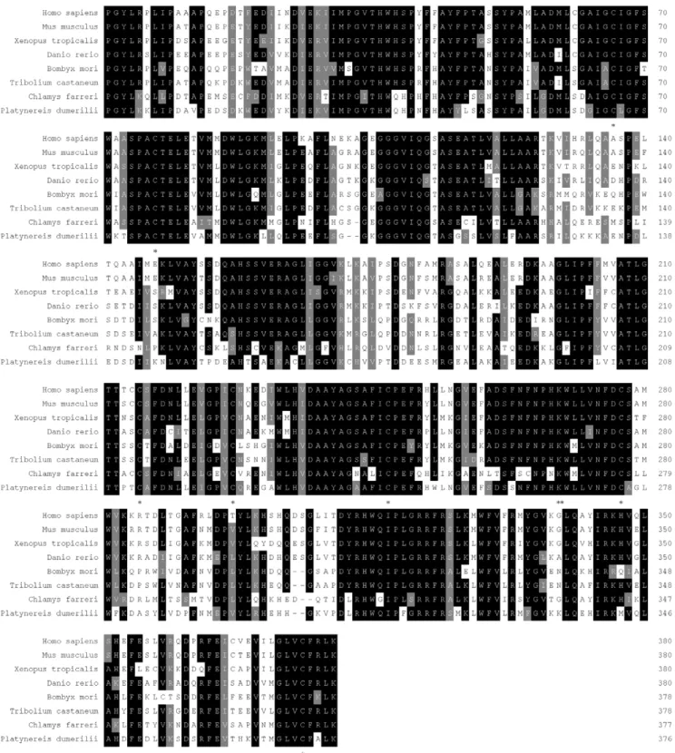
(AAP35655). Multiple alignments of the functional Pyridoxal_deC domain in CfDDC and other DDCs revealed seven conserved cysteine residues (Cys66, Cys77, Cys214, Cys226, Cys246, Cys276 and Cys373) (Fig. 2).
The phylogenic tree constructed based on all amino acid sequences of DDCs was shown in Fig. 3. Three distinct groups were separated in the tree. The CfDDC was first clustered with DDC from clamworm P. dumerilii (CAJ38793) to form the first group, and then gathered together with DDCs from arthropods in the second group, including beetle T. castaneum (ABU25222), mosquito Aedes aegypti (AAC31639), moth Manduca sexta
(AAC46604), silkwormB. mori(AAK48988) and silkwormAntheraea pernyi (AAR23825). All the DDCs from invertebrates were clustered together and formed a sister clade to those from vertebrates, including cattleBos Taurus(ABG66984), pigSus scrofa
(ABO15741), human H. sapiens (AAP35655), rat M. musculus
(CAI23994) and mouse Rattus norvegicus (AAA41087), frog X. tropicalis(NP_001011289, fishOryzias latipes(BAH37023) and fish
D. rerio(AAH56292).
The distribution of CfDDC mRNA in different tissues
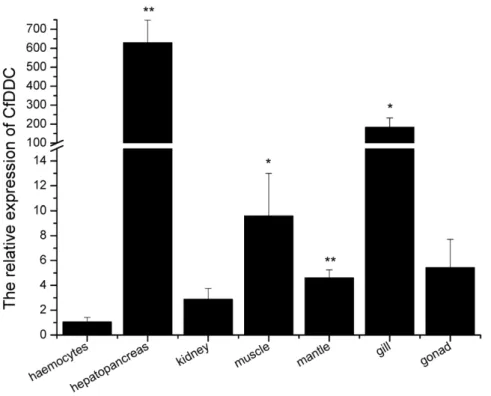
internal control. For CfDDC andb-actin gene, there was only one peak at the corresponding melting temperature in the dissociation curve analysis, indicating that the PCR was specifically amplified (data not shown). The CfDDC transcript was ubiquitously detectable in all seven tested tissues, including haemocytes, hepatopancreas, kidney, adductor muscle, mantle, gill and gonad. It was relatively abundant in hepatopancreas, adductor muscle, mantle, and gill. The highest expression of CfDDC mRNA was found in hepatopancreas, which was 629.4-fold (P,0.01) of that in haemocytes. In addition, the CfDDC expression level in adductor muscle, mantle, and gill was 9.6-fold (P,0.05), 4.6-fold (P,0.01) and 183.8-fold (P,0.05) of that in haemocytes, respectively. The CfDDC mRNA expression level in kidney and gonad was relatively low, and there were no significant differences with that in haemocytes (P.0.05) (Fig. 4).
The temporal expression of CfDDC mRNA in haemocytes after LPS stimulation
The mRNA expression of CfDDC in haemocytes of scallops after LPS stimulation was quantified by quantitative real-time RT-PCR (Fig. 5). The expression level of CfDDC mRNA began to increase significantly at 3 h (5.5-fold, P,0.05), and reached the highest expression level at 12 h (9.8-fold, P,0.01). Then it was down-regulated to 1.6-fold (P.0.05) at 24 h and 0.43-fold (P.0.05) at 48 h after LPS stimulation, respectively. Furthermore, there was no significant difference in CfDDC mRNA expression between the blank and control group during the whole experiment.
Expression and enzyme activity of CfDDC recombination protein
After IPTG induction, the whole cell lysate of E. coli BL21 (DE3)-Transetta with pEASY-E1-CfDDC was analyzed by SDS-PAGE. A distinct band with a molecular weight of ,65 kDa
(Fig. 6) was revealed, which was consistent with the predicted molecular mass. The concentration of the purified rCfDDC was determined to be 367mg ml21.
Dopamine ELISA kit (USCNLIFE) was used to determine the concentration of dopamine in the enzyme reaction mixture. No dopamine was detected in the control reactions, whereas there was detectable dopamine in the reactions with the addition of rCfDDC, and the enzyme activity of rCfDDC was estimated to be 1.65160.22 ng h21mg21(N = 3).
Western blotting of rCfDDC
The specificity of the polyclonal antibody of rCfDDC was examined by western blotting. There was only a distinct band in the nitrocellulose membrane, whose molecular weight was in accordance with that of rCfDDC (Fig. 6). Meanwhile, no distinct band was shown in the negative control.
Haemocyte encapsulation
Haemocyte encapsulation was observed by encapsulation assay
in vitro. When scallop haemocytes were incubated with the agarose beads, more agarose beads coated with rCfDDC were encapsu-lated (Fig. 7). About 70% agarose beads coated with rCfDDC were encapsulated by haemocytes of scallops within 6 h, while 23% of the beads coated with BSA were encapsulated (Fig. 8a, 8c). After
24 h of incubation, the encapsulation rate of beads coated with rCfDDC increased to 93%, but the encapsulation rate of the beads coated with BSA was only increased to 25% (Fig. 8b, 8d). However, the enhancement of rCfDDC to encapsulation was repressed by the addition of rCfDDC. After adding anti-rCfDDC, the encapsulation rate of beads coated with rCfDDC decreased significantly to the 13% at 6 h and 12% at 24 h of incubation (Fig. 8e, 8f).
ROS change in haemocytes after the injection of DDC inhibitor methyldopa
The level of ROS in haemocytes of scallops after the injection of DDC inhibitor methyldopa was determined. In the inhibitor stimulation group, the level of ROS in haemocytes was decreased significantly in comparison with that in the control group (injected with PBS) (Fig. 9). It was declined to 0.41-fold of the blank group (P,0.05) at 12 h, and maintained the low level of 0.47-fold (P,0.05) at 24 h after the inhibitor stimulation.
Discussion
DDC is a crucial metabolic enzyme responsible for catalyzing the conversion of L-Dopa to dopamine, and subsequently involved in the modulation of the immune response against pathogenic microorganism infection. In the present study, a DDC gene was identified from the scallop C. farreri. The full-length cDNA of CfDDC was of 2348 bp, containing a 59UTR of 9 bp, a 39UTR of 656 bp with a poly (A) tail, and an ORF of 1683 bp encoding a polypeptide of 560 amino acids. The deduced amino acids sequence of CfDDC shared high similarity (50.2–52.7%) with those of other reported DDCs. SMART program revealed that CfDDC contained a conserved Pyridoxal_deC domain, and this domain shared 70.0% similarity with that of rat M. musculus
(CAI23994). In this functional domain, there was a PLP-binding motif and a glycine-rich region which were considered to be important for the activity of DDC [11], suggesting that CfDDC possessed the similar structure and function with previously reported DDCs. In NJ phylogenic tree, CfDDC was first clustered with DDCs from invertebrates such as clamworm and insects, and then gathered together with DDCs from vertebrates. The amino acid sequence similarity of CfDDC with other DDCs, together with its conserved functional domain and evolutionary relation-ship, strongly suggested it was a homologue of DDC in scallopC. farreri.
As an important enzyme in the neuroendocrine-immune regulatory network, DDC mainly occurred in the nervous and immune tissues in insect and chordate animals. To understand the possible function of CfDDC in the neuroendocrine-immune regulatory network of scallops, the tissue distribution of CfDDC mRNA transcript was detected by quantitative real-time RT-PCR. CfDDC mRNA transcript was detected in all the examined tissue, including haemocytes, hepatopancreas, kidney, muscle, mantle, gill and gonad. The universal distribution implied that CfDDC might be involved in the modulation of respiration, reproduction and other physiological activity of scallops by synthesizing dopamine [33,34,35,36,37,38,39]. It was notable that CfDDC mRNA distributed in both haemocytes and hepatopancreas, which were considered the main immune tissue of scallops [40,41], indicating CfDDC might be implicated in the immune
Figure 1. Nucleotide and deduced amino acid sequences of CfDDC.The nucleotides and amino acids are numbered along the left margin. The asterisk (*) indicates the stop codon. Polyadenylation signal is bolded and italicized. The predicted PLP-binding motif and the predicted N-glycosylation sites are underlined and double-underlined respectively.
response of scallops and played important roles in the neuroen-docrine-immune regulatory network. The higher mRNA expres-sion of CfDDC in hepatopancreas might be partly due to the
abundant distribution of catecholaminergic neurons in the hepatopancreas of scallops, which was also reported in mussels [42].
Figure 2. Multiple alignment of the Pyridoxal_deC domain of CfDDC with other DDCs deposited in GenBank.The black shadow region indicates positions where all sequences share the same amino acid residue. Similar amino acids are shaded in grey. Gaps are indicated by dashes to improve the alignment. The asterisk (*) indicates the conserved cysteine residue. The species and the GenBank accession numbers are as follows:H. sapiens(AAP35655),M. musculus(CAI23994),X. tropicalis(NP_001011289),D. rerio(AAH56292),B. mori(AAK48988),T. castaneum(ABU25222),P. dumerilii(CAJ38793).
DDC in immunocytes is capable of modulating the immune response in vertebrate and insect. In mollusc, circulating haemocytes are important immunocytes responsible for the
immune responses including recognition and elimination of pathogens, mainly by phagocytosis, encapsulation and nodulation, and oxidative killing [43]. To determine the response of CfDDC in
Figure 4. Tissues distribution of the CfDDC transcript detected by SYBR Green real-time RT-PCR. CfDDC transcript level in hepatopancreas, kidney, adductor muscle, gonad, gill and mantle of six adult scallops is normalized to that in haemocytes. Vertical bars represent the mean6SE (N = 6).
doi:10.1371/journal.pone.0018596.g004
Figure 3. Consensus neighbour-joining tree based on the sequences of DDC from different animals.The numbers at the forks indicate the bootstrap. The protein sequences used for phylogenetic analysis include:B. Taurus(ABG66984),S. scrofa(ABO15741),H. sapiens(AAP35655),M. musculus(CAI23994),R. norvegicus(AAA41087),X. tropicalis(NP_001011289),O. latipes(BAH37023),D. rerio(AAH56292),T. castaneum(ABU25222),A. aegypti(AAC31639),M. sexta(AAC46604),A. pernyi(AAR23825),B. mori(AAK48988) andP. dumerilii(CAJ38793).
scallop haemocytes to immune stimulation, the temporal expres-sion of its mRNA was investigated in scallops stimulated by LPS. In the stimulation group, the expression level of CfDDC mRNA was increased significantly during 3–12 h in comparison with that in the control and blank group, and the peak appeared at 12 h after LPS stimulation. It suggested that LPS stimulation up-regulated the expression of CfDDC in haemocytes. And the high
mRNA level might be induced by the immune mediators produced by the activated immune system. In previous studies, some immune mediators including cytokines had been reported to induce the DDC expression in insectPseudaletia separata [11,44]. The higher level of CfDDC would catalyze the decarboxylation and produce more dopamine, and these dopamine would be further converted to another catecholamine norepinephrine by dopamine beta hydroxylase in scallops [45]. These catecholamines could be released to modulate the immunological competence of scallops [4,46,47,48,49,50,51], and help shifting immune function into the most optimal configuration depending on the physiolog-ical context [52]. The present result indicated LPS stimulation could induce the CfDDC expression in haemocytes to produce more dopamine to modulate the immune response of scallops.
Figure 6. SDS-PAGE analysis of rCfDDC and the western blotting of anti-rCfDDC.Lane M: protein molecular standard (kDa). Lane 1: negative control for rCfDDC (without induction). Lane 2: IPTG induced rCfDDC. Lane 3: purified rCfDDC. Lane 4: the western blotting of anti-rCfDDC.
doi:10.1371/journal.pone.0018596.g006
Figure 7. Encapsulated beads (%) of scallop haemocytes to agarose beads coated with rCfDDC and BSA at 6 h and 24 h.
Vertical bars represent the mean6SE (N = 3). doi:10.1371/journal.pone.0018596.g007
Figure 5. Temporal expression of CfDDC mRNA detected by real-time RT-PCR in scallop haemocytes at 3, 6, 12, 24 and 48 h after LPS stimulation.b-actin gene is used as an internal control to calibrate the cDNA template for all the samples. Each values are shown as mean6SE (N = 6). Significant difference between challenged group and blank group is indicated by asterisks (P,0.05).
To further verify the enzyme activity of CfDDC, the recombinant protein of CfDDC (rCfDDC) was expressed in E. coli BL21 (DE3)-Transetta and the activity of rCfDDC was examined in vitro by determination of the end production dopamine in the reaction catalyze by rCfDDC. The enzyme activity of rCfDDC was 1.65160.22 ng h21mg21(N = 3), clearly demonstrating that CfDDC had the same function to catalyze dopamine synthesis as other reported DDCs [10]. The result was in correspondence with the reports that dopamine could be determined in scallop C. farreri and other mollusc [53,54], and
revealed that CfDDC was involved in the complex neuroendo-crine-immune regulatory network of scallops.
DDC accomplishes immunomodulation by regulating the metabolism of dopamine. For instance, DDC modulates phago-cytosis, nodulation and other cellular immune response through melanization reaction with the involvement of dopamine and phenol oxidase in insect [21,22,55]. In the present study, the effect of surface-associated rCfDDC on haemocyte encapsulation was investigated to survey the cellular immunomodulation of CfDDC. The haemocyte encapsulation to the agarose beads coated with rCfDDC was significantly higher than that to the agarose beads coated with BSA. The antibody specific for rCfDDC significantly repressed the haemocyte encapsulation to the agarose beads coated with rCfDDC. It suggested surface-associated rCfDDC in haemocytes modulated the haemocyte encapsulation of scallops, and this modulation could be similar with that in insect implemented by a pathway in which melanin was synthesized from dopamine [14,21]. The result indicated that the haemocyte encapsulation of scallops could be improved by the surface-associated CfDDC which was induced by the immune response. However, the downstream of DDC modulation pathway needs the involvement of other enzymes and intermediate substances, such as phenol oxidase and quinonoid [56]. Although the phenol oxidase activities in haemolymph of bivalve mollusc had been reported to be detectable [57], the molecule components of this DDC modulation pathway in mollusc as well as their respective function still needs to be further investigated.
To examine the modulation of CfDDC to the humoral immune response, DDC inhibitor methyldopa was injected to inhibit the CfDDC activityin vivo, and then the ROS level in haemocytes was detected in the present study. The ROS level in haemocytes was repressed significantly at 12 h (0.41-fold, P,0.05) and 24 h (0.41-fold, P,0.05) after the methyldopa injection, suggesting that CfDDC activity was necessary to keep an optimal ROS level in haemocytes. The decreased ROS level in haemocytes could result from the modulation of dopamine through the dopamine receptor or the degradation of dopamine. Dopamine could bind the dopamine receptor in the cell membrane of immunocytes to modulate the expression of antioxidases, such as superoxide
Figure 8. The haemocyte encapsulation promoted by rCfDDC.Agarose beads coated with rCfDDC (a, b), BSA (c, d, as a control protein), or anti-rCfDDC (e, f) were incubated with scallop haemocytes.
doi:10.1371/journal.pone.0018596.g008
Figure 9. ROS level in haemocytes of scallops at 6, 12 and 24 h after the injection of DDC inhibitor methyldopa.Each values are shown as mean6SE (N = 6). Significant difference between challenged group and blank group is indicated by asterisks (P,0.05).
dismutase [4,25]. Furthermore, less dopamine produced by the inhibited CfDDC was transformed to hydrogen peroxide through its catabolism to decrease the ROS level in haemocytes [15]. The oxidative deamination of CfDDC to aromatic amines was also suspected to be repressed to produce less ROS [58]. Considering ROS was an important weapon of immune response against invasive pathogen [59], the decline of its level in haemocytes resulted from the inhibition of CfDDC activity by methyldopa suggested that CfDDC might depress the immune capability by terms of the change of ROS level, and consequently lead the increasing susceptibility to pathogen for scallops.
In conclusion, a DDC gene was cloned from scallopC. farreri
and its expression in haemocytes was up-regulated by LPS stimulation. The recombinant CfDDC protein could catalyze the decarboxylation of L-Dopa to dopamine in vitro. And CfDDC could modulate the haemocyte encapsulation and the ROS level
in haemocytes through the action of dopamine. These results indicated that CfDDC was involved in the modulation of immune response in scallops, and therefore provided evidences for CfDDC as an immunomodulating enzyme in the neuroendocrine-immune regulatory network.
Acknowledgments
The authors were grateful to all the laboratory members for continuous technical advice and helpful discussion.
Author Contributions
Conceived and designed the experiments: LS LW ZZ. Performed the experiments: ZZ JY HZ YG XS MW PK LQ. Analyzed the data: ZZ LW LS. Contributed reagents/materials/analysis tools: ZZ LW HZ LQ. Wrote the paper: ZZ LS LW.
References
1. Rahman MK, Nagatsu T, Kato T (1981) Determination of aromatic L-amino acid decarboxylase in serum of various animals by high-performance liquid chromatography with electrochemical detection. Life Sci 28: 485–492. 2. Rahman MK, Nagatsu T, Kato T (1981) Aromatic L-amino acid decarboxylase
activity in central and peripheral tissues and serum of rats with DOPA and L-5-hydroxytryptophan as substrates. Biochem Pharmacol 30: 645–649. 3. Lovenberg W, Weissbach H, Udenfriend S (1962) Aromatic L-amino acid
decarboxylase. J Biol Chem 237: 89–93.
4. Sarkar C, Basu B, Chakroborty D, Dasgupta PS, Basu S (2010) The immunoregulatory role of dopamine: an update. Brain Behav Immun 24: 525–528.
5. Malashkevich VN, Burkhard P, Dominici P, Moore PS, Borri Voltattorni C, et al. (1999) Preliminary X-ray analysis of a new crystal form of recombinant pig kidney DOPA decarboxylase. Acta Crystallogr D Biol Crystallogr 55: 568–570. 6. Burkhard P, Dominici P, Borri-Voltattorni C, Jansonius JN, Malashkevich VN (2001) Structural insight into Parkinson’s disease treatment from drug-inhibited DOPA decarboxylase. Nat Struct Biol 8: 963–967.
7. Bertoldi M, Voltattorni CB (2009) Multiple roles of the active site lysine of Dopa decarboxylase. Arch Biochem Biophys 488: 130–139.
8. Kitahama K, Ikemoto K, Jouvet A, Araneda S, Nagatsu I, et al. (2009) Aromatic L-amino acid decarboxylase-immunoreactive structures in human midbrain, pons, and medulla. J Chem Neuroanat 38: 130–140.
9. Adam WR, Culvenor AJ, Hall J, Jarrott B, Wellard RM (1986) Aromatic L-amino acid decarboxylase: histochemical localization in rat kidney and lack of effect of dietary potassium or sodium loading on enzyme distribution. Clin Exp Pharmacol Physiol 13: 47–53.
10. Kokkinou I, Nikolouzou E, Hatzimanolis A, Fragoulis EG, Vassilacopoulou D (2009) Expression of enzymatically active L-DOPA decarboxylase in human peripheral leukocytes. Blood Cells Mol Dis 42: 92–98.
11. Noguchi H, Tsuzuki S, Tanaka K, Matsumoto H, Hiruma K, et al. (2003) Isolation and characterization of a dopa decarboxylase cDNA and the induction of its expression by an insect cytokine, growth-blocking peptide inPseudaletia separata. Insect Biochem Mol Biol 33: 209–217.
12. Ferdig MT, Taft AS, Smartt CT, Lowenberger CA, Li J, et al. (2000)Aedes aegypti dopa decarboxylase: gene structure and regulation. Insect Mol Biol 9: 231–239. 13. Han Q, Ding H, Robinson H, Christensen BM, Li J (2010) Crystal Structure and Substrate Specificity ofDrosophila3,4-Dihydroxyphenylalanine Decarbox-ylase. PLoS ONE 5: e8826.
14. Hodgetts RB, O’Keefe SL (2006) Dopa decarboxylase: a model gene-enzyme system for studying development, behavior, and systematics. Annu Rev Entomol 51: 259–284.
15. Flierl MA, Rittirsch D, Huber-Lang M, Sarma JV, Ward PA (2008) Catecholamines-crafty weapons in the inflammatory arsenal of immune/ inflammatory cells or opening pandora’s box? Mol Med 14: 195–204. 16. Kvetnansky R, Sabban EL, Palkovits M (2009) Catecholaminergic systems in
stress: structural and molecular genetic approaches. Physiol Rev 89: 535–606. 17. Kvetnansky R, Pacak K, Fukuhara K, Viskupic E, Hiremagalur B, et al. (1995)
Sympathoadrenal system in stress. Interaction with the hypothalamic-pituitary-adrenocortical system. Ann N Y Acad Sci 771: 131–158.
18. Bergmann M, Sautner T (2002) Immunomodulatory effects of vasoactive catecholamines. Wien Klin Wochenschr 114: 752–761.
19. JIANGJian-Lan QY-H, PENGYu-Ping, WANGJian-Jun (2006) Immunoregu-latory role of endogenous catecholamines synthesized by immune cells. Acta Physiologica Sinica 58: 309–317.
20. Flierl MA, Rittirsch D, Nadeau BA, Sarma JV, Day DE, et al. (2009) Upregulation of phagocyte-derived catecholamines augments the acute inflam-matory response. PLoS One 4: e4414.
21. Tang HP (2009) Regulation and function of the melanization reaction in Drosophila. Fly 3: 105–111.
22. Paskewitz SM, Andreev O (2008) Silencing the genes for dopa decarboxylase or dopachrome conversion enzyme reduces melanization of foreign targets in Anopheles gambiae. Comp Biochem Physiol B Biochem Mol Biol 150: 403–408. 23. Sideri M, Tsakas S, Markoutsa E, Lampropoulou M, Marmaras VJ (2008)
Innate immunity in insects: surface-associated dopa decarboxylase-dependent pathways regulate phagocytosis, nodulation and melanization in medfly haemocytes. Immunology 123: 528–537.
24. Chang CC, Wu ZR, Kuo CM, Cheng W (2007) Dopamine depresses immunity in the tiger shrimpPenaeus monodon. Fish Shellfish Immunol 23: 24–33. 25. Li JT, Lee PP, Chen OC, Cheng W, Kuo CM (2005) Dopamine depresses the
immune ability and increases susceptibility toLactococcus garvieaein the freshwater giant prawn,Macrobrachium rosenbergii. Fish Shellfish Immunol 19: 269–280. 26. Butts CL, Sternberg EM (2008) Neuroendocrine factors alter host defense by
modulating immune function. Cell Immunol 252: 7–15.
27. Wang L, Song L, Zhao J, Qiu L, Zhang H, et al. (2009) Expressed sequence tags from the zhikong scallop (Chlamys farreri): discovery and annotation of host-defense genes. Fish Shellfish Immunol 26: 744–750.
28. Zhang H, Kong P, Wang L, Zhou Z, Yang J, et al. (2010) Cflec-5, a pattern recognition receptor in scallopChlamys farreriagglutinating yeast Pichia pastoris. Fish Shellfish Immunol.
29. Zhang Y, Wang L, Zhao J, Song L, Gai Y, et al. (2010) The effects of CpG C oligodeoxynucleotides on innate immune responses inEriocheir sinensis(H. Milne Edwards, 1853). Aquaculture Research.
30. Fragoulis EG, Sekeris CE (1975) Purification and characteristics of DOPA-decarboxylase from the integument of Calliphora vicina larve. Arch Biochem Biophys 168: 15–25.
31. Cheng S, Zhan W, Xing J, Sheng X (2006) Development and characterization of monoclonal antibody to the lymphocystis disease virus of Japanese flounder Paralichthys olivaceusisolated from China. J Virol Methods 135: 173–180. 32. Ao J, Ling E, Yu XQ (2007) Drosophila C-type lectins enhance cellular
encapsulation. Mol Immunol 44: 2541–2548.
33. Chang C-C, Wu Z-R, Chen C-S, Kuo C-M, Cheng W (2007) Dopamine modulates the physiological response of the tiger shrimp Penaeus monodon. Aquaculture 270: 333–342.
34. Chiu H-T, Yeh S-P, Huang S-C, Chang C-C, Kuo C-M, et al. (2006) Dopamine induces transient modulation of the physiological responses of whiteleg shrimp, Litopenaeus vannamei. Aquaculture 251: 558–566.
35. KhotimchenkoYu S (1991) Biogenic monoamines in oocytes of echinoderms and bivalve molluscs. A formation of intracellular regulatory systems in oogenesis. Comp Biochem Physiol C 100: 671–675.
36. De Luca M, Roshina NV, Geiger-Thornsberry GL, Lyman RF, Pasyukova EG, et al. (2003) Dopa decarboxylase (Ddc) affects variation in Drosophila longevity. Nat Genet 34: 429–433.
37. R. Beiras JW (1995) Effect of the neurotransmitters dopamine, serotonin and norepinephrine on the ciliary activity of mussel (Mytilus edulis) larvae. Marine Biology 122: 597–603.
38. Pavlova GA (2001) Effects of serotonin, dopamine and ergometrine on locomotion in the pulmonate molluscHelix lucorum. J Exp Biol 204: 1625–1633. 39. Carroll MA, Catapane EJ (2007) The nervous system control of lateral ciliary activity of the gill of the bivalve mollusc, Crassostrea virginica. Comparative Biochemistry and Physiology a-Molecular & Integrative Physiology 148: 445–450.
40. Wootton EC, Dyrynda EA, Ratcliffe NA (2003) Bivalve immunity: comparisons between the marine mussel (Mytilus edulis), the edible cockle (Cerastoderma edule) and the razor-shell (Ensis siliqua). Fish Shellfish Immunol 15: 195–210. 41. Zhao J, Song L, Li C, Zou H, Ni D, et al. (2007) Molecular cloning of an
42. Voronezhskaya EE, Nezlin LP, Odintsova NA, Plummer JT, Croll RP (2008) Neuronal development in larval musselMytilus trossulus(Mollusca : Bivalvia). Zoomorphology 127: 97–110.
43. Costa MM, Prado-Alvarez M, Gestal C, Li H, Roch P, et al. (2009) Functional and molecular immune response of Mediterranean mussel (Mytilus galloprovincia-lis) haemocytes against pathogen-associated molecular patterns and bacteria. Fish Shellfish Immunol 26: 515–523.
44. Sternberg EM (2006) Neural regulation of innate immunity: a coordinated nonspecific host response to pathogens. Nat Rev Immunol 6: 318–328. 45. Zhou Z, Wang L, Yang J, Zhang H, Kong P, et al. (2010) A dopamine beta
hydroxylase from Chlamys farreri and its induced mRNA expression in the haemocytes after LPS stimulation. Fish Shellfish Immunol 30: 154–162. 46. Besser MJ, Ganor Y, Levite M (2005) Dopamine by itself activates either D2, D3 or
D1/D5 dopaminergic receptors in normal human T-cells and triggers the selective secretion of either IL-10, TNFalpha or both. J Neuroimmunol 169: 161–171. 47. Ghosh MC, Mondal AC, Basu S, Banerjee S, Majumder J, et al. (2003)
Dopamine inhibits cytokine release and expression of tyrosine kinases, Lck and Fyn in activated T cells. Int Immunopharmacol 3: 1019–1026.
48. Basu S, Dasgupta PS (2000) Dopamine, a neurotransmitter, influences the immune system. J Neuroimmunol 102: 113–124.
49. Gospe SM, Jr. (1983) Studies of dopamine pharmacology in molluscs. Life Sci 33: 1945–1957.
50. Flierl MA, Rittirsch D, Nadeau BA, Chen AJ, Sarma JV, et al. (2007) Phagocyte-derived catecholamines enhance acute inflammatory injury. Nature 449: 721–725.
51. Drummond AH, Bucher F, Levitan IB (1980) Distribution of serotonin and dopamine receptors in Aplysia tissues: analysis by [3H]LSD binding and adenylate cyclase stimulation. Brain Res 184: 163–177.
52. Adamo SA (2010) Why should an immune response activate the stress response? Insights from the insects (the cricket Gryllus texensis). Brain Behavior and Immunity 24: 194–200.
53. Chen M, Yang H, Xu B, Wang F, Liu B (2008) Catecholaminergic responses to environmental stress in the hemolymph of Zhikong scallopChlamys farreri. J Exp Zool A Ecol Genet Physiol 309: 289–296.
54. Pani AK, Croll RP (1995) Distribution of catecholamines, indoleamines, and their precursors and metabolites in the scallop,Placopecten magellanicus(Bivalvia, Pectinidae). Cell Mol Neurobiol 15: 371–386.
55. Huang CY, Chou SY, Bartholomay LC, Christensen BM, Chen CC (2005) The use of gene silencing to study the role of dopa decarboxylase in mosquito melanization reactions. Insect Mol Biol 14: 237–244.
56. Bertoldi M, Cellini B, Maras B, Voltattorni CB (2005) A quinonoid is an intermediate of oxidative deamination reaction catalyzed by Dopa decarbox-ylase. FEBS Lett 579: 5175–5180.
57. Luna-Gonzalez A, Maeda-Martinez AN, Vargas-Albores F, Ascencio-Valle F, Robles-Mungaray M (2003) Phenoloxidase activity in larval and juvenile homogenates and adult plasma and haemocytes of bivalve molluscs. Fish Shellfish Immunol 15: 275–282.
58. Bertoldi M, Cellini B, Montioli R, Borri Voltattorni C (2008) Insights into the mechanism of oxidative deamination catalyzed by DOPA decarboxylase. Biochemistry 47: 7187–7195.