Lentinula edodes
-Derived Polysaccharide
Alters the Spatial Structure of Gut Microbiota
in Mice
Xiaofei Xu, Xuewu Zhang*
College of Light Industry and Food Sciences, South China University of Technology, Guangzhou, China
Abstract
Lentinula edodes-derived polysaccharides possess many therapeutic characteristics, includ-ing anti-tumor and immuno-modulation. The gut microbes play a critical role in modulation of immune function. However, the impact ofLentinula edodes-derived polysaccharides on the gut microbes have not yet been explored. In this study, high-throughput pyrosequencing technique was employed to investigate the effects of a new heteropolysaccharide L2 from
Lentinula edodeson microbiota diversity and composition of small intestine, cecum, colon and distal end of colon (feces) in mice. The results demonstrated that along mouse intestine the microbiota exhibit distinctly different space distribution. L2 treatment reduced the diversity and evenness of gut microbiota along the intestine, especially in the cecum and colon. In the fecal microbial communities, the decrease of Bacteroidetes by significantly increasing Proteobacteria were observed, which were characterized by the increasedHelicobacteraceae
and reduced S24-7 at family level. Some OTUs, corresponding toBacteroides acidifaciens,
AlistipesandHelicobacter suncus, were found to be significantly increased in L2 treated-mice. In particular, 4 phyla Chloroflexi, Gemmatimonadetes, Nitrospirae and Planctomycetes are exclusively present in L2-treated mice. This is helpful for further demonstrating healthy action mechanism ofLentinula edodes-derived polysaccharide L2.
Introduction
Lentinula edodesis the second most popular edible mushroom in the world. With the enor-mous development in the field of purification and structure determination, many types of polysaccharides have been obtained from the fruit body ofLentinula edodes[1]. Most of the Lentinula edodes-derived polysaccharides have been shown to possess many therapeutic appli-cations, such as cancer, depressed immune function, and hyperlipidemia [2]. However, these polysaccharides, known as nondigestible carbohydrates, are not fully digested in the upper gut. It has become clear that the healthy benefits of nondigestible carbohydrates are attributed to the contribution of gut microbiota, i.e. fermenting nondigestible carbohydrates to produce gut-absorbable metabolites and to stimulate proliferation of certain bacteria [3,4]. Recent evidence OPEN ACCESS
Citation:Xu X, Zhang X (2015)Lentinula edodes -De-rived Polysaccharide Alters the Spatial Structure of Gut Microbiota in Mice. PLoS ONE 10(1): e0115037. doi:10.1371/journal.pone.0115037
Academic Editor:Emiko Mizoguchi, Massachusetts General Hospital, UNITED STATES
Received:June 23, 2014
Accepted:November 18, 2014
Published:January 21, 2015
Copyright:© 2015 Xu, Zhang. This is an open ac-cess article distributed under the terms of the Creative Commons Attribution License, which permits unrestricted use, distribution, and reproduction in any medium, provided the original author and source are credited.
Data Availability Statement:All relevant data are within the paper and its Supporting Information files.
Funding:The authors have no support or funding to report.
has shown that several types of non-digestible carbohydrates have a major influence on micro-bial community composition both in the short and long term [5]. For example, Martinez et al. [6] demonstrated that resistant starches types 2 and 4 exhibit functional differences in their ef-fect on human fecal microbiota composition. Marín-Manzano et al. [7] assessed the modulato-ry influence of novel galacto-oligosaccharides derived from lactulose (GOS-Lu) in rat gut microbiota.
On the other hand, dysbiosis of gut microbiota is closely related to human health and diseases [8]. Numerous researches have showed that the gut microbes play a critical role in the development of immune system and modulation of immune function [9,10]. So, it is important to understand the community profiles and system characteristics of gut microbiota after administration of non-digestible polysaccharides. As the first medicinal mushroom in clinical field, the immune-modulating and anti-tumor characteristics of polysaccharides derived from
Lentinula edodeshave been intensively investigated [1]. However, it is less clear about how
Lentinula edodes-derived polysaccharides impact the characteristics and distribution of gut microbiota. In our previous study [11], a new heteropolysaccharide L2 with immunostimulating activity was isolated from the fruit body ofLentinula edodes. Chemical characterization indicat-ed that L2 consists of glucose (87.5%), galactose (9.6%), and arabinose (2.8%), with a molecular weight of 26 KDa. The primary linkages are the 1!or 1!6 glycosidic linkages accounted for 44.5% and 1!2 or 1!4 glycosidic linkages accounted for 17.9%. Other linkages include
(1!3)-linked, (1!2, 3)-linked, (1!2, 4)-linked, (1!3, 4)-linked, (1!3, 6)-linked, and (1!2, 3, 4)-linked through glucose, which account for about 37.6% of all linkages in the molecule. Especially, L2 does not possess a triple-helical conformation by Congo red assay [11]. The goal of this study is to investigate the effects of L2 on microbiota diversity and composition along the mouse intestine using a high-throughput pyrosequencing technique. It is expected to provide foundation forLentinula edodes-derived polysaccharide L2 in understanding healthy action mechanism and discovering potential side effects.
Materials and Methods
Reagents and experimental design
Lentinula edodes-derived polysaccharide L2 was prepared as described before [8]. Specific pathogen-free male C57BL/6 mice (8-week old) were obtained from Vital River Laboratory Animal Technology Co. Ltd. (Beijing, China). All chemical reagents were at analytical grade.
The mice were kept at a temperature of 22°C and 12-h light/dark cycles environment for at least two weeks before use, and fed on the same batch of standard laboratory diet to minimize the variation of environment factors. The experiments were approved by the Animal Care Wel-fare Committee of Guangzhou University of Chinese Medicine. Adequate measures were taken to minimize pain of experimental animals. Mice were divided into two groups for 28 consecu-tive days: (1)Lentinula edodes-derived polysaccharide L2 treatment groups (gavage adminis-tration of L2 40mg/kg body weight, n = 7) (Na groups), which were reared in the same cage; (2) normal groups (gavage administration with the same volume of sterile physiological saline, 8-week old, n = 7) (N groups), which were reared in the same cage.
Fresh fecal samples were collected on the last day and immediately frozen in liquid nitrogen before storage at−80°C for further analysis. Then, mice were weighted and peripheral blood
DNA Extraction and Pyrosequencing
Six fecal samples and 3 intestinal content samples were randomly selected from each group for 16S rRNA gene pyrosequencing. Genomic DNA was extracted from small intestine, cecum and colon contents as well as fecal samples by using the Soil DNA kit (Omega Bio-Tek, Inc., GA, USA) according to the manufacturer’s instructions.
Pyrosequencing was carried out according to previously described [12–13]. PCR amplifica-tion of the V1-V3 region of bacterial 16 S rRNA gene was performed using universal primers (533R 50-TTACCGCGGCTGCTGGCAC-30, 27F 50-AGAGTTTGATCCTGGCTCAG-30) in-corporating the FLX Titanium adapters and a sample barcode sequence. The forward primer (B-27F) was 50-TATCCCCTGTGTGCCTTGGCAGT CGACT AGAGTTTGATCCTGGCTCAG-30, where the sequence of the B adaptor is shown in italics and underlined. The reverse primer (A-533R) was 50-ATCTCATCCCTGCGTGTCTC CGACGACTNNNNNNNNTTACCGCGG CTGCTGGCAC-30, where the sequence of the A adaptor is shown in italics and underlined and the Ns represent an eight-base sample specific barcode sequence. Briefly, Each 20μL PCR reaction included 4μL of 5FastPfu Buffer, 2μL of 2.5 mM dNTPs, 0.4μL of Forward Primer (5 mM), 0.4μL of Reverse Primer (5 mM),0.5μL of DNA template, 0.4μL of Fastfu Polymer-ase, and added ddH2O to make up the final volume to 20μL. The cycling parameters were as follows: 95°C for 2 min; 25 cycles of 95°C for30 s, 56°C for 30 s, 72°C for 30 s with a final exten-sion at 72°C for 5 min. Duplicate PCR products were pooled. Then they were visualized on aga-rose gels (2% in TBE buffer) containing ethidium bromide, and purified using the AXYGEN gel extraction kit (Axygen, USA). Amplicon DNA concentrations were measured using the Quant-iT PicoGreen dsDNA reagent and kit (Invitrogen, Germany) and was quality controlled on an Agilent 2100 bioanalyzer (Agilent, USA). Following quantitation, the amplicon from each reaction mixture were pooled in equimolar ratios based on concentration and subjected to emulsion PCR to generate amplicon libraries, as recommended by 454 Life Sciences. Pyrosequencing was performed by a 454 Life Sciences Genome Sequencer FLX Titanium instrument (Roche) (Shanghai Majorbio, Shanghai, China).
All pyrosequencing reads were filtered according to barcode and primer sequences. The re-sulting sequences were further screened and filtered for quality and length. Sequences that were less than 200 bp, contained ambiguous characters, contained over two mismatches to the primers, had an average quality score below 25 or contained mononucleotide repeats of over 8 bp were removed using Mothur software package (version 1.28.0) (command trim.seqs)(http://
www.mothur.org/wiki/Main_Page) [14,15]. A total of 69,700 high quality sequences were
ob-tained after the filtering process.
Bioinformatic Analysis
The high-quality sequences were assigned to samples according to barcodes. Sequences were aligned in accordance with SILVA alignment (Bacterial SILVA database, SILVA version 111,
http://www.arb-silva.de/) using kmer searching (http://www.mothur.org/wiki/Align.seqsin
(PCA) based on weighted Unifac distance, Jaccard tree clustering analysis, and heatmap were performed using Mothur and R software package (http://www.R-project.org).
Statistical Analysis
The Mann-Whitney test and student’s-test were performed using SPSS19.0. P-values<0.05 were considered significant unless otherwise stated.
Results
Diversity of the bacterial communities along the gastrointestinal tract in
normal and L2-treated mice
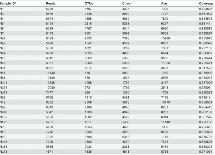
Multiplex pyrosequencing of covering the V1-V3 hypervariable regions of 16S rRNA gene was employed to characterize the bacterial community diversity along the mice gastrointestinal tract. Following all denoising and filtering steps, a dataset consisting of 117497 (mean±S.D., 7833±1682) reads from N groups and 106268 (7084±2056) reads from Na groups were used in the final analysis (Table 1). Based on a sequence similarity of greater than 97%, an average of 1862 and 1611 OTUs were defined for N and Na groups, respectively (Table 1).
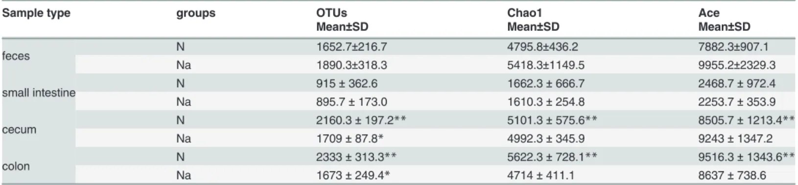
Table 2showed that cecum and colon displayed higher diversity of microbiota than that of
small intestine in N groups (p<0.01). For the estimated richness (Chao1 and Ace) of fecal, small intestine, cecum or colon microbiota, no significant differences were observed in N and Na groups. But L2 treatment significantly decreased the amount of OTUs in cecum or colon micro-biota (p<0.05), compared with N groups.Fig. 1demonstrated that the shannon and evenness indices are similar in N and Na groups for small intestine or fecal microbiota. However, L2 treat-ment significantly increased the simpson index in cecum, colon or fecal microbiota, compared with N groups (p<0.05 or p<0.01), i.e. higher degree of convergence in Na groups than in N groups.
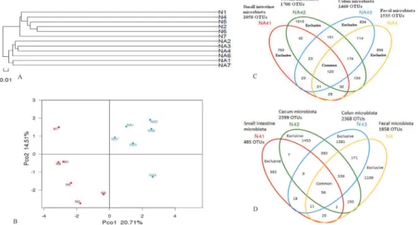
For fecal microbiota, clustering analysis displayed the formation of two major clusters corre-sponding to normal and L2-treated groups, respectively (Fig. 2A). Weighted Unifrac analysis revealed that each mouse was obviously different from all others and a high degree of variation between individuals existed (Fig. 2B). The first principal coordinate (PC1), which accounted for 20.71% of variance in the data, can completely separate N from L2-treated (Na) groups
(Fig. 2B). The second principal coordinate (PC2, 14.51% of variance in the data) can separate
N from L2-treated (Na) groups with>80% of accuracy, except for the mice N7 in normal groups and Na4 in L2-treated groups (Fig. 2B). By random selection of an individual mouse in each group (Na4 and N4), the diversity of bacterial populations along the intestinal tract was compared. The amount of OTUs existed exclusively in small intestine, cecum, colon and fecal microbiota were 769, 1016, 834,896 for Na4, and 362, 1453, 1381, 1109 for N4, respectively; whereas the amount of their common OTUs were 120 and 56, respectively (Fig. 2C;Fig. 2D).
Spatial structures of the bacterial communities from small intestine,
cecum and colon contents in normal and L2-treated mice
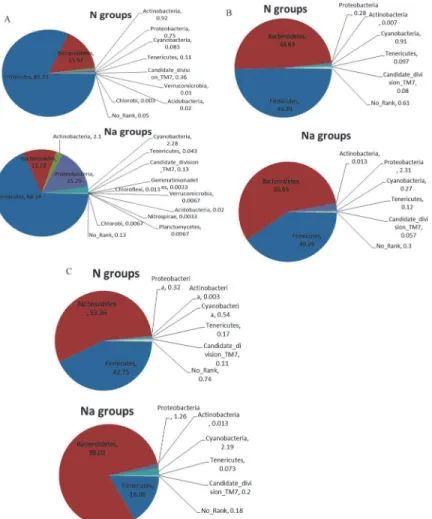
For cecum microbiota (Fig. 3B), the prevalent bacterial communities are also 2 phyla (in total>96%): Firmicutes (F) and Bacteroidetes (B), with F/B = 1.02 and F/B = 0.71 in N and L2-treated groups, respectively. Specifically, L2 induced the decrease of Firmicutes and the increase of Bacteroidetes. Especially, L2 remarkably increased the proportion of Proteobacteria (2.31% vs. 0.28%, P = 0.08), and significantly decreased the abundance of Cyanobacteria (0.27% vs. 0.91%, P<0.05), compared with normal mice. In cecum microbial cummunities, the domi-nant bacteria groups were Bacteroidaceae, Rikenellaceae, Ruminococcaceae, Lachnospiraceae, and S24-7. Microbial communities from 6 mice were clustered into 2 groups: Na42, Na62; N22, N42, N62, Na72, according to the similarity in relative abundance at family level (Figure A in S1 File).
Similar to cecum microbiota, the most abundant bacterial communities in colon microbiota
(Fig. 3C) remains 2 phyla (in total>96%): Firmicutes (F) and Bacteroidetes (B), but the ratio
F/B is greatly changed as 0.77 and 0.20 in N and L2-treated groups, respectively. In other words, L2 significantly elevated the proportions of Bacteroidetes (80.03% vs. 55.36%, p<0.05),
Table 1. 16S rRNA gene sequencing statistics from normal and L2-treated C57BL/6 mice.
Sample ID* Reads OTUs Chao1 Ace Coverage
N1 6785 1687 4277 7309 0.823876
N2 9879 2145 5039 7777 0.867902
N4 6574 1858 4659 7829 0.814573
N5 8494 2218 5351 9217 0.83141
N6 9019 1757 4344 6630 0.880585
N7 6443 2051 5099 8532 0.788297
Na1 6439 2352 7326 13286 0.735673
Na2 5726 1547 4589 8637 0.806322
Na3 5860 1831 5267 10311 0.777133
Na4 6290 1535 4005 6474 0.836089
Na6 6212 2009 5396 9665 0.770444
Na7 6951 2068 5927 11358 0.793411
N21 8851 1372 2519 3608 0.917523
N41 11130 485 893 1232 0.976999
N61 10120 888 1575 2566 0.952273
Na41 12305 1058 1789 2457 0.957009
Na61 10030 973 1792 2548 0.95005
Na71 7177 656 1250 1756 0.950258
N22 5766 1916 4491 7176 0.78616
N42 6582 2399 5873 10110 0.756001
N62 6019 2166 4940 8231 0.764413
Na42 5547 1708 4949 8267 0.785109
Na62 5908 1602 4592 8314 0.807549
Na72 5791 1817 5436 11148 0.775168
N23 6188 1933 4625 7860 0.795895
N43 7715 2368 5899 9538 0.802074
N63 7932 2698 6343 11151 0.776727
Na43 7295 1460 4270 7674 0.863605
Na63 9866 2023 5261 9469 0.860328
Na73 4871 1536 4611 8768 0.771094
*N×1 (Na×1), N×2 (Na×2) and N×3 (Na×3) correspond to small intestine, cecum and colon samples in mice Nx (Nax), respectively.
and reduced the level of Firmicutes (16.06% vs. 42.75%, p<0.05), compared with N groups. Moreover, more Proteobacteria were observed in L2-treated groups than in N groups (1.26% vs. 0.32%, p<0.05). However, the dominant bacterial populations were consisted of S24-7, Lachnospiraceae, Rikenellaceae, Bacteroidaceae, Ruminococcaceae, and Prevotellaceae. Micro-bial communities from 6 mice were clustered into 2 groups: Na43, Na63; N43, N63, N23, Na73, which could be further clustered into 3 subgroups: Na43, Na63; N43, N63; N23, Na73, according to the similarity in relative abundance at family level (Figure B inS1 File).
L2 treatment shifts the composition of fecal microbiota
Next, we focus on the impacts of L2 treatment on fecal microbiota in mice. Phylum-level com-parison of fecal microbiota in normal and L2-treated mice showed that Firmicutes (F) and Bacteroidetes (B) are prevalent communities, in total 97.8%, F/B = 0.5, in N groups; while in
Table 2. Diversity of gut microbiota in normal mice (N) and L2-treated mice (Na).
Sample type groups OTUs Chao1 Ace
Mean±SD Mean±SD Mean±SD
feces N 1652.7±216.7 4795.8±436.2 7882.3±907.1
Na 1890.3±318.3 5418.3±1149.5 9955.2±2329.3
small intestine N 915±362.6 1662.3±666.7 2468.7±972.4
Na 895.7±173.0 1610.3±254.8 2253.7±353.9
cecum N 2160.3±197.2** 5101.3±575.6** 8505.7±1213.4**
Na 1709±87.8* 4992.3±345.9 9243±1347.2
colon N 2333±313.3** 5622.3±728.1** 9516.3±1343.6**
Na 1673±249.4* 4714±411.1 8637±738.6
*P<0.05
**, p<0.01, student t-test
doi:10.1371/journal.pone.0115037.t002
Figure 1. Alpha diversity of fecal microbiota in normal (N) and L2-treated mice (Na).*, p<0.05;**p<0.01.The index of evenness (J indices) was calculated with the formula E = H/ln(S), where H is the Shannon index and S is the number of OTU in that animal.
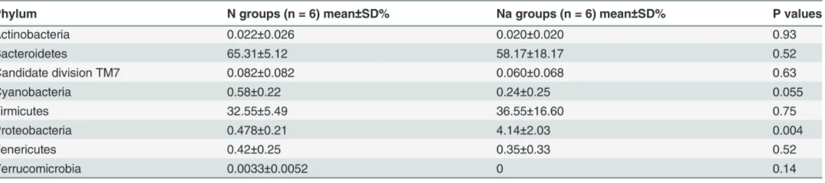
L2-treated groups the prevalent communities include 3 phyla: Firmicutes, Bacteroidetes and Proteobacteria, in total 98.86%, F/B = 0.63 (Fig. 4A). It is noted that Proteobacteria is very sig-nificantly increased (4.14% vs. 0.043%, p<0.01), and Cyanobacteria is decreased (0.24% vs. 0.58%, p = 0.055) in L2-treated groups, compared with N groups (Table 3).
At class level (Fig. 4B), the bacterial communities in N groups are consisted of two major classes: Bacteroidia (B) (65.2%) and Clostridia (C) (32%), B+C = 97.2% and B/C = 2; while the bacterial populations in L2-treated mice primarily include 3 phyla: Bacteroidia (58.1%), Clostridia (35.5%) and Epsilonproteobacteria (3.6%), in total 97.2%, B+C = 93.6% and B/C = 1.6. The major differences compared with normal groups are the significant increases of Epsilonproteobacteria (p<0.05) and moderate increase of Bacili (0.35% vs 0.75%, p = 0.1).
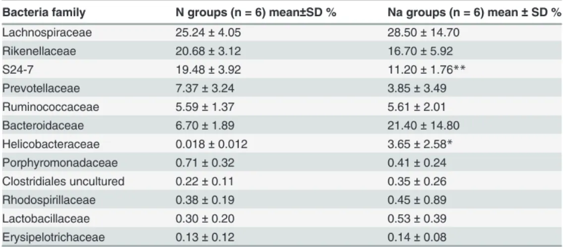
The composition of fecal microbiota at family level in N and L2-treated groups and the heatmap analysis of their relative abundance are displayed inFig. 4CandFig. 4D, respectively. In N groups, the most abundant families are composed of 6 populations: Lachnospiraceae, Rikenellaceae, S24-7, Prevotellaceae, Ruminococcaceae and Bacteroidaceae, totally>80%. In L2-treated groups, the top populations adds up to 7 families: Lachnospiraceae, Rikenellaceae, S24-7, Prevotellaceae, Ruminococcaceae, Bacteroidaceae and Helicobacteraceae, totally>90%. The major variations are: Helicobacteraceae (3.6% vs 0.02%, p<0.05) are significantly in-creased, while S24-7 is significantly reduced (11.2% vs 19.5, p<0.01) in L2-treated groups, compared with N groups (Table 4). Strikingly, the speciesHelicobacter suncuswas significantly increased from 0 to 2.34% after L2 treatment.
Furthermore, top 80 OTUs (Table A inS1 File) of relative abundance presented in N and L2-treated groups were selected for comparison.Fig. 4Edemonstrated that the top 80 OTUs exhibited two clusters and can separate fecal microbiota of both groups, consistent with cluster-ing analysis above. In particular, the microbial communities of Na4, Na6, Na7, N2, N4, N6 were also clustered into 2 subgroups: Na4, Na6; N2, N4, N6, Na7, which were consistent with the results in cecum and colon (Figure A and Figure B inS1 File). Compared with N groups, L2 can significantly (p<0.05) facilitate the growth of 15 OTUs (Table B inS1 File), such as OTU1 (0.004% vs 7.71%), OTU4 (0.004% vs 4.09%), OTU5 (0.024% vs 2.54%), OTU18 (0% vs
Figure 2. Clustering (A) and weighted unifrac PCA (B) analysis of fecal microbiota, as well as OTUs overlaps of a L2-treated mice Na4 (C) and a normal mice N4 (D) along the intestinal tract.
2.31%), OTU19 (0.024% vs 2.70%). Among them, OTU1 and OTU4 are attibuted to species of
Bacteroides acidifaciens, OTU5 and OTU19 belong to species ofAlistipes uncultured Bacteroi-daceae bacterium, while OTU18 is the speciesHelicobacter suncus. On the other hand, com-pared with N groups, 34 OTUs are significantly (p<0.05) repressed in L2-treated groups (Table B inS1 File). The representative OTUs include: OTU3 (2.1% vs 0.58%), OTU11 (1.1% vs 0.133%), OTU13 (1.52% vs 0.014%), OTU30 (1.19% vs 0.024%), OTU31 (1.68% vs 0.17%), OTU33 (1.85% vs 0.008%), OTU42 (2.16% vs 0.003%), OTU47 (2.18% vs 0.017%), OTU48 (2.29% vs 0.027%), OTU49(1.31% vs 0%), OTU61 (0.98% vs 0.023%), OTU100 (0.522% vs 0.033%). Except for OTU49, OTU133, OTU139 belonging to Lachnospiraceae (Phylum of Firmicutes), other OTUs belong to the order Bacteroidales, and the majority of them are from the genusAlistipes(especially in the species ofAlistipes uncultured Bacteroidaceae bacterium) and the family S24-7 (especially in the species ofS24-7 mouse gut metagenome).
Discussion
Gut microbiota has been recognized as being implicated in human health and disease, but feces do not fully reflect microbial ecology in the intestine [19]. It is essential to investigate the char-acteristics and distribution of the microbial community along the mouse gastrointestinal tract [20]. Non-digestible carbohydrates have great impact on the gut microbiota [4,5]. More and
Figure 3. Comparison of small intestine (A), cecum (B) and colon (C) microbiota at phylum level.
more evidences indicated that non-digestible carbohydrates affect the profiles of intestinal mi-crobial community depending on the structure of polysaccharides such as glycosidic linkage and monosaccharide composition [4–5,21–23]. Mushroom polysaccharides have been pro-posed as new prebiotic resource and have attractive attentions from researchers [3,24].Letinula edodes-derived polysaccharides have been intensively investigated for their potentially thera-peutic applications [1,25]. However, the influence ofLetinula edodes-derived polysaccharides
Figure 4. Composition of fecal microbiota at phylum (A), class (B), family (C, D), and OTUs (E) levels.
doi:10.1371/journal.pone.0115037.g004
Table 3. Phylum-level comparison of fecal microbiota in normal (N) and L2-treated mice (Na).
Phylum N groups (n = 6) mean±SD% Na groups (n = 6) mean±SD% P values
Actinobacteria 0.022±0.026 0.020±0.020 0.93
Bacteroidetes 65.31±5.12 58.17±18.17 0.52
Candidate division TM7 0.082±0.082 0.060±0.068 0.63
Cyanobacteria 0.58±0.22 0.24±0.25 0.055
Firmicutes 32.55±5.49 36.55±16.60 0.75
Proteobacteria 0.478±0.21 4.14±2.03 0.004
Tenericutes 0.42±0.25 0.35±0.33 0.52
Verrucomicrobia 0.0033±0.0052 0 0.14
Mann Whitney test.
on gut microbiota was rarely reported. The present study, explored the impact of a mushroom polysaccharide,Lentinula edodes-derived polysaccharide L2, on microbiota diversity and com-position along the mouse intestine using a high-throughput pyrosequencing technique.
Overall, as described above, along the small intestine, cecum, colon and distal colon (feces), the microbiota composition changes from Firmicutes (F)-dominant to Bacteroidetes (B)-domi-nant structure in normal and L2-treated mice. The most significantly difference happens in the colon, the ratio F/B varies from 0.77 to 0.2. This suggests the massive presence of responsive bacteria to polysaccharide L2 in colon, and these responders primarily belong to Bacteroidetes population, which is consistent with previous findings in African children consuming the high-plant polysaccharide diets [26]. It is well known that plant polysaccharides are not di-gested by human enzymes, but are processed to absorbable short chain fatty acids (SCFA) by gut bacteria. Bacteroidetes can use a series of membrane protein complexes, termed Sus-like systems, to metabolize many complex carbohydrates [27]. For example, B. thetaiotaomicron and B. ovatus were found to have capability of utilizing nearly all of the major plant and host glycans [28]. Interestingly, a previous study revealed that non-digestible polysaccharide (oat β-glucan) was not degraded while passing the small intestine in human [29], suggesting L2 could reach colon where L2 was fermented by gut microbiota. The phylum Bacteroidetes were enriched for carbohydrate metabolic pathways, whereas the phylum Firrmicutes possessed a disproportionately fewer number of polysaccharide-degrading enzymes [30,31]. So, the enrich-ment of Bacteroidetes in the colon in L2-treated mice could be related to degradation of L2 in mouse intestine.
Another common feature is that Proteobacteria is significantly increased in all microbiota from small intestine, cecum, colon and distal colon (feces), up to 20.4, 8.3, 3.9 and 8.6 folds, re-spectively, speculating that this might be associated with the immuno-stimulating activity of polysaccharide L2. For example, Proteobacteria can induce specific IgA response to regulate the maturation of intestinal microbiota [32].Bacteroides acidifacienswas found to promote IgA production in the large intestine by inducing germinal center formation and increasing the number of IgA+ B cells [33]. However, in contrast to the previous results that plant-derived non-digestible carbohydrates increased the diversity of fecal microbiota [26,34–35], L2
Table 4. Family-level comparison of fecal microbiota in normal (N) and L2-treated mice (Na). Bacteria family N groups (n = 6) mean±SD % Na groups (n = 6) mean±SD %
Lachnospiraceae 25.24±4.05 28.50±14.70
Rikenellaceae 20.68±3.12 16.70±5.92
S24-7 19.48±3.92 11.20±1.76**
Prevotellaceae 7.37±3.24 3.85±3.49
Ruminococcaceae 5.59±1.37 5.61±2.01
Bacteroidaceae 6.70±1.89 21.40±14.80
Helicobacteraceae 0.018±0.012 3.65±2.58*
Porphyromonadaceae 0.71±0.32 0.41±0.24
Clostridiales uncultured 0.22±0.11 0.35±0.26
Rhodospirillaceae 0.38±0.19 0.45±0.89
Lactobacillaceae 0.30±0.20 0.53±0.39
Erysipelotrichaceae 0.13±0.12 0.14±0.08
Mann Whitney test.
*, P<0.05 **, P<0.01.
reduced the richness, diversity, and evenness of microbial communities in cecum and colon. The possible reasons could be due to the direct stimulation of L2 on intestinal epithelial cells (IECs) that can secret cytokines and regulate the host immune responses to the gut microbiota upon activated by fungal polysaccharides [36–38], which finally shaped the communities of gut microbiota [39]. The supernatant of illeostomic content from ileostomic patients taking oat β-glucan showed immuno-stimulating effects both in small intestinal (INT407) and colon (HT29) cell lines [29], indicating that the TLR2-involved L2 might have immuno-stimulating effects on enterocytes because enterocytes expressed toll-like receptors [11,40]. In SPF mice, the clusters of low richness and diversity in fecal microbiota characterized by the decreased Firmicutes and the increased Bacteroidetes, Proteobacteria were observed recently, which were found to be associated with low grade intestinal inflammation [41]. Similarly, Bacteroidaceae, Lachnospiraceae, Rikenellaceae, Prevotellaceae as well as Ruminococcaceae were correlated to low grade intestinal inflammation [41]. L2 could activate pro-inflammatory cytokines secre-tion in immune cells in vitro [11]. In intestine, two important components of the intestinal immune system are the IECs that form a physical barrier and the gut-associated lymphoid tissue (GALT) system consisting of various immune cells (T-cells, B-cells, and intestinal macrophages)[42]. After oral administration, IECs might transduced signals from L2 to adja-cent immune cells of the intestinal immune system via pattern recognition receptors (PRRs), like toll-like receptors (TLRs), and induced the transcription of pro-inflammatory cytokines. Taken together, the shifts of gut microbiota in L2-treated mice might be attributed in part to the immuno-stimulating activity of L2. The results emphasized the need for further research to explain effects of L2 on IECs and gut bacteria groups.
In particular, by comparing top 80 OTUs presented in N and L2-treated groups, several spe-cies or OTUs, including spespe-cies ofBacteroides acidifaciensand genus ofAlistipes, were found to be significantly increased after L2 treatment. The more abundantBacteroides acidifaciens
andAlistipesmight be linked to L2 degradation in mouse intestine. Such as,Alistipes putredinis
can degrade fiber and glucosinolates [43].Alistipes finegoldiiwere suggested to be involved in the metabolism of glycans [44]. Different resources of non-digestible polysaccharides have dif-ferent chemical structure, which might influenced the profiles of populations and metabolites of bacteria groups. Hull-less barley cultivars, barley cultivars with hulls, oat cultivars, oat groats differed inβ-glucan, non-starch polysaccharide, and resistant starch have been used to explore their effects on microbial communities and SCFA profiles in vitro and in vivo. The results re-vealed a complex interactions between different polysaccharides and the intestinal bacteria, in-dicating that the glycosidic linkage type might influence the intestinal microbial communities and metabolites [21–23,45–49]. The characteristics that some intestinal microbial groups might prefer to utilize specific polysaccharides and produce different metabolites profiles are of fundamental importance. Understanding the mechanism would provide opportunity for de-signing non-digestible polysaccharides formula to manipulate the intestinal microbial commu-nities and their metabolisms.
Fungal polysaccharides, especially mushroom polysaccharides, mostly have anti-inflammatory activity [50]. Mizuno et al demonstrated that Lentinan,Lentinula edodes-derivedβ-1!3,1!
6-glucan, showed anti-inflammatory activity on an in vitro gut inflammation model[37]. Further-more, Lentinan significantly ameliorated DSS-induced colitis, indicating the potential application in treatment of gut inflammatory diseases such as ulcerative colitis and Crohn’s disease [51]. In-flammatory bowel disease (IBD) is characterized by the dysbiosis of gut microbiota [52]. L2 showed great influences on the gut microbiota and immune stimulating activity. Further studies are urgently needed to elucidate the possibility in therapeutic application of L2 to IBD [53–54].
Firmicutes (F)-dominant to Bacteroidetes (B)-dominant structure, with F/B values as 5.12, 1, 0.77, 0.50 in normal mice and 5.82, 0.71, 0.20, 0.63 in L2-treated mice. L2 treatment decreased the diversity and evenness of gut microbiota along the intestine, especially in the cecum and colon. Other significantly changed populations in response to L2 treatment include: Proteobac-teria,Bacteroides acidifaciens, AlistipesandHelicobacter suncus. In particular, 4 phyla Chloro-flexi, Gemmatimonadetes, Nitrospirae and Planctomycetes are exclusively present in L2-treated mice. It warrants further study on the association of these structural features with healthy benefits of polysaccharide L2, such as the treatment of dysbiosis-related diseases, IBD.
Supporting Information
S1 File. Table A. The taxonomical information of the top 80 OTUs in fecal microbiota.
Table B. Alterations in the top 80 OTUs responding to L2 treatment. Figure A. Comparison of cecum microbial communities at family level. Figure B. Comparison of colon microbial com-munities at family level.
(DOC)
Acknowledgments
Authors express thanks for the help of providing the fruit body ofLentinula edodesfrom Infinitus (China) company.
Author Contributions
Conceived and designed the experiments: XFX XWZ. Performed the experiments: XFX. Ana-lyzed the data: XFX XWZ. Contributed reagents/materials/analysis tools: XFX. Wrote the paper: XFX XWZ. Performed the experiments and wrote the manuscript: XFX. Designed the experiments and revised the manuscript: XWZ.
References
1. Xu XF, Yan HD, Tang J, Chen J, Zhang X (2014) Polysaccharides inLentinus edodes: isolation, struc-ture, immunomodulating activity and future prospective. Critical Reviews in Food Science and Nutrition 54(4):474–487. doi:10.1080/10408398.2011.587616PMID:24236998
2. Bisen PS, Baghel RK, Sanodiya BS, Thakur GS, Prasad GB (2010)Lentinula edodes: a macrofungus with pharmacological activities. Curr Med Chem 17(22):2419–30.
3. Chou WT, Sheih I, Fang TJ (2013) The applications of polysaccharides from various mushroom wastes as prebiotics in different systems. J Food Sci 78(7): M1041–M1048. doi:10.1111/1750-3841.12160 PMID:23701736
4. Flint HJ, Scott KP, Louis P, Duncan SH (2012) The role of the gut microbiota in nutrition and health. Nat Rev Gastroenterol Hepatol 9(10):577–89. PMID:22945443
5. Jeffery IB, O’Toole PW Diet-microbiota interactions and their implications for healthy living. Nutrients 2013 5(1):234–52. PMID:23344252
6. Martínez I, Kim J, Duffy PR, Schlegel VL, Walter J (2010) Resistant starches types 2 and 4 have differ-ential effects on the composition of the fecal microbiota in human subjects. PLoS One 5(11):e15046. doi:10.1371/journal.pone.0015046PMID:21151493
7. Marín-Manzano MC, Abecia L, Hernández-Hernández O, Sanz ML, Montilla A, et al. (2013) Galacto-oligosaccharides derived from lactulose exert a selective stimulation on the growth of Bifidobacterium animalis in the large intestine of growing rats. J Agric Food Chem 61(31):7560–7. doi:10.1021/ jf402218zPMID:23855738
8. Xu X, Xu P, Ma C, Tang J, Zhang X (2013) Gut microbiota, host health, and polysaccharides. Biotechnol Adv 31(2):318–37. PMID:23280014
10. Kamada N, Seo SU, Chen GY, Núñez G (2013) Role of the gut microbiota in immunity and inflammatory disease. Nat Rev Immunol 13(5): 321–335. PMID:23618829
11. Xu XF, Yan HD, Zhang XW (2012) Structure and immuno-stimulating activities of a new heteropolysac-charide from Lentinula edodes. J Agric Food Chem 60 (46): 11560–11566. doi:10.1021/jf304364c PMID:23106232
12. Chen W, Liu F, Ling Z, Tong X, Xiang C (2012) Human intestinal lumen and mucosa-associated micro-biota in patients with Colorectal Cancer. PLoS One 7(6):e39743. doi:10.1371/journal.pone.0039743 PMID:22761885
13. Wu W, Yang L, Wang J (2013) Denitrification performance and microbial diversity in a packed-bed bio-reactor using PCL as carbon source and biofilm carrier. Appl microbial biotechnol 97(6):2725–2733. doi:10.1007/s00253-012-4110-4PMID:22555916
14. Hamady M, Walker JJ, Harris JK, Gold NJ, Knight R (2008) Error-correcting barcoded primers for pyrosequencing hundreds of samples in multiplex. Nat Methods 5(3):235–237. doi:10.1038/nmeth. 1184PMID:18264105
15. Schloss PD, Westcott SL, Ryabin T, Hall JR, Hartmann M, et al. (2009) Introducing mothur: open-source, platform-independent, community-supported software for describing and comparing microbial communities. Appl Environ Microbiol 75: 7537–7541. doi:10.1128/AEM.01541-09PMID:19801464
16. Pruesse E, Quast C, Knittel K, Fuchs BM, Ludwig W, Peplies J, Glöckner FO (2007) SILVA: a compre-hensive online resource for quality checked and aligned ribosomal RNA sequence data compatible with ARB. Nucleic Acids Res 35(21):7188–7196. PMID:17947321
17. Edgar RC, Haas BJ, Clemente JC, Quince C, Knight R (2011) UCHIME improves sensitivity and speed of chimera detection. Bioinformatics 27: 2194–2200. doi:10.1093/bioinformatics/btr381PMID: 21700674
18. Wang Q, Garrity GM, Tiedje JM, Cole JR (2007) Naive Bayesian classifier for rapid assignment of rRNA sequences into the new bacterial taxonomy. Appl Environ Microbiol 73: 5261–5267. PMID: 17586664
19. Lavelle A, Lennon G, Docherty N, Balfe A, Mulcahy HE, et al. (2013) Depth-dependent differences in community structure of the human colonic microbiota in health. PLoS One. 8(11):e78835.14. doi:10. 1371/journal.pone.0078835PMID:24223167
20. Gu S, Chen D, Zhang JN, Lv X, Wang K, et al. (2013) Bacterial community mapping of the mouse gas-trointestinal tract. PLoS One 8(10):e74957. doi:10.1371/journal.pone.0074957PMID:24116019
21. Tachon S, Zhou J, Keenan M, Martin R, Marco ML. The intestinal microbiota in aged mice is modulated by dietary resistant starch and correlated with improvements in host responses. FEMS microbiology ecology, 2013, 83(2): 299–309. doi:10.1111/j.1574-6941.2012.01475.xPMID:22909308
22. Pieper R, Bindelle J, Rossnagel B, Van Kessel A, Leterme P. (2009) Effect of carbohydrate composition in barley and oat cultivars on microbial ecophysiology and proliferation of Salmonella enterca in an in vitro model of the porcine gastrointestinal tract. Appl Environ Microbiol 75:7006–16. doi:10.1128/AEM. 01343-09PMID:19783749
23. Abell GC, Christophersen CT, McOrist AL, Clarke JM. (2011) Dietary resistant and butyrylated starches have different effects on the faecal bacterial flora of azoxymethane-treated rats. Br Nutr 105(10):1480– 5. doi:10.1017/S0007114510005349PMID:21255474
24. Aida F, Shuhaimi M, Yazid M, Maaruf AG. (2009) Mushroom as a potential source of prebiotics: a re-view. Trends Food Sci Technol 20(11): 567–575. PMID:22135894
25. Finimundy TC, Dillon AJP, Henriques JAP, Ely MR (2014) A review on general nutritional compounds and pharmacological properties of theLentinula edodesmushroom. Food Nutri Sci doi:10.4236/ fns.2014.512119
26. De Filippo C, Cavalieri D, Di Paola M, Ramazzotti M, Poullet JB, et al. (2010) Impact of diet in shaping gut microbiota revealed by a comparative study in children from Europe and rural Africa. Proc Natl Acad Sci U S A 107(33):14691–6. doi:10.1073/pnas.1005963107PMID:20679230
27. Martens EC, Koropatkin NM, Smith TJ, Gordon JI (2009) Complex glycan catabolism by the human gut microbiota: the Bacteroidetes Sus-like paradigm. J Biol Chem 284(37):24673–7. doi:10.1074/jbc. R109.022848PMID:19553672
28. Martens EC, Lowe EC, Chiang H, Pudlo NA, Wu M, et al. (2011) Recognition and degradation of plant cell wall polysaccharides by two human gut symbionts. PLoS Biol. 9(12):e1001221. doi:10.1371/ journal.pbio.1001221PMID:22205877
30. Turnbaugh PJ, Ridaura VK, Faith JJ, Rey FE, Knight R, et al. (2009) The effect of diet on the human gut microbiome: A metagenomic analysis in humanized gnotobiotic mice. Sci Transl Med 1:6–14. doi:10. 1126/scitranslmed.3000322PMID:20368178
31. Bolam DN, Sonnenburg JL (2011) Mechanistic insight into polysaccharide use within the intestinal microbiota.Gut Microbes 2(2):86–90. PMID:21637023
32. Mirpuri J, Raetz M, Sturge CR, Wilhelm CL, Benson A, et al. (2013) Proteobacteria-specific IgA regu-lates maturation of the intestinal microbiota. Gut Microbes 5(1):19–18. doi:10.4161/gmic.26489PMID: 24637807
33. Yanagibashi T, Hosono A, Oyama A, Tsuda M, Suzuki A, et al. (2013) IgA production in the large intes-tine is modulated by a different mechanism than in the small intesintes-tine: Bacteroides acidifaciens pro-motes IgA production in the large intestine by inducing germinal center formation and increasing the number of IgA+ B cells. Immunobiology. 218(4):645–51. doi:10.1016/j.imbio.2012.07.033PMID: 22940255
34. Wu GD, Chen J, Hoffmann C, Bittinger K, Chen Y, Keilbaugh SA, et al. (2011) Linking long-term dietary patterns with gut microbial enterotypes. Science 334:105–8. doi:10.1126/science.1208344PMID: 21885731
35. Hu JH, Nie SP, Wu QM, Li C, Fu ZH, et al. (2014) Polysaccharide from seeds ofPlantago asiatica L. af-fects lipid metabolism and colon microbiota of mouse. J Agric Food Chem 62 (1), pp 229–23420. doi: 10.1021/jf4040942PMID:24341731
36. Volman JJ, Helsper JPFG, Wei S, Baars JJP, van Griensven LJLD, et al. (2010) Effects of mushroom— derivedβ—glucan—rich polysaccharide extracts on nitric oxide production by bone marrow—derived macrophages and nuclear factor—κB transactivation in Caco—2 reporter cells: Can effects be explained by structure?. Molecular Nutrition & Food Research, 54(2): 268–276.
37. Mizuno M, Nishitani Y, Hashimoto T, Kanazawa K (2009) Different suppressive effects of fucoidan and lentinan on IL-8 mRNA expression in in vitro gut inflammation. Biosci Biotechnol Biochem 73:2324–5 PMID:19809164
38. Wells JM, Rossi O, Meijerink M, van Baarlen P. (2011) Epithelial crosstalk at the microbiota-mucosal in-terface. Proc Natl Acad Sci USA 108(suppl 1):4607–14. doi:10.1073/pnas.1000092107PMID: 20826446
39. Hooper LV, Littman DR, Macpherson AJ ( 2012) Interactions between the microbiota and the immune system. Science 336(6086): 1268–1273.
40. Gribar SC, Richardson WM, Sodhi CP, Hackam DJ (2008) No longer an innocent bystander: epithelial toll-like receptor signaling in the development of mucosal inflammation. Molecular medicine 14(9–10): 645. doi:10.2119/2008-00035.GribarPMID:18584047
41. Hildebrand F, Nguyen T, Brinkman B, Yunta RG, Cauwe B, et al. (2013) Inflammation-associated enterotypes, host genotype, cage and inter-individual effects drive gut microbiota variation in common laboratory mice. Genome Biol 14:R4 doi:10.1186/gb-2013-14-1-r4PMID:23347395
42. Abreu MT (2010). Toll-like receptor signalling in the intestinal epithelium: how bacterial recognition shapes intestinal function. Nat Rev Immunol 10(2):131–144. doi:10.1038/nri2707PMID:20098461
43. Li F, Hullar MA, Schwarz Y, Lampe JW (2009) Human gut bacterial communities are altered by addition of cruciferous vegetables to a controlled fruit- and vegetable-free diet. J Nutr. 139(9):1685–91. doi:10. 3945/jn.109.108191PMID:19640972
44. Nihira T, Suzuki E, Kitaoka M, Nishimoto M, Ohtsubo K, et al. (2013) Discovery ofβ -1,4-D-mannosyl-N-acetyl-D-glucosamine phosphorylase involved in the metabolism of N-glycans. J Biol Chem 288 (38):27366–74. doi:10.1074/jbc.M113.469080PMID:23943617
45. Neyrinck AM, Possemiers S, Druart C, Van de Wiele T, De Backer F, et al. (2011) Prebiotic effects of wheat arabinoxylan related to the increase in Bifidobacteria, Roseburia and Bacteroides/Prevotella in diet-induced obese mice. PLoS ONE 6(6): e20944. doi:10.1371/journal.pone.0020944PMID: 21695273
46. Martınez I, Kim J, Duffy PR, Schlegel VL, Walter J (2010) Resistant starches types 2 and 4 have
differ-ential effects on the composition of the fecal microbiota in human subjects. PLoS ONE 5(11): e15046. doi:10.1371/journal.pone.0015046PMID:21151493
47. Haenen D, Zhang J, da Silva CS, Bosch G, van der Meer IM, et al. A diet high in resistant starch modu-lates microbiota composition, SCFA concentrations, and gene expression in pig intestine. The Journal of nutrition, 2013, 143(3): 274–283. doi:10.3945/jn.112.169672PMID:23325922
49. Jha R, Rossnagel B, Pieper R, Van Kessel A, Leterme P (2010) Barley and oat cultivars with diverse carbohydrate composition alter ileal and total tract nutrient digestibility and fermentation metabolites in weaned piglets. Animal 4: 724–731. doi:10.1017/S1751731109991510PMID:22444125
50. Wang C, Cui H, Wang Y, Wang Z, Li Z, et al. (2014) Bidirectional immunomodulatory activities of poly-saccharides purified from Pleurotus nebrodensis. Inflammation 37(1): 83–93. doi: 10.1007/s10753-013-9714-zPMID:23963923
51. Nishitani Y, Zhang L, Yoshida M, Azuma T, Kanazawa K, et al. (2013) Intestinal anti-inflammatory activ-ity of lentinan: influence on IL-8 and TNFR1 expression in intestinal epithelial cells. PloS One 8(4): e62441. doi:10.1371/journal.pone.0062441PMID:23630633
52. Manichanh C, Borruel N, Casellas F, Guarner F (2012) The gut microbiota in IBD. Nat Rev Gastroen-terol Hepatol 9(10): 599–608.
53. Mizoguchi A, Mizoguchi E (2010) Animal models of IBD: linkage to human disease. Curr Opin Pharma-col. 10(5):578–87. PMID:20860919