Regionally Distinct Alterations in the
Composition of the Gut Microbiota in Rats
with Streptozotocin-Induced Diabetes
Roland Wirth1., Nikolett Bo´di2., Gergely Maro´ti3
, Ma´ria Bagya´nszki2, Petra Talapka2, E´ va Fekete2, Zolta´n Bagi1, Korne´l L. Kova´cs1,4,5*
1.Department of Biotechnology, University of Szeged, Szeged, Hungary,2.Department of Physiology, Anatomy and Neuroscience, University of Szeged, Szeged, Hungary,3.Institute of Biochemistry, Biological Research Centre, Hungarian Academy of Sciences, Szeged, Hungary,4.Institute of Biophysics, Biological Research Centre, Hungarian Academy of Sciences, Szeged, Hungary,5.Department of Oral Biology and Experimental Dentistry, University of Szeged, Szeged, Hungary
.These authors contributed equally to this work.
Abstract
The aim of this study was to map the microbiota distribution along the gut and establish whether colon/faecal samples from diabetic rats adequately reflect the diabetic alterations in the microbiome. Streptozotocin-treated rats were used to model type 1 diabetes mellitus (T1D). Segments of the duodenum, ileum and colon were dissected, and the microbiome of the lumen material was analysed by using next-generation DNA sequencing, from phylum to genus level. The intestinal luminal contents were compared between diabetic, insulin-treated diabetic and healthy control rats. No significant differences in bacterial composition were found in the luminal contents from the duodenum of the experimental animal groups, whereas distinct patterns were seen in the ileum and colon, depending on the history of the luminal samples. Ileal samples from diabetic rats exhibited particularly striking alterations, while the richness and diversity obscured some of the
modifications in the colon. Characteristic rearrangements in microbiome
composition and diversity were detected after insulin treatment, though the normal gut flora was not restored. TheProteobacteria displayed more pronounced shifts than those of the predominant phyla (FirmicutesandBacteroidetes) in the rat model of T1D. Diabetes and insulin replacement affect the composition of the gut microbiota in different, gut region-specific manners. The luminal samples from the ileum appear more suitable for diagnostic purposes than the colon/faeces. The
Proteobacteriashould be at the focus of diagnosis and potential therapy.Klebsiella
are recommended as biomarkers of T1D. OPEN ACCESS
Citation:Wirth R, Bo´di N, Maro´ti G, Bagya´nszki M, Talapka P, et al. (2014) Regionally Distinct Alterations in the Composition of the Gut Microbiota in Rats with Streptozotocin-Induced Diabetes. PLoS ONE 9(12): e110440. doi:10.1371/ journal.pone.0110440
Editor:Markus M. Heimesaat, Charite´, Campus Benjamin Franklin, Germany
Received:July 2, 2014
Accepted:September 18, 2014
Published:December 3, 2014
Copyright:ß2014 Wirth et al. This is an open-access article distributed under the terms of the
Creative Commons Attribution License, which permits unrestricted use, distribution, and repro-duction in any medium, provided the original author and source are credited.
Data Availability:The authors confirm that all data underlying the findings are fully available without restriction. All DNA sequence files are available from the SRA database under the accession number SRX642704.
Funding:Funding from TA´MOP-4.2.2/B-10/1-2010-0012 and GOP-1.1.1-11-2012-0128 grants is gratefully acknowledged. N.B. was supported by the Hungarian Scientific Research Fund, OTKA grant PD 108309 and by the European Union and the State of Hungary, co-financed by the European Social Fund in the framework of TA´MOP 4.2.4.A/2-11-1-2012-0001 ‘National Excellence Program’. M.B. was supported by the Ja´nos Bolyai Research Scholarship of the Hungarian Academy of Sciences. The funders had no role in study design, data collection and analysis, decision to publish, or preparation of the manuscript.
Introduction
Type 1 diabetes (T1D) is an autoimmune disease that results from the T cell-mediated destruction of insulin-producing beta cells [1]. Recent studies suggest that there might be an inflammation-triggering effect of the intestinal microbiota in the development of autoimmune diabetes [2–5]. The link between the gut microbiota and the development of autoimmune diabetes can be explained by the shared lymphocyte-homing receptors in the gut and inflamed pancreas [6]. T1D is frequently associated with a variety of gastrointestinal (GI) motility abnormalities in which selective nitrergic myenteric neuropathy has been well documented both in humans [7–9] and in rodent models [10–12]. Nevertheless, diabetes-related enteric neuropathy as an immune-mediated disease has received less attention. We earlier demonstrated [10–11] that myenteric neurones and microvessels adjacent to the myenteric ganglia are direct targets of diabetes, and the rate and extent of their damage depend strictly on the intestinal segment in which the particular neurones or capillaries are located. Moreover, their responsiveness to insulin replacement is also gut region-dependent. These observations indicated a diabetes-related pathological microenvironment, allowing neurones to survive in a strictly intestinal segment-specific way, even under appropriate glycaemic control. Several studies have indicated that, in addition to being targets of inflammation, the peptidergic enteric neurones modulate the immune cell function and can therefore stimulate pro-inflammatory cytokine production and result in neurodegeneration [13–15]. These data suggest that the pathogenic cascade which leads to the development of autoimmune diabetes through secreted lymphokines might also result in altered neuro-immune interactions and provoke myenteric neuropathy. Of the potential environmental triggers implicated in the development of diabetes-related myenteric neuropathy, the intestinal microbiome is regarded as primary candidate.
The microbiome of the gut has been extensively studied, particularly in humans, during the past few years, including major metagenomic projects in the USA [16–19] and Europe [20–22]. A major limitation of these mega efforts is the fact that the overwhelming majority of the samples used to study microbial events in the gut were taken from stools, i.e. material from the very end of food processing. The underlying assumption that all microbes thriving in the gut are equally represented in the stools is at best only partially correct [23–27]. The relative simplicity and non-invasive nature of this sampling method remain the main justifications of this approach.
In consequence of the difficulties in sample collection, much less is known about the composition of the microbiota in the duodenum, jejunum and ileum in healthy or various disease states. Although the available data indicate a gut region-specific composition of the microbiota associated with health and GI disorders, this situation is frequently disregarded [23–28].
attempt to search for a causal relationship between the prevalence of bacteria in the specific parts of the GI tract and the region-specific pathological
microenvironments, we investigated the spatial distribution of the microbes along the gut of diabetic and insulin-treated diabetic rats relative to healthy controls.
Materials and Methods
Animal model
Adult male Wistar rats (Crl:WI BR; Toxi-Coop Zrt.) weighing 290–300 g, kept on standard laboratory chow (BioplanKft.) and with free access to drinking water, were used throughout the experiments. The rats were divided randomly into three groups: animals with STZ-induced diabetes (n58), animals with insulin-treated diabetes (n58), and sex- and age-matched controls (n56). Hyperglycaemia was induced by a single intraperitoneal injection of STZ (Sigma, USA) [10]. Forty-eight hours later, the nonfasting blood glucose concentration was determined in blood obtained from the cut tip of the tail by the glucose oxidase method, using a portable blood glucose monitoring device (D-Cont Personal, 77 Elektronika Kft, Hungary). The animals were considered diabetic if the non-fasting blood glucose concentration was .18 mM [10–11]. From this time on one group of
hyperglycaemic rats received a subcutaneous injection of insulin (Humulin M3; Eli Lilly Nederland) each morning (4 U) and afternoon (2 U). The non-fasting blood glucose concentration and weight of each animal were measured weekly. In all procedures involving experimental animals, the principles of laboratory animal care (NIH Publication No. 85-23, revised 1985) were strictly followed, and all the experiments were approved in advance by the Local Ethics Committee for Animal Research Studies at the University of Szeged.
Tissue handling and collection of intestinal contents
Ten weeks after the onset of diabetes, the animals were killed by cervical
dislocation under chloral hydrate anaesthesia (375 mg/kg i.p). The gut segments of the rats in the control, STZ-induced diabetes and insulin-treated diabetes groups were dissected and rinsed in sterile distilled water (Milli-Q). Samples were taken from the duodenum (10-cm-long samples from distal to the pylorus), the ileum (10-cm-long samples from proximal to the ileo-caecal junction), and the entire colon and processed for metagenomic studies. For the collection of intestinal contents, the dissected gut segments were washed thoroughly twice with a strong jet of sterile distilled water (2610 ml, Milli-Q). This combined solution
was shaken in sterile Falcon tubes and divided into 2 ml aliquots, which were and frozen at 280
˚
C until DNA extraction.DNA isolation for metagenomic studies
The lumen contents of each gut segments (362 ml) were used to prepare the total
enzymatic (lysozyme), chemical (cetyltrimethylammonium bromide, CTAB) and physical (heat or mechanical) cell wall disintegration methods were employed. The lysis conditions were optimized for each GI segment. Samples from the duodenum and ileum were incubated at 37
˚
C for 1 h to complete cell disruption in each lysis solution (A, B and C in Table 1); the more concentrated colon samples were treated with bead disintegration. After lysis, samples from the duodenum and ileum were mixed with 125 ml (duodenum) or 200 ml (ileum) Qiagen QIAamp Stool AL (Qiagen, 51504) buffer and 25 ml proteinase K (Panreac Applichem GmbH, A3830), and were further incubated at 56˚
C for 1 h. The DNA samples were next centrifuged (12,000 rpm, 2 min) and the DNA from the supernatant was precipitated with 400 ml chilled ethanol. In the following steps, the manufacturers’ instructions were followed as given in the Qiagen QIAamp DNA Stool DNA (duodenum and ileum) and Zymo Research Fecal DNA miniprep (Zymo Research Europe) (colon) kits, respectively. Finally, the DNA isolated from the various GI segments with each lysis method was combined. The DNA preparations from the intestinal segments of 3 rats for each condition were isolated separately and parallel samples were pooled for sequencing.The quantity of DNA was determined in a NanoDrop ND-1000 spectro-photometer (NanoDrop Technologies) and Qubit 2.0 Fluorometer (Life Technologies). DNA purity was tested by agarose gel electrophoresis and by Agilent 2200 Tape Station (Agilent Technologies).
Next-generation DNA sequencing and data handling
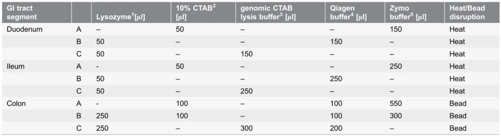
The sample preparation for total metagenome sequencing of the pooled samples was carried out following the recommendations of Ion Torrent PGM sequencing platform (Life Technologies). Sequencing was performed using Ion Torrent PGM 316 chips [29]. The reads were analysed and quality values were determined for Table 1.Cell lysis conditions applied to obtain maximum DNA for the next-generation sequencing of the microbial community in the various segments of the GI tract.
GI tract
segment Lysozyme1[
ml]
10% CTAB2
[ml]
genomic CTAB lysis buffer3[
ml]
Qiagen buffer4[
ml]
Zymo buffer5[
ml]
Heat/Bead disruption
Duodenum A – 50 – – 150 Heat
B 50 – – 150 – Heat
C 50 – 150 – – Heat
Ileum A - 50 – – 250 Heat
B 50 – – 250 – Heat
C 50 – 250 – – Heat
Colon A - 100 – 100 550 Bead
B 250 100 – 100 300 Bead
C 250 – 300 200 – Bead
1100 mg/ml (Applycehm).2Cetyltrimethylammonium bromide (w/v).31 M Tris-HCl 100 ml, 500 mM EDTA 50 ml, 5 M NaCl 300 ml, 10% CTAB, 20% SDS,
pH5829.4ASL buffer from Qiagen QIAamp DNA Stool miniprep kit (Qiagen, 51504).5From Zymo Research Fecal DNA kit (Zymo Research, D6010).
each nucleotide. The 100–200 nucleotide long individual sequences were further analysed by using the MG-RAST software package, which is a modified version of RAST (Rapid Annotations based on Subsystem Technology). The MG-RAST server initially runs a quality control test. If the data appear reliable, the system automatically screens for sequences of potential protein encoding regions via a BLASTX search against the comprehensive non-redundant database compiled from various publicly available sequencing centres and other sources. These databases include several rDNA datasets, e.g. GREENGENES, RDP II and European 16S RNA, among other information sources. The generated matches to external databases were used to compute the derived data. Details of the statistical calculations were published [29].
Results
Weight and glycaemic characteristics of experimental animals
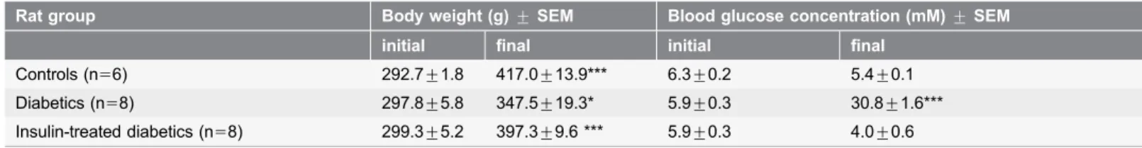
Ten weeks after the onset of diabetes, the diabetic rats were characterized by a reduced body weight and an increased blood glucose concentration as compared with the age- and sex-matched controls. The insulin-treated diabetic rats did not differ significantly from the control animals in weight or blood glucose
concentration (Table 2).
The composition of the microbiome along the gut
Two sets of metagenomic data were evaluated throughout the present study. The distributions at phylum, class and order levels (Figures 1–3) included all DNA reads comprising the eukaryotic sequences and unassigned ones. The latter indicated the number of sequences that could not be assigned to any known prokaryotic genome uploaded onto publicly available databases. In this way, we intended to present both the variations due to unrelated or unidentifiable data and the distribution of abundances among the bacterial taxa. The most important results are indicated at a genus level for the control, the diabetic and insulin-treated ileum in figure 2 and for diabetic colon in Figure 3.
The microbiome of the duodenum
The microbiome of the ileum
The microbiome of the control ileum
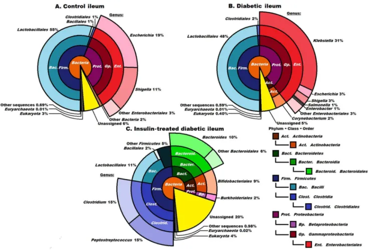
Only 3% of the DNA sequences originated from eukaryotic cells in this segment. The ileum was populated byBacteria, but representatives of Archaea(0.01%) also occurred (Figure 2A). The majority of the domainBacteriabelonged in the phyla Table 2.Weight and glycaemic characteristics of the three experimental groups of rats.
Rat group Body weight (g)¡SEM Blood glucose concentration (mM)¡SEM initial final initial final
Controls (n56) 292.7¡1.8 417.0¡13.9*** 6.3¡0.2 5.4¡0.1 Diabetics (n58) 297.8¡5.8 347.5¡19.3* 5.9¡0.3 30.8¡1.6*** Insulin-treated diabetics (n58) 299.3¡5.2 397.3¡9.6 *** 5.9¡0.3 4.0¡0.6
Initial vs. final: *p,0.05; ***p,0.001.
doi:10.1371/journal.pone.0110440.t002
Figure 1. Compositions of the control, diabetic and insulin-treated diabetic bacterial microbiomes in the duodenum at domain, phylum and class levels.Only the predominant orderLactobacillalesis indicated. The abbreviated and colour-coded taxa are indicated on the right side in systematic sequence. ‘‘Other sequences’’ are probably those of viruses not covered in the databases used. The majority of the identified DNA sequences related to the Eukaryota, indicating the overall low abundance of microbes. Among theBacteriatheLactobacillalespredominated. No significant differences were observed between the control and the diabetic and insulin-treated diabetic samples.
Firmicutes (57%) andProteobacteria (about 30%). The majority of theFirmicutes were identified in the class Bacilli, order Lactobacillales (55%) and genus
Lactobacillus (54%). The genera Escherichia (19%) andShigella (11%) pre-dominated in the Proteobacteria.
The microbiome of the diabetic ileum
Striking differences were observed in the composition of the microbial community of the ileal lumen between the diabetic rats and the controls (
Figures 2A and 2B). Among the Firmicutes (50%), the genus Lactobacillus predominated (48%) and Streptococcus (2%) was also detected, the distribution strongly resembling that in the control ileum. TheActinobacteria(not observed in the controls) appeared with representatives of the order Corynebacterium (2%). The total ratio ofProteobacteria was not changed greatly (42% as compared with 33% in the control ileum), but a massive invasion by the genus Klebsiella(31%) Figure 2. Compositions of the control, diabetic and insulin-treated diabetic microbiomes in the ileum at domain, phylum, class and order levels. The ordersEnterobacteriales(2B),ClostridialesandBacteroidetes(2C), displaying the most striking differences, are shown in higher resolution. The abbreviated and colour-coded taxa are indicated on the right side in systematic sequence. ‘‘Other sequences’’ are probably those of viruses not covered in the databases used. Taxa with very low (,1%) abundances are indicated to two decimal places. A striking invasion of the genusKlebsiellawas apparent in diabetes. It is noteworthy that the insulin treatment eliminated theKlebsiellainvasion, but did not restore the ‘‘healthy’’ microbiome, and the representation of the classesClostridiaandBacteroidiaincreased massively.
led to its predominance in the bacterial sequences in the diabetic ileum (
Figure 2B).
The microbiome of the insulin-treated diabetic ileum
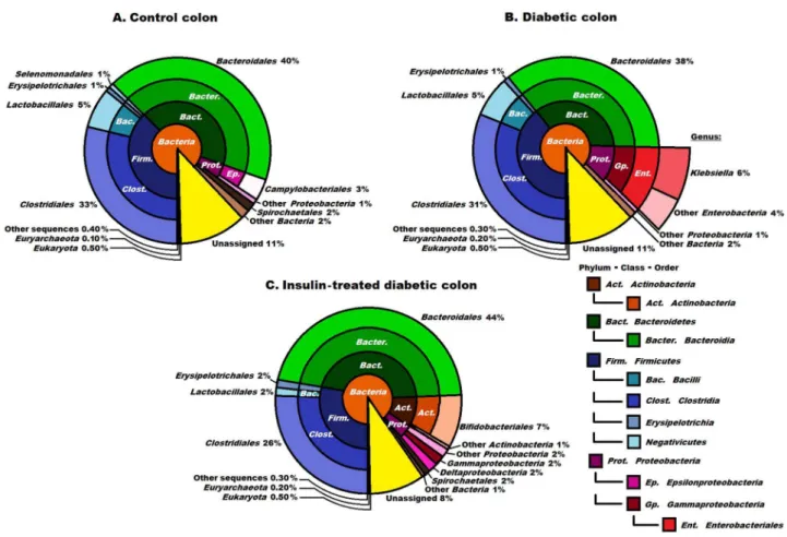
Insulin treatment resulted in a major reorganization of the microbial community of the ileum (Figure 2C). The ratio of Firmicutes did not change (48%), but the Bacteroidetes (scarce in the controls) made up 16% of the reads, accounting almost exclusively for the members of the genusBacteroides(data not shown). The Actinobacteria were represented twice as strongly as in the diabetic samples (the order Bifidobacteriales 9% replacing the orderCorynebacterium in the diabetic ileum). At the level of lower taxonomic units, characteristic signs of increasing microbial diversity and rearrangement of the microbiota were evident. Within the Firmicutes, the orderClostridiales(hardly detectable in the controls) was enriched to an astonishing 30% relative abundance. This order consisted essentially of two genera in equal representation, Clostridium andPeptostreptococcus. Insulin Figure 3. Compositions of the control, diabetic and insulin- treated diabetic microbiomes in the colon at domain, phylum, class and order levels. The abbreviated and colour-coded taxa are indicated on the right side in systematic sequence. Two minor classes belonging in the phylumFirmicutesare marked only with colours. ‘‘Other sequences’’ are probably those of viruses not covered in the databases used. Taxa with very low (,1%) abundances are indicated to two decimal places. The diversity of the microbial community was increased significantly in the insulin-treated diabetic animals, members of the orderBacteroidalespredominating. The orderEnterobacterialesis shown at higher resolution in diabetes (3B). The presence of theKlebsiellain the diabetic samples was still clearly seen although not as markedly as in the case of the ileum. Insulin promoted the abundance of the orderBifidobacteriales.
treatment was apparently deleterious for the order Lactobacillales in the class Bacilli: their number decreased 4-fold relative to the control and the diabetic ileum. The Proteobacteria, including the genusKlebsiella, the indicative signature of diabetes, were practically eradicated by insulin from the ileum of the diabetic rats. Only the order Burkholderiales represented theProteobacteria, in 2% abundance. A notably high proportion of the microbial community (20%) remained unassigned in these samples. Although rationalization of the consider-able changes in the various taxa as a result of insulin medication requires further study, it is clear that the microbial community of the ileum was rearranged and diversified substantially.
The microbiome of the colon
The microbiome of the control colon
The number of Archaea (0.1%) found in the colon was still small relative to the domain Bacteria, but was 10-fold higher relative to that in the ileum. The phyla Firmicutes andBacteroidetes predominated in the colon of the control rats (
Figure 3A). These two phyla each comprised 40% of all the identified sequences, Proteobacteria (4% representation) and Spirochaetes (2%) were also present. The other microbial phyla displayed significantly lower relative abundances. At the genus level, the order Clostridiales consisted primarily of the genera Clostridium (11%), Eubacteria (6%),Ruminococcus (5%) and Lactobacillus(4%). The most abundant bacteria in the order Bacteroidales were categorized in the genera Bacteroides (18%),Prevotella (12%) andAlistipes (5%).
The microbiome of the diabetic colon
The microbiome of the insulin-treated colon
Insulin treatment of the diabetic rats resulted in significant differences in the colon microbiome, which rather resembled that in the ileum, although the differences relative to the controls were less pronounced (Figures 2C,3A and 3C). The most obvious of the insulin effects on the microbiota was a general escalation of the diversity of the microbial community. The phyla Bacteroidetes and
Firmicutes still ruled over the microbial landscape, totalling 44% and 30% of the population, respectively. Among the other phyla, the Actinobacteria were
appreciably represented (8%), followed by the Proteobacteria (6%) and
Spirochaetes(2%). It should be noted that the insulin treatment was accompanied by fundamental changes within the phylum Proteobacteria: their overall
abundance decreased spectacularly, and the genus Klebsiella was basically
eradicated, being replaced by the generaDesulfovibrio andBifidobacterium, with a combined abundance of 4% (not shown).
Discussion
The overwhelming majority of studies published to date on the role of enteric pathogens in the development of autoimmune diabetes [30–32] were carried out on faecal samples. Community-level investigations were recently extended to metagenomic [33], meta-proteomic [34] and metabolomics [35] descriptions of pathological alterations in the GI microbiome, but all involved faecal samples. Thus, these data do not necessarily reflect the microbiological events that take place in the various segments of the GI system under the pathological conditions leading to T1D and related enteric neuropathies. In order to explore the possible correlation between the diabetes-related gut region-dependent nitrergic myenteric neuropathy and the altered mesenteric capillaries [10–11] and the spatially-restricted distribution of the gut microbiota, we therefore carried out a
metagenomic analysis of the luminal contents of the duodenum, ileum and colon of rats with STZ-induced diabetes and insulin-treated diabetes in comparison with control rats.
The proximal part of the GI tract does not harbour a rich microbial
community: around 104–105 cells/g are found there [36]. The predominance of Lactobacillus strains, which characteristically inhabit the upper gut, [37] was not markedly different in the duodenal luminal contents of the diabetic and the insulin-treated diabetic rats. Accordingly, the composition of the duodenal microbiota did not indicate the development of a pathological enteric
In the ileum, where a significant decrease in the total number of myenteric neurones is accompanied by severe structural damage of the mesenteric capillaries in rats with STZ-induced diabetes, [10–11] the diabetes is characterized by a massive Klebsiellainvasion. The Klebsiellaare among the most common Gram-negative bacteria that cause severe intestinal inflammation in humans [38–39]. The mucosal inflammation results in a leaky epithelium; this allows the easier passage of bacteria through the intestinal epithelium, initiating a pathological cascade disturbing the intestinal immunology, a critical element in the development of the autoimmune T1D [40–42]. The aberrant microbiota that develops in the ileum in STZ-induced diabetes and the characteristic upsurge of the Gram-negative Klebsiellacould therefore be directly associated with the inflammation and the development of the pathological microenvironment, leading to both autoimmune diabetes and diabetes-related enteric neuropathy. It is not clear whether theKlebsiellacontribute directly to the advance of the disease, or whether the weakened intestinal barrier facilitates the growth of theKlebsiella, i.e. whether the Klebsiella are the cause or the consequence of the autoimmune disease. It is noteworthy that a close relative from the same family, the pathogenic Escherichia coli, has been identified as the main culprit in various GI tract diseases, including autoimmune diabetes, in humans [43–48]. We found that insulin treatment brought about a drastic reorganization of the microbial community in the luminal content of the ileum. The microbial diversity increased enormously. The Protobacteriawere practically eliminated and the genus Klebsiella, the indicative signature of diabetes in the ileum, diminished. The Proteobacteriawere represented only by the genusBordatella(orderBurkholderiales). These changes in the composition of microbiota may correlate with the glycaemic control and the restoration of the enteric microenvironment, which prevented the neuronal cell loss and the structural damage of the capillary wall. Insulin treatment rearranged the microbiota of the ileum so that it became very distinct from both the control and the diabetes populations. The microbiome of the ileum in insulin-treated diabetes resembled the colon community and insulin could not restore the control microbiota in either GI segment.
in the generaLactobacillusandBifidobacteriumwere more abundant in genetically diabetes-resistant rats, which correlated well with the appearance of the order Bifidobacteriales in insulin-treated animals in our case. Their abundance was 9% and 7% in the insulin-treated ileum and colon, respectively. However, in mice a pro-diabetogenic, gluten-containing diet increased the relative numbers of the Bifidobacterium, theBarnesiellaand the Tanerella[32].
Insulin appropriately controlled the hyperglycaemia in the colon and prevented the myenteric neuronal loss. Unlike the ileum, the structural alterations of the microvessels remained unchanged relative to the diabetic counterparts [11]. This suggests that, although hyperglycaemia can be controlled by appropriate insulin medication, some of the associated system irregularities persist. The fact that insulin did not restore normal conditions in the capillary endothelium or in the microbiota of the ileum or the colon further indicated that the main location of hyperglycaemia-dependent events is in the ileum, and studies involving only the colon or the stools are unlikely to give an overall picture.
Conclusions
irregula-rities persist in the different intestinal segments and influence the outcome of the therapies.
Acknowledgments
The authors thank Ms. Netta Bozo´ki for technical assistance.
Author Contributions
Conceived and designed the experiments: E´F KLK ZB. Performed the experiments: RW NB PT GM. Analyzed the data: RW NB GM MB KLK.
Contributed reagents/materials/analysis tools: GM E´F ZB KLK. Wrote the paper: E´F KLK.
References
1. Boerner BP, Sarvetnick NE(2011) Type 1 diabetes: role of intestinal microbiome in humans and mice. Ann NY Acad Sci 1243: 103–118.
2. Brugman S, Klatter FA, Visser JTJ, Wildeboer-Veloo ACM, Harmsenet HJM, et al.(2006) Antibiotic treatment partially protects against type 1diabetes in the bio-breeding diabetes-prone rat. Is the gut flora involved in the development of type 1 diabetes? Diabet 49: 2105–2108.
3. Sokol H, Pigneur B, Watterlot L, Lakhdari O, Bermu´dez-Humara´n LG, et al.(2008)Faecalibacterium prausnitziiis an anti-inflammatory commensal bacterium identified by gut microbiota analysis of Crohn disease patients. Proc Nat Acad Sci USA 105: 16731–16736.
4. Valladares R, Sankar D, Li N, Williams E, Lai K-K, et al.(2010)Lactobacillus johnsoniiN6.2 mitigates the development of type 1 diabetes in BB-DP rats. PloS ONE 5: e10507.
5. Lau K, Benitez P, Ardissone A, Wilson TD, Collins EL, et al. (2011) Inhibition of type 1 diabetes correlated to aLactobacillus johnsoniiN6.2-mediated Th17 bias. J Immun 186: 3538–3546.
6. Ha¨nninen A, Nurmela R, Maksimow M, Heino J, Jalkanen S, et al.(2007) Islet beta-cell-specific T cells can use different homing mechanisms to infiltrate and destroy pancreatic islets. Am J Pathol 170(1): 240–250.
7. Bytzer P, Talley NJ, Leemon M, Young LJ, Jones MP, et al.(2001) Prevalence of gastrointestinal symptoms associated with diabetes mellitus: a population-based survey of 15,000 adults. Ach Inter Med 161: 1989–1996.
8. Roszto´czy A, Ro´ka R, Va´rkonyi TT, Lengyel C, Izbe´ki F, et al. (2004) Regional differences in the manifestation of gastrointestinal motor disorders in type 1 diabetic patients with autonomic neuropathy. Z Gastroenterol 42: 1295–1300.
9. O¨ rdo¨g T, Hayashi Y, Gibbons SJ(2009) Cellular pathogenesis of diabetic gastroenteropathy. Minerva Gastroenterol Dietol 55: 315–343.
10. Izbe´ki F, Wittman T, Roszto´czy A, Linke N, Bo´di N, et al.(2008) Immediate insulin treatment prevents gut motility alterations and loss of nitrergic neurons in the ileum and colon of rats with streptozotocin-induced diabetes. Diab Res Clin Pract 80: 192–198.
11. Bo´di N, Talapka P, Poles P, Hermesz E, Jancso´ Z, et al.(2012) Gut region-specific diabetic damage to the capillary endothelium adjacent to the myenteric plexus. Microcirc 19: 316–326.
12. Demendts I, Masaoka T, Kindt S, De Hertogh G, Geboes K, et al.(2013) Gastrointestinal motility changes and myenteric plexus alterations in spontaneously diabetic biobreeding rats.
J Neurogastroenterol Motil 19: 161–170.
14. Wheway J, Mackay CR, Newton RA, Sainsbury A, Boey D, et al.(2005) A fundamental bimodal role for neuropeptide Y1 receptor in the immune system. JEM 202: 1527–1538.
15. Gutierrez-Canas I, Juarraz Y, Santiago B, Arranz A, Martinez C, et al.(2006) VIP down-regulates TLR4 expression and TLR4-mediated chemokine production in human rheumatoid synovial fibroblasts. Rheum 45: 527–532.
16. NIH HMP Working Group, Peterson J, Garges S, Giovanni M, McInnes P, et al. (2009) The NIH human microbiome project. Genome Res 19: 2317–2323.
17. Neish AS(2009) Microbes in gastrointestinal health and disease. Gastroenterol 136: 65–80.
18. Prakash S, Rodes L, Coussa-Charley M, Tomaro-Duchesneau C(2011) Gut microbiota: next frontier in understanding human health and development of biotherapeutics. Biologics 5: 71–86.
19. Hoffmann C, Dollive S, Grunberg S, Chen J, Li H, et al.(2013) Archaea and fungi of the human gut microbiome: correlations with diet and bacterial residents. PloS ONE 8: e66019.
20. Qin J, Li R, Raes J, Arumugam M, Burgdorf KS, et al.(2010) The human gut microbial gene catalogue established by metagenomic sequencing. Nature 464: 59–65.
21. Lepage P, Leclerc MC, Joossens M, Mondot S, Blottie`re HM, et al.(2013) A metagenomic insight into our gut’s microbiome. Gut 62: 146–158.
22. Le Chatelier E, Nielsen T, Qin J, Prifti E, Hildebrand F, et al. (2013) Richness of human gut microbiome correlates with metabolic markers. Nature 500: 541–546.
23. Zoetnedal EG, von Wright A, Vilpponen-Salmela T, et al.Mucosa-associated bacteria in the human gastrointestinal tract are uniformly distributed along the colon and differ from the community recovered from feces. Appl Environ Microbiol 2002; 68: 3401–3407.
24. Echburg PB, Bik EM, Barnstein CN, Purdom E, Dethlefsen L, et al.(2005) Diversity of the human intestinal microbial flora. Science 308: 1635–1638.
25. Lepage P, Seksik P, Sutren M, de la Cochetie`re MF, Jian R, et al.(2005) Biodiversity of the mucosa-associated microbiota is stable along the distal digestive tract in healthy Individuals and patients with IBD. Imflamm Bowel Dis 11: 473–480.
26. Zoetendal EG, Rajilic-Stonajovic M, de Vos MW (2013) High-throughput diversity and functionality analysis of the gastrointestinal tract microbiota. Gut 57: 1605–1615.
27. Gu S, Chen D, Zhang J-N, Lv X, Wang K, et al.(2013) Bacterial community mapping of the mouse gastrointestinal tract. PloS ONE 8: e74957.
28. Wang Z-K, Yang Y-S(2013) Upper gastrointestinal microbiota and digestive diseases. WJG 19: 1541– 1550.
29. Wirth R, Kova´cs E, Maro´ti G, BagiZ, Ra´khely G, et al.(2012) Characterization of a biogas-producing microbial community by short-read next generation DNA sequencing. Biotechnol Biofuels 5: 41.
30. Macpherson AJ, Guenking MB, McCoy KM (2005) Immune responses that adapt the intestinal mucosa to commensal intestinal bacteria. Immunol 115: 153–162.
31. Giongo A, Gano KA, Crabb DB, Mukherjee N, Novelo LL, et al. (2011) Toward defining the autoimmune microbiome for type 1 diabetes. ISME 5: 82–91.
32. King C, Sarvetnick N(2011) The incidence of type-1 diabetes in NOD mice Is modulated by restricted flora not germ-free conditions. PloS ONE 6: e17049.
33. Marietta EV, Gomez AM, Yeoman C, Tilahun AY, Clark CR, et al. (2013) Low incidence of spontaneous type 1 diabetes in non- obese diabetic mice raised on gluten-free diets is associated with changes in the intestinal microbiome. PloS ONE 8: e78687.
34. Kinross JM, Darzi AW, Nicholson JK(2011) Gut microbiome-host interactions in health and disease. Genome Medicine 3: 14.
35. Ugarte M, Brown M, Hollywood KA (2012) Metabolomic analysis of rat serum in streptozotocin-induced diabetes and after treatment with oral triethylenetetramine (TETA). Genome Medicine 4: 35.
37. Almiro´n M, Traglia G, Rubio A, Sanjuan N.(2013) Colonization of the mouse upper gastrointestinal tract byLactobacillus murinus: a histological, immunocytochemical, and ultrastructural study. Curr Microbiol 67(4): 395–398.
38. Rashid T, Ebringer A, Tiwana, M Fielder(2009) Role of Klebsiella and collagens in Crohn’s disease: a new prospect in the use of low-starch diet. Eur J Gastroentrerol 21(8): 843–849.
39. Vasseur MV, Laurentie M, Rolland JG, Perrin-Guyomard A, Henri J, et al.(2014) Low or high doses of cefquinome targeting low or high bacterial inocula cureKlebsiella pneumoniaelung infections but differentially impact the levels of antibiotic resistance in fecal flora. Antimicrob Agents Chemother 58(3): 1744–1748.
40. Vaarala O, Atkinson MA, Neu J.(2008) The ‘‘perfect storm’’ for type 1 diabetes. The complex interplay between intestinal microbiota, gut permeability, and mucosal immunity. Diabetes 57: 2555–2562.
41. Vaarala O, Ilonen J, Ruohtula T, Pesola J, Virtanen SM, et al.(2012) Removal of bovine insulin from cow’s milk formula and early initiation of beta-cell autoimmunity in the FINDIA pilot study. Arch Pediatr Adolesc Med 166(7): 608–614.
42. Vaarala O(2013) Human intestinal microbiota and type 1 diabetes. Curr Diab Rep 13: 601–607.
43. Nadal I, Donat E, Ribes-Koninckx C, Calabuig M, Sanz Y(2007) Imbalance in the composition of the duodenal microbiota of children with coeliac disease. J Med Microbiol 56: 1669–1674.
44. Sartor RB(2008) Microbial influences in inflammatory bowel diseases. Gastroenterol 134: 577–594.
45. Neish AS(2009) Microbes in gastrointestinal health and disease. Gastroenterol 136: 65–80.
46. Willing B, Halfvarson J, Dicksved J, Rosenquist M, Ja¨rnerot G, et al.(2009) Twin studies reveal specific imbalances in the mucosaassociated microbiota of patients with ileal Crohn’s disease. Inflamm Bowel Dis 15: 653–660.
47. Sekirov I, Russell SL, Antunes CM, Finlay BB(2010) Gut microbiota in health and disease. Physiol Rev 90: 859–904.
48. Krause DO, Little AC, Dowd SE, Bernstein CN(2011) Genome announcements. J Bacteriol 193: 583.