Co-Culture of α TC-6 Cells and β TC-1 Cells: Morphology
and Function
Sung Man Kim1, Eun Ju Lee1, Hye Sook Jung2, Na Han1, You Jeong Kim1, Tae Kyoon Kim1, Tae Nyun Kim1, Min Jeong Kwon1, Soon Hee Lee1, Jeong Hyun Park1,2, Byoung Doo Rhee1, Mi-Kyung Kim1,2
1Department of Internal Medicine, Inje University College of Medicine; 2Moelcular Therapy Lab, Paik Memorial Institute for Clinical Research, Inje University, Busan, Korea
Background:In vitro experiments using only β-cell lines instead of islets are limited because pancreatic islets are composed of four different types of endocrine cells. Several recent studies have focused on cellular interactions among these cell types, espe
-cially α- and β-cells. Because islet isolation needs time and experience, we tested a simple co-culture system with α- and β-cells. Their morphology and function were assessed by comparison to each single cell culture and pancreatic islets.
Methods: α TC-6 cells and β TC-1 cells were maintained in Dulbecco’s Minimal Essential Medium containing 5 mM glucose and 10% fetal bovine serum. Cells were mixed at a 1:1 ratio (5×10⁵) in 6-well plates and cultured for 24, 48, and 72 hours. After
culture, cells were used for insulin and glucagon immunoassays and tested for glucose-stimulated insulin secretion (GSIS). Results: α TC-6 and β TC-1 cells became condensed by 24 hours and were more strongly compacted after 48 hours. β TC-1 cells showed both β-β and β-α cell contacts. GSIS increased with increasing glucose concentration in co-cultured cells, which showed lower secreted insulin levels than β TC-1 cells alone. The increase in the secreted insulin/insulin content ratio was significantly lower for co-cultured cells than for β-cells alone (P=0.04). Compared to islets, the α-/β-cell co-culture showed a higher ratio of GSIS to insulin content, but the difference was not statistically significant (P=0.09).
Conclusion: α TC-6 and β TC-1 cells in the co-culture system showed cell-to-cell contacts and a similar stimulated insulin secre
-tion pattern to islets. The co-culture system may be used to better mimic pancreatic islets in in vitro assessments.
Keywords: α-Cell; β-Cell; Insulin
INTRODUCTION
In mammals, the endocrine pancreas comprises a majority of the islets of Langerhans that are dispersed within the pancreas [1]. There are approximately 1,000 islets in a mouse pancreas and 1 million islets in a human pancreas [2,3]. Each islet is a
round or ovoid mass, 50 to 600 µm in diameter and comprising ~50 to 3,000 cells [1,3]. Pancreatic islets are composed of four different endocrine cell types. Highly specialized cells called β-cells represent about 60% of the cells in human islets. The remaining cells are α, δ, polypeptide-producing, and epsilon cells, which produce glucagon, somatostatin, pancreatic poly
-Received: 7 April 2014, Revised: 29 May 2014, 14 July 2014 Accepted: 5 August 2014
Corresponding author: Mi-Kyung Kim
Department of Internal Medicine, Inje University Haeundae Paik Hospital, Inje University College of Medicine, 875 Haeun-daero, Haeundae-gu, Busan 612-862, Korea
Tel: +82-51-797-0636, Fax: +82-51-797-2070, E-mail: kmkdoc@hanmail.net
Copyright © 2015 Korean Endocrine Society
This is an Open Access article distributed under the terms of the Creative Com-mons Attribution Non-Commercial License (http://creativecomCom-mons.org/ licenses/by-nc/3.0/) which permits unrestricted non-commercial use, distribu-tion, and reproduction in any medium, provided the original work is properly
Copyright © 2015 Korean Endocrine Society peptide, and ghrelin, respectively [1,2].
The endocrine pancreas alone is responsible for insulin se
-cretion in mammals and plays a prominent role in controlling circulating glucose levels and glucose metabolism [2,4]. Insu
-lin is produced by β-cells in the pancreas [2,5-7]. Pancreatic β-cells produce, store, and finally secrete insulin in response to increased glycemia [1]. However, the modulation of insulin se
-cretion as a function of changing metabolic demands and envi
-ronmental conditions, especially levels of circulating glucose, is not completely performed by β-cells alone. It is a highly complex process, and many factors affect β-cells through vas
-cular, paracrine, autocrine, neural, and cellular pathways [8]. Within pancreatic islets, each cell communicates in a variety of ways, including through indirect and direct mechanisms [1]. Indirect cell communication does not require cell contact but is instead mediated by an extracellular flux of molecules that si
-multaneously signal to multiple cells. In addition, various mo
-dalities of direct cell-to-cell communication operate within pancreatic islets. Therefore, the segregation of islets into single cells leads to a rapid loss of insulin regulation. This regulation can be restored by reestablishing cell contacts [1,9-12]. Be
-cause the integrated secretory responses of islet cells are essen
-tial for the maintenance of normal patterns of insulin secretion, several recent studies have focused on interactions between different islet cell types, especially α- and β-cells [13].
Research on pancreatic β-cell function and regulation in many cases still relies on clonal cell line monolayers, which have limited ability to mimic endogenous pancreatic islets. Us
-ing pancreatic islets themselves is the best approach. However, islet isolation is complex and time consuming. The number of islets that can be obtained by that process largely depends on the experience of the experimenters. In addition, isolated islets cannot survive longer than 2 weeks or be stockpiled. Therefore, we sought to determine whether it would be viable to use co-cultured α and β clonal cell lines instead of islets. We assessed their morphology and function and compared them to single cell cultures and pancreatic islets.
METHODS
Cell lines
Mouse α TC-6 cells and β TC-1 cells were obtained from the American Type Culture Collection (Manassas, VA, USA). Cells were grown at 37.0°C in a 5% carbon dioxide incubator in Dul
-becco’s Modified Eagle’s Medium (DMEM) containing 4 mM L-glutamine, 5 mM glucose, 44 mM sodium bicarbonate, 10%
heat-inactivated fetal bovine serum (FBS), 15 mM 4-(2-hydroxyethyl)-1-piperazineethanesulfonic acid (HEPES), and 0.1 mM nones
-sential amino acids.
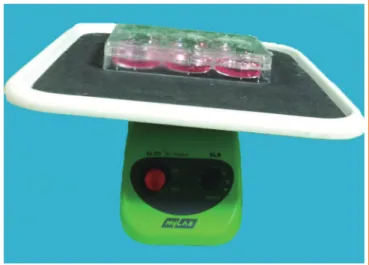
α TC-6 cell and β TC-1 cell co-culture system
Cells were cultured in culture plates in Dulbecco’s Minimal Es
-sential Medium containing 5 mM glucose and supplemented with 44 mM sodium bicarbonate, 10% FBS, and 15 mM HEPES. Initially the cells were mixed at various α:β ratios: 1:4, 1:2, and 1:1. However, we found it difficult to observe the α-cells at ratios of 1:4 and 1:2.
Therefore, cells were mixed at a 1:1 ratio (5×10⁵
) in 6-well
plates. The plates were shaken on a gyratory shaker at 37°C (Fig. 1).
Rat islet isolation
Hank’s balanced salt solution (HBSS) was used for perfusion and preservation of pancreas. Pancreatic islets were isolated us
-ing Lacy and Kostianovsky’s method with modifications. Pan
-creases were distended by intraductal injection of cold collage
-nase solution (0.8 mg/mL in HBSS) into the common bile duct after occlusion of the distal end, close to the duodenum. Diges
-tion was performed in a water bath at 37°C for 14 minutes 20 seconds and was stopped by the addition of cold HBSS. Each pancreas was broken up by vortexing for 15 seconds. After cen
-trifugation for 10 seconds at 1,500 rpm, the supernatant was re
-moved. The pancreatic tissue was resuspended in 30 mL HBSS and passed through a 400-µm mesh. The filtered tissue was washed with HBSS and centrifuged on a Bicoll density gradient (1.085, 1.069, and 1.037 g/mL). The islets were cultured with
M199 medium containing 10% FBS in a noncoated plate.
Copyright © 2015 Korean Endocrine Society Morphological examination and immunostaining
The aggregates were observed at different time points with a phase-contrast microscope. α TC-6 and β TC-1 cells were plat
-ed onto cover slides. After incubation under appropriate condi
-tions, the cells were fixed and permeabilized with Cytofix/Cy
-toperm (BD, San Diego, CA, USA) for 15 minutes and washed with perm/wash buffer (0.1% saponin, 1 mM CaCl2∙2H2O, 1
mM MgSO4, 0.05% NaNa3, 10 mM HEPES, 1% bovine serum
albumin). Cells were incubated with an anti-insulin or antiglu
-cagon antibody overnight at 4°C and washed with phosphate-buffered saline (PBS). Then the cells were incubated with sec
-ondary antibody (antirabbit immunoglobulin G [IgG] Alexa Fluor 488 conjugate or Alexa Fluor 555 conjugate [Cell signal
-ing, Danvers, MA, USA]) for 1 hour at room temperature and washed with PBS. We used anti-insulin and antiglucagon anti
-bodies isolated from rabbits instead of rats because we used mouse α TC-6 and β TC-1 cells. However, the antirabbit IgG secondary antibody stained both insulin and glucagon. There
-fore, we could not use confocal microscopy to distinguish be
-tween insulin and glucagon, and we stained each of them sepa
-rately. Fluorescent in situ hybridization analysis was performed using a BX-51 green fluorescence microscope (Olympus, To
-kyo, Japan). Then we calculated the cellular composition of the co-culture aggregates at 24, 48, and 72 hours. The results are presented as cell number/area (%).
Quantification of secreted insulin protein by enzyme-linked immunosorbent assay
After incubation, cells were washed with PBS. To measure
glucose-stimulated insulin secretion (GSIS), we starved cells for 5 hours in DMEM containing 5 mM glucose and 2% FBS. Then the medium was replaced with KRBB solution (4.74
mM KCl, 1.19 mM KH2PO4, 1.19 mM MgCl2∙6H2O, 35 mM
NaHCO3, 10 mM HEPES) containing 5 mM glucose or 25
mM glucose and the cells were incubated for 1 hour. To sepa
-rate total insulin protein, we washed cells with PBS and lysed them with mammalian tissue lysis/extraction reagent contain
-ing a protease inhibitor. Insulin protein in the medium was measured using a Rat/Mouse Insulin ELISA Kit (Linco Re
-search, St. Charles, MO, USA).
RESULTS
Formation of aggregates in the co-culture system and morphologic examination
Cells were mixed at a 1:1 ratio (5×10⁵) in 6-well plates and the
plates were sealed and shaken on a gyratory shaker to form ag
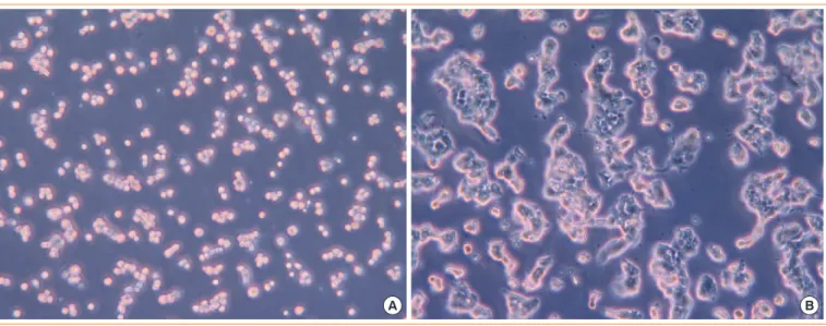
-gregates. After culture, we observed the morphology with an optical microscope (×200) at 2 hours (Fig. 2A) and 48 hours (Fig. 2B). Cells were scattered at 2 hours (Fig. 2A), but nu
-merous aggregates of heterogeneous size had formed from α TC-6 and β TC-1 cells at 48 hours (Fig. 2B). They became condensed and more strongly compacted at 72 hours. They sur
-vived well in the co-culture system, and good cell-to-cell con
-tacts were formed using the gyratory shaker.
Immunohistochemical staining of co-cultured cells
Immunohistochemical staining for insulin and glucagon at 24
Fig. 2. Morphology in the co-culture at (A) 2 hours and (B) 48 hours, as observed using an optical microscope (×200).
Copyright © 2015 Korean Endocrine Society
hours (Fig. 3A), 48 hours (Fig. 3B), and 72 hours (Fig. 3C) is shown in Fig. 3. β TC-1 cells showed both β-β and β-α cell con
-tacts, whereas α TC-6 cells showed both α-α and β-α cell con
-tacts. α TC-6 cells became compact and more spherical by 72 hours, but β TC-1 cells did not show a specific pattern in the
co-culture system.
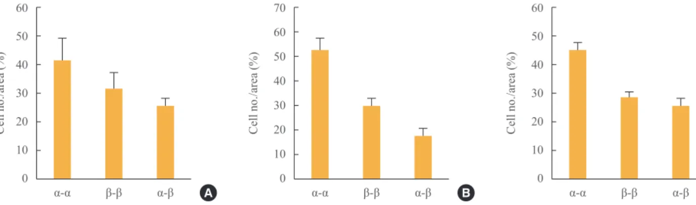
We also analyzed the cellular composition of the aggregates (Fig. 4). Of cells mixed at a 1:1 ratio (5×10⁵), ~45% showed
α-α cell contacts and ~25% showed β-α cell contacts. The per
-centage of β-α cell contacts was similar through 72 hours, al
-Fig. 3. Cellular composition of the aggregates, as studied by immunohistochemical staining for insulin and glucagon at (A) 24 hours, (B)
48 hours, and (C) 72 hours. Red, glucagon; green, insulin; blue, DAPI.
A B C
Fig. 4. Cellular composition of the aggregates in co-culture at (A) 24, (B) 48, and (C) 72 hours.
60
50
40
30
20
10
0
70
60
50
40
30
20
10
0
60
50
40
30
20
10
0
Cell
no./area
(%)
Cell
no./area
(%)
Cell
no./area
(%)
α-α β-β α-β A α-α β-β α-β B α-α β-β α-β C
Fig. 5. Glucose-stimulated insulin secretion from β TC-1 cells and
co-cultured cells.
0.6
0.5
0.4
0.3
0.2
0.1
0
Insulin
concentration
(pg/mL)
3 mM 15 mM
β-cell
Glucose α/β-cell
3 mM 15 mM
Fig. 6. Increase in secreted insulin/insulin content in β TC-1 cells, co-cultured cells, and islets.
4
3.5
3
2.5
2
1.5
1
0.5
0
Increase
of
secreted
insulin/insulin
β α/β Islet
Copyright © 2015 Korean Endocrine Society though the total cell population increased.
Glucose-stimulated insulin secretion
GSIS from β TC-1 cells and co-cultured cells is shown in Fig. 5. Insulin secretion was increased by glucose stimulation in both cultures. The degree of GSIS was higher in β-cells cul
-tured alone than in the α-/β-cell co-culture.
We also compared the ratio of GSIS to insulin contents in β TC-1 cells, co-cultured cells, and islets. As shown in Fig. 6, the increase in secreted insulin/insulin content was significantly lower for co-cultured cells than for β-cells only (P=0.04). Compared to islets, the α-/β-cell co-culture showed a higher ra
-tio of GSIS to insulin content, but the difference was not statis
-tically significant (P=0.09).
DISCUSSION
The present study demonstrated that α TC-6 cells and β TC-1 cells formed aggregates that looked like pseudoislets and sur
-vived well in our co-culture system. They showed similar GSIS to pancreatic islets. Although our morphologic study did not show a particular topography similar to the three-dimen
-sional architecture of native islets, the co-cultured cells formed multiple aggregates with cell contacts and showed similar GSIS to islets isolated from rats.
In previous studies, different cell types showed a particular intraislet topography: β-cells were located in the central core, whereas non-β-cells surrounded the β-cells to form a mantle layer [8,13].
Most researchers study pancreatic β-cells with clonal cell line monolayers. However, the normal pattern of insulin secre
-tion is facilitated by the maintenance of islet architecture, and the integrated secretory response of islet cells is essential for the normal pattern [14]. There have been many studies of in
-teractions between and within pancreatic islets. It is now known that intraislet cell communication is important for ade
-quate release of insulin and is largely achieved through gap junction-medicated communication [15]. Although the exact mechanism is unknown, several mechanisms depend on dif
-ferent families of integral membrane proteins, which ensure direct (cadherins, neural cell adhesion molecule, occludin, and claudins) and paracrine (pannexins) communication between β-cells and between these cells and the other islet cell types [1]. Also, other proteins (integrins) provide communication between the different islet cell types and the materials that form the islet basal laminae and extracellular matrix. The dis
-sociation of islet cells is associated with a dramatic change in insulin secretion in response to glucose. The formation of cel
-lular aggregates called pseudoislets marks a significant im
-provement in insulin secretion [1,2,8]. In the early work of Timble and others, the cellular glucagon content of dorsal is
-lets was associated with insulin secretion [16]. Another study showed that an increase in the percentage of glucagon-secret
-ing cells to 50% had a detrimental effect on insulin release from neighboring β-cells [17]. These findings suggest that the interaction of insulin-secreting cells and α-cells and changes in their relative proportions have marked effects on insulin re
-lease. Pseudoislets comprising insulin-secreting cell lines and α-cell lines could be a useful model for examining islet inter
-actions and insulin secretion.
Most pseudoislet research has been conducted on clonal β-cell lines only and has isolated β-cell lines without other islet cells such as α-cells. There have been few studies of heterotypic pseudoislets containing α- and β-cell lines [8,13,18]. One study used a model containing low concentrations of α TC1 and MIN6 cells. α TC1 cells had no effect on insulin secretion from MIN6 cells in that study [18]. However, in another study, co-cultured pancreatic α- and β-cell lines exhibited contact-depen
-dent inhibition of insulin secretion [13]. In that study, insulin-secreting β TC cells and glucagon-insulin-secreting α TC cells formed pseudoislets. Our present study showed comparable results: the co-cultured cells formed pseudoislet-like aggregates and showed decreased GSIS compared to β-cells cultured alone. It has not been clearly explained how insulin secretion from β-cells is in
-hibited via contact with α-cells. Previous studies have suggested several hypotheses. An increase in the α TC/β TC cell ratio in aggregate cultures was accompanied by a decrease in secreted insulin and an increase in the intracellular insulin content of the β TC component [13]. Because the intracellular insulin content of β TC cells was not affected by α TC cells in a Transwell (Cat. No. 3413, Costar, Cambridge, MA, USA) co-culture system in which direct cell-to-cell contacts were prevented by a semiper
-meable membrane that permitted chemical communication via secreted metabolites, the authors guessed that insulin secretion from β TC cells may possibly be inhibited as a result of contact with α TC cells. However, the authors did not completely rule out the possible effects of soluble factors. The possibility of apoptosis of β TC cells due to cell-to-cell contacts has been sug
-gested [13]. We think that further investigation is needed to de
-termine the precise mechanism.
Copyright © 2015 Korean Endocrine Society
cells have similar GSIS to pancreatic islets. Although the use of isolated islets is the best way to study pancreatic function, this method is problematic for many reasons. We think that our pseudoislets, created from α- and β-cell lines using a sim
-ple method, may mimic pancreatic islets, reducing the limita
-tions of in vitro experiments.
CONFLICTS OF INTEREST
No potential conflict of interest relevant to this article was re -ported.
REFERENCES
1. Meda P. Protein-mediated interactions of pancreatic islet
cells. Scientifica (Cairo) 2013;2013:621249.
2. Scharfmann R, Rachdi L, Ravassard P. Concise review: in
search of unlimited sources of functional human pancreatic beta cells. Stem Cells Transl Med 2013;2:61-7.
3. Seino S, Bell GI, eds. Pancreatic beta cell in health and dis
-ease. Berlin: Springer; 2008. Chapter 1, Architecture of pan
-creatic islets; p. 3-11.
4. Ravassard P, Hazhouz Y, Pechberty S, Bricout-Neveu E, Armanet M, Czernichow P, Scharfmann R. A genetically
engineered human pancreatic beta cell line exhibiting
glu-cose-inducible insulin secretion. J Clin Invest 2011;121:
3589-97.
5. Bergsten P, Hellman B. Glucose-induced amplitude regu
-lation of pulsatile insulin secretion from individual pancre
-atic islets. Diabetes 1993;42:670-4.
6. Gilon P, Ravier MA, Jonas JC, Henquin JC. Control mech
-anisms of the oscillations of insulin secretion in vitro and in vivo. Diabetes 2002;51 Suppl 1:S144-51.
7. Tengholm A, Gylfe E. Oscillatory control of insulin secre -tion. Mol Cell Endocrinol 2009;297:58-72.
8. Kelly C, McClenaghan NH, Flatt PR. Role of islet struc
-ture and cellular interactions in the control of insulin secre
-tion. Islets 2011;3:41-7.
9. Bosco D, Haefliger JA, Meda P. Connexins: key mediators
of endocrine function. Physiol Rev 2011;91:1393-445.
10. Renold AE, Zahnd GR, Wollheim CB. Contributions of
physiology to the understanding of diabetes. Berlin: Spring
-er; 1997. Chapter 3, Intercellular communication and insu
-lin secretion; p. 24-42.
11. Bavamian S, Klee P, Britan A, Populaire C, Caille D, Can
-cela J, Charollais A, Meda P. Islet-cell-to-cell communica
-tion as basis for normal insulin secre-tion. Diabetes Obes
Metab 2007;9 Suppl 2:118-32.
12. Potolicchio I, Cigliola V, Velazquez-Garcia S, Klee P, Valje
-vac A, Kapic D, Cosovic E, Lepara O, Hadzovic-Dzuvo A, Mornjacovic Z, Meda P. Connexin-dependent signaling in neuro-hormonal systems. Biochim Biophys Acta 2012;
1818:1919-36.
13. Hamaguchi K, Utsunomiya N, Takaki R, Yoshimatsu H,
Sakata T. Cellular interaction between mouse pancreatic alpha-cell and beta-cell lines: possible contact-dependent inhibition of insulin secretion. Exp Biol Med (Maywood)
2003;228:1227-33.
14. Hopcroft DW, Mason DR, Scott RS. Structure-function re
-lationships in pancreatic islets: support for intraislet modula
-tion of insulin secre-tion. Endocrinology 1985;117:2073-80.
15. Meda P, Halban P, Perrelet A, Renold AE, Orci L. Gap junc
-tion development is correlated with insulin content in the
pancreatic B cell. Science 1980;209:1026-8.
16. Trimble ER, Halban PA, Wollheim CB, Renold AE. Func
-tional differences between rat islets of ventral and dorsal pancreatic origin. J Clin Invest 1982;69:405-13.
17. Leite AR, Carvalho CP, Furtado AG, Barbosa HC, Bosche
-ro AC, Collares-Buzato CB. Co-expression and regulation of connexins 36 and 43 in cultured neonatal rat pancreatic islets. Can J Physiol Pharmacol 2005;83:142-51.
18. Brereton H, Carvell MJ, Persaud SJ, Jones PM. Islet
alpha-cells do not influence insulin secretion from beta-alpha-cells