Cells for Clinical Application
Maria Margarida de Carvalho Negrão Serra
Dissertation presented to obtain a Ph.D degree in Engineering and
Technology Sciences, Biomedical Engineering at the Instituto de
Tecnologia Química e Biológica, Universidade Nova de Lisboa
Supervisor: Paula Marques Alves
Oeiras, February 2011
Second edition: February 2011
Front cover: Composite image of the main stem cell models and 3-D culture
strategies used for the development of novel stem cell bioprocesses; phase contrast and immunofluorescence microscopy images of rPSCs immobilized on microcarriers, NT2 cell aggregates and alginate microencapsulated hESCs immobilized on microcarriers; immunofluorescence microscopy images of rPSCs labelled for nestin (green), NT2 neurosphere stained with nestin (red) and
β‐tubulin‐III protein (green), hESCs labelled for oct-4 (green), nuclei were stained with dapi (blue).
Back cover: Neuronal differentiation of NT2 cells. Immunofluorescence microscopy images of NT2 cultures composed by non-neuronal cells (stained with nestin, red) and neurons (labelled with β‐tubulin‐III protein, green), nuclei were stained with dapi (blue); phase contrast image of a pure population of neurons.
By Margarida Serra and Teresa Serra
ITQB-UNL/IBET Animal Cell Technology Unit
Instituto de Tecnologia Química e Biológica-Universidade Nova de Lisboa/ Instituto de Biologia Experimental e Tecnológica
Av. da República EAN, 2780-157 Oeiras, Portugal Fax: +351 21 442 11 61; Phone: +351 21 446 91 00 http://www.itqb.unl.pt
http://www.ibet.pt
Copyright © 2011 by Maria Margarida Serra All rights reserved
Supervisor
Dr. Paula Maria Marques Leal Sanches Alves, Principal Investigator and Head of the Animal Cell Technology Unit at ITQB-UNL and Executive Director of IBET, Oeiras, Portugal.
President of the jury
Dr. Carlos Crispim Romão, Professor at Instituto de Tecnologia Química e Biológica, Universidade Nova de Lisboa (ITQB-UNL), Oeiras, Portugal.
Jury
Dr. Marc Peschanski, Scientific Director of the Institute for Stem Cell Therapy and Exploration of Monogenic diseases (I-STEM, INSERM/UEVE), Evry, France.
Dr. Lino Ferreira, Principal Investigator at Centro de Neurosciências e Biologia Celular, Universidade de Coimbra, Coimbra, Portugal.
Foreword
This thesis dissertation represents four years of research undertaken at the Animal Cell Technology Unit of the Instituto de Tecnologia Química e
Biológica from the Universidade Nova de Lisboa/Instituto de Biologia
Experimental e Tecnológica under the supervision of Dr. Paula Alves.
À memória dos meus avós Clara e Camilo
Aos meus pais
thesis, which supported and helped me during this long, but rewarding, journey.
To, Dr Paula Alves, my supervisor, from whom I learned so much. For her courage and enthusiasm to start this area of research at ITQB-UNL/IBET and for her efforts to create the excellent conditions that we have at the Animal Cell Technology Unit. Her outstanding personality, scientific attitude, truly dedication and professionalism have made me grow as a scientist and as a person. For her guidance, encouragement, confidence and friendship. For allowing me the freedom and opportunities to evolve in this very exciting area.
To Prof. Manuel Carrondo, for his endless support, valuable suggestions and guidance throughout these years. For his enthusiasm, inspiration, and for his sharp view of science and technology, that contributed also for the expansion of this area of research. This thesis is also yours.
To the “Stem Cell Bioengineering team”; to Dr Catarina Brito for helpful discussions and critical suggestions during this project, for her promptness to help and for all the excitement that we shared when we started with hESC cultures at the “stem cell lab”; Eng. Marcos Sousa for his valuable input to
“move” stem cells to bioreactors, for the his expertise and fruitful advices in the field of bioprocess; Cláudia Correia, Sofia Leite, Rui Tostões, Eunice Costa and Rita Malpique for all the valuable support with stem cell cultures, on the implementation of analytical methods and for our challenging discussions. A special acknowledge to Cláudia for her commitment, enthusiasm and perseverance in bringing microencapsulation technology to stem cells.
To Dr Hagen von Briesen, for the fruitful visit to his laboratory in St Ingbert, where I had my first contact with stem cells; Dr Erwin Gorjup for the valuable support in pancreatic stem cell cultures and challenging discussions.
To Dr. Julia Costa and Glycobiology group for their interest in this work, ensuring the smooth transfer of know‐how of NT2 cell culture between our laboratories; a special acknowledge to Catarina Brito and Ricardo Gouveia, who taught me how to “make neurons”.
To Dr. Pedro Cruz and Dr. Helena Vieira, for the optimism, the helpful discussions and advices during this project.
To all present members of the Animal Cell Technology Unit; to Ana Sofia, Ana Teixeira, Cristina Peixoto, Nuno Carinhas, Tiago Vicente, Ricardo Perdigão, Carina Brilha, Hélio, Paulo, Daniel for their promptness to help and for creating a stimulating and great working environment; to former members Tiago Ferreira, Isabel Marcelino, José Bragança and Ana Mendes. A special thanks to Nita’s
memory, who in 2004 showed me the lab for the first time...I will never forget you.
I am deeply grateful to António, Marcos, Cláudia, Carina and Sofia for all the support, loyalty, encouragement, laugh, friendship and for always taking care of me. Thank you for being with me and for being as you are.
I would like to acknowledge the financial support from Fundação para a Ciência e Tecnologia (PTDC/BIO/72755/2006, SFRH/BD//42176/2007), and European Commission (CellPROM: NMP4‐CT‐2004‐500039, Vitrocellomics: LSHB-CT-2006-018940 and Clinigene: LSHB-CT-2006-018933) without which this thesis would not have been possible.
Aos meus amigos, em especial ao Pedro, Vera, Vanessa, Sofia, Ricardo, Diana e Joana por perceberem as minhas ausências e mesmo assim estarem sempre tão perto.
garnered a lot of attention owing to their inherent self-renewal ability and pluripotency. These characteristics have opened opportunities for potential stem cell-based regenerative medicines, for development of drug discovery platforms and as unique in vitro models for the study of
early human development.
With “large-scale” applications of hESCs in the horizon, the
establishment of scalable and well defined culture methods that must preserve their proliferation capacity and differentiation potential is still a challenge. Currently, 2-D culture systems are well established for routine hESC cultivation. However, the inherent variability, lack of environment control and the low productions yields associated with these 2-D culturing approaches are the main drawbacks limiting their use in clinical or industrial scale.
In Chapter 1 the recent advances in stem cell bioprocessing are
reviewed, with particular relevance given to specific environmental factors impacting on stem cell fate decisions and culture outcome. A special focus is given to the current drawbacks of standard protocols for hESC cultivation and the potential of novel culture strategies and bioreactor systems to overcome them.
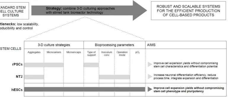
In Chapter 2, a scalable and controlled strategy was developed for the
expansion of undifferentiated rat pancreatic stem cells (rPSCs) which are anchorage-dependent cells that present high proliferation capacity and differentiation potential. This was done by combining microcarrier technology with environmentally controlled stirred tank bioreactors. The use of microcarrier supports overcame the main drawbacks of aggregate culture namely by avoiding the cell clumping that prevented pluriferation. Although the two microcarriers tested were suitable for PSC culture, Cytodex 3 provided a better matrix to promote cell attachment and growth. At the end, the controlled bioprocess allows the efficient expansion of rPSCs, without compromising stem cell characteristics and differentiation potential, representing an efficient starting point towards the development of novel protocols for other stem cell lines including hESCs.
The main focus of Chapters 3 and 4 was the development of a robust
cultivation of NT2 cells as 3-D cell aggregates (“neurospheres”) in stirred
tank bioreactors was the strategy adopted aiming to accomplish three main objectives: i) up-scale ii) accelerate and iii) enhance neuronal differentiation of stem cells.
Chapter 3 describes in detail the three-step protocol developed for NT2
differentiation. It was shown that, both cell-cell interactions and retinoic acid treatment presented in the 3-D neurosphere system contributed to a more efficient (4-fold) and rapid (approximately 50%) neuronal differentiation process than in the conventional cell monolayer cultures. Efforts were also directed in the optimization of cell harvesting procedures. The highest percentage of recovered neurons was achieved when intact neurospheres were transferred directly to treated surfaces, indicating that both 3-D neurosphere dynamics and the extracellular matrix surfaces are sufficient to provide an optimal system for the harvesting of NT2-N neurons.
The expansion step of NT2 aggregates was further investigated in
Chapter 4. Different bioprocess variables were tested and the best
compromise was obtained using an inoculum concentration of 4x10⁵
In Chapters 5 and 6, we step into the complexity of hESC cultivation.
The potential of bioreactor technology was explored in Chapter 5 to
improve the expansion of pluripotent hESC on microcarriers. The importance of controlling dissolved oxygen at 30% air saturation and the impact of incorporating an automated continuous perfusion system on cell growth and metabolism were discussed, demonstrating to be critical for the production of relevant cell numbers without compromising their pluripotency. At the end an improvement of 12-fold in the final cell yield was obtained when compared to static 2-D cultures, yielding almost 7×10⁸ of pluripotent hESCs
per 300mL bioreactor run.
Cell microencapsulation in alginate was investigated in Chapter 6 as the
main strategy to improve further hESC expansion and facilitate bioprocess integration with cryopreservation protocols. For this purpose three different 3-D culture strategies were evaluated and compared: microencapsulation of hESCs as single cells, aggregates and immobilized on microcarriers. The combination of cell microencapsulation and microcarrier technology resulted in an optimum protocol for the production and storage of pluripotent hESCs. This strategy ensures high expansion ratios (approximately 19-fold increase in cell concentration) and high cell recovery yields after cryopreservation. hESCs-microcapsules were cultured in stirred tank bioreactors and, after expansion, cryopreserved in cryovials, aiming to implement a scalable and straightforward integrated bioprocess.
Chapter 7 consists of a general discussion, where main achievements
(hESCs) despertaram muita atenção devido à sua capacidade de auto-renovação e pluripotência. Estas propriedades conferem às hESCs uma enorme aplicabilidade em medicina regenerativa, no rastreio de novos fármacos e em investigação científica por constituírem modelos celulares únicos para o estudo e compreensão dos processos de desenvolvimento embrionário inicial.
Contudo, a aplicação crescente das hESCs requer ainda o desenvolvimento de metodologias de cultura bem definidas e reproduzíveis em maior escala, que garantam a manutenção das propriedades de auto-renovação e diferenciação das células após o processo. Os métodos mais correntemente utilizados para cultura de hESCs são as monocamadas bi-dimensionais (2-D). No entanto, a baixa reprodutibilidade, a falta de controlo ambiental e os baixos rendimentos celulares associados a estas abordagens de cultura 2-D limitam a utilização destes sistemas numa escala clínica ou industrial.
implementar bioprocessos robustos para a produção de hESCs pluripotentes. Foi desenvolvida uma abordagem integrada através da combinação de diferentes estratégias de cultura tri-dimensionais (3-D) (tais como agregados celulares e células imobilizadas em microsuportes) com a tecnologia de bioreactores de tanque agitado, e através da manipulação de diversas variáveis críticas ao bioprocesso.
No Capítulo 1, é apresentada uma introdução geral ao tema de
bioprocessamento de células estaminais, incidindo no impacto de determinados factores ambientais na cultura de células estaminais. É dada particular relevância ao estado da arte relativamente às limitações dos sistemas tradicionais de cultura em 2-D, bem como ao potencial de novas estratégias de cultura 3-D e à tecnologia de biorreatores para ultrapassar essas limitações.
No Capítulo 2, foi desenvolvida uma estratégia escalonável e robusta
O principal objectivo dos Capítulos 3 e 4 foi o desenvolvimento de uma
plataforma robusta para produção de neurónios humanos derivados de células estaminais. O racional por detrás da selecção da linha celular de células estaminais de um teratocarcinoma (NT2) baseou-se, não só pelo facto destas células apresentarem características importantes e semelhantes às hESCs (expressão de marcadores de células estaminais, capacidade de auto-renovação e pluripotência), mas também por constituírem um bom modelo celular para a diferenciação neuronal in vitro; os neurónios derivados desta linha celular, NT2-N, têm
sido usados em ensaios de terapia celular e no desenvolvimento de novos fármacos. Nestes capítulos, as células NT2 foram cultivadas como agregados 3-D de células ("neurosferas") em biorreatores de tanque agitado com o objectivo de: i) aumentar a escala ii) acelerar e iii) melhorar o processo de diferenciação neuronal de células estaminais.
O Capítulo 3 descreve em pormenor o protocolo 3-D estabelecido para
melhor compromisso foi obtido utilizando uma concentração de inóculo de 4x10⁵ célula/mL e a mudança de meio como modo de operação da
cultura. Estas condições garantiram a produção rápida de números elevados de células sem comprometer o seu fenótipo e potencial de diferenciação. Ao integrar as etapas de expansão e diferenciação, o bioprocesso desenvolvido permitiu obter neurónios diferenciados em apenas duas semanas de diferenciação. Para além disso, no final da terceira semana, o número de neurónios produzidos foi significativamente superior quando comparado com o protocolo de cultura em sistema estático (aumento de 10 vezes da eficiência de diferenciação). No fim, o bioprocesso foi reproduzido e validado em bioreactores de tanque agitado e em ambiente controlado, conferindo ao processo automatização, escalabilidade e reprodutibilidade, requisitos importantes no bioprocessamento de células estaminais.
Os Capítulos 5 e 6, descrevem já um nível de complexidade superior,
utilizando culturas de hESCs. O potencial da tecnologia de bioreactores, adquirido nos Capítulos 3 e 4, foi explorado no Capítulo 5 com o
objectivo de melhorar a expansão de hESCs pluripotentes em microsuportes. A importância do controlo de oxigénio dissolvido (30% de ar saturado) e o impacto da perfusão contínua da cultura no crescimento celular e no metabolismo foram estudados, e demonstraram ser parâmetros fundamentais para a produção de hESCs em elevadas quantidades sem comprometer a sua pluripotência. No final, o rendimento celular foi melhorado 12 vezes relativamente aos métodos de culturas 2-D em sistema estático, garantindo cerca de 7×10⁸ hESCs
expansão de hESCs e desenvolver um bioprocesso integrado com protocolos de criopreservação. Para este efeito, foram avaliadas e comparadas diferentes estratégias de cultura 3-D, nomeadamente a microencapsulação de hESCs como: células individualizadas, agregados de células e células imobilizadas em microsuportes. A combinação da microencapsulação de células com a tecnologia microsuportes resultou num protocolo eficiente para a produção e armazenamento de hESCs pluripotentes. Esta estratégia garantiu rendimentos de expansão celular elevados (aumento de cerca de 19 vezes na concentração de células) e percentagens de viabilidade celular altas após a criopreservação. As culturas de hESCs microencapsuladas foram cultivadas em biorreactores de tanque agitado e, após a expansão, criopreservadas em criotubos, visando a implementação de um bioprocesso simples integrado e possível de aumento de escala.
O Capítulo 7 consiste numa discussão geral, onde são apresentadas os
principais resultados, conclusões e as perspectivas futuras do trabalho.
Serra, M., Leite, S.B., Brito, C., Costa, J., Carrondo, M.J.T., Alves, P.M., 2007.
Novel culture strategy for human stem cell expansion and neuronal differentiation. J Neurosc Res 85(16), 3557-3566.
Serra, M., Brito, C., Costa, E.M., Sousa, M.F. and Alves, P.M., 2009. Integrating
human stem cell expansion and neuronal differentiation in bioreactors. BMC Biotechnol. 9, 82.
Serra, M., Brito, C., Leite, S.B., Gorjup, E., von Briesen, H., Carrondo, M.J. and
Alves, P.M., 2009. Stirred bioreactors for the expansion of adult pancreatic stem cells. Ann. Anat. 191, 104-115.
Serra, M., Brito, C., Sousa, M.F., Jensen, J., Tostões, R., Clemente, J., Strehl,
R., Hyllner, J., Carrondo, M.J. and Alves, P.M., 2010. Improving expansion of pluripotent human embryonic stem cells in perfused bioreactors through oxygen control. J Biotechnol. 148(4), 208-15.
Serra, M.,Brito, C. and Alves, P.M., 2010. Bioengineering strategies for stem cell expansion and differentiation. Canal Bioquímica 7, 30-38.
Serra, M., Correia, C., Malpique, R., Brito, C., Jensen, J., Bjorquist, P.,
Abbreviation Full form
µ apparent growth rate
2-D two-dimensional
3-D three-dimensional
ANOVA analysis of variance (statistics)
AP alkaline phosphatase
AraC cytosine arabinoside ASCs adult stem cell
BDNF brain-derived neurotrophic factor bFGF basic fibroblast growth facto BSA bovine serum albumin
cDNA complementary deoxyribonucleic acid DAPI 4',6-diamidino-2-phenylindole DMEM Dulbecco's modified Eagle medium
DMEM-HG Dulbecco's modified Eagle medium with high glucose concentration
EBs embryoid body
EC embryonal carcinoma
ECM extracellular matrix
ELISA enzyme linked immuno sorbent assay ESC embryonic stem cell
FBS foetal bovine serum
FDA Food and Drug Administration FI fold increase in cell expansion FOX A2 forkheadbox A2
FSG fish skin gelatin Fudr fluorodeoxyuridine GFAP glial fibrillary acidic protein
GLC glucose
GLN glutamine
GMP good manufacturing practices
GRNOPC1 geron’s oligodendrocyte progenitor cells derived from hESCs
hFF human foreskin fibroblasts
hFFCellectTM antibody with high specificity against surface epitopes in undifferentiated hFFs
HGF hepatocyte growth factors
ICM inner cell mass
IgG immunoglobulin G
IgM immunoglobulin M
IPATIMUP Instituto de Patologia e Imunologia Molecular da Universidade do Porto
iPSC induced pluripotent stem cell
kd apparent death rate
Ki67 protein that is encoded by the MKI67 gene (antigen identified by monoclonal antibody Ki-67); marker to determine the growth fraction of a given cell population
KO-DMEM knock out Dulbecco's modified Eagle medium KO-SR knock out serum replacement
LAC Lactate
LDH lactate dehydrogenase
MAP2a&b microtubule associated protein-2 mESC mouse embryonic stem cell
MG Matrigel
MSC mesenchymal stem cell
NF200 heavy chain neurofilament, NF-L light chain neurofilament
NH4+ Ammonia
NT2 human embryonal carcinoma stem cell line NTera-2/cl.D1 NT2-N neurons derived from NT2 cells
O4 oligodendrocyte marker O4 P/S penicillin-streptomycin
PAM pharmacologically active microcarriers PBS phosphate buffer saline
PCR polymerase chain reaction
PDL poly-D-Lysine
PFA Paraformaldehyde
qLDH specific rate of LDH release
qRT-PCR quantitative real time polymerase chain reaction
RA retinoic acid
RCC rotary cell culture
RNA ribonucleic acid
rPSCs rat pancreatic stem cells
SCED™461 hESC line; feeder-cell based culture system developed for the easy propagation of hES cells by enzymatic digestion (SCED- single cell enzymatic dissociation)
SSEA-1 stage specific embryonic antigen-1 SSEA-4 stage specific embryonic antigen-4 STLV slow turning lateral vessel
td doubling time
TGFβ transforming growth factor beta TRA-1-60 tumor related antigen-1-60 TRA-1-60 tumor related antigen-1-60
TRAP telomeric repeat amplificationprotocol TX-100 triton X-100
UK United Kingdom
Urd uridine
VEGF vascular endothelial growth factor Xmax maximum cell concentration
YLAC/GLC yields of lactate production from glucose consumption
α-SMA α-smooth muscle actin
Figure 1.2 Page 9 Stem cell sources and characteristics
Figure 1.3 Page 12 Environmental factors and bioprocessing parameters impacting stem cell fate decisions
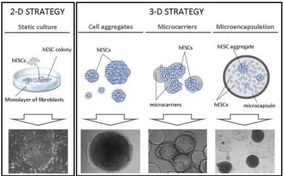
Figure 1.4 Page 21 2-D and 3-D strategies for cultivation of human embryonic stem cells
Figure 1.5 Page 33 Bioreactors used for stem cell cultivation
Figure 1.6 Page 36 Schematic representation of stirred tank bioreactor system for stem cell cultivation
Figure 1.7 Page 40 Diagram of the main aims proposed for this thesis
Figure 2.1 Page 63 Phase contrast photomicrographs of rPSCs cultured as small aggregates in spinner vessels
Figure 2.2 Page 64 Phase contrast photomicrographs of rPSCs cultured in
Cytodex 3 microcarriers under stirred suspension conditions
Figure 2.3 Page 64 Growth curves and viability of rPSCs cultured in microcarriers using stirred suspension systems
Figure 2.4 Page 66 Glucose uptake and lactate release of rPSCs cultured in Cytodex 1 and Cytodex 3 microcarriers, using spinner vessels and a 250 mL bioreactor
Figure 2.5 Page 69 Phase contrast photomicrographs of rPSCs cultured in Cytodex 3 microcarriers in a 250 mL bioreactor
Figure 2.6 Page 71 Characterization of rPSCs cultured in Cytodex 3 microcarriers
Figure 3.1 Page 89 Phase contrast photographs of NT2 cells cultured in stirred conditions (spinner)
Figure 3.2 Page 90 Effect of neurosphere dissociation protocol in concentration of viable and non viable cells
Figure 3.3 Page 91 Phase contrast micrographs showing the neurosphere dissociated cultures
Figure 3.4 Page 93 Characterization of NT2-N cultures
Figure 4.2 Page 119 Effect of inoculum concentration in NT2 cell expansion as 3D-aggregates
Figure 4.3 Page 122 Effect of culture operation mode on NT2 cell expansion as 3D-aggregates
Figure 4.4 Page 125 Characterization of NT2 cells expanded as 3D-aggregates
Figure 4.5 Page 127 Neuronal differentiation of NT2 cells in a fully controlled bioreactor
Figure 5.1 Page 143 Effect of inoculum concentration in the expansion of hESCs
adherent to microcarriers
Figure 5.2 Page 145 Effect of pO2 in the expansion of hESCs in bioreactors
Figure 5.3 Page 148 Metabolic performance of hESCs cultured in bioreactors
Figure 5.4 Page 149 Impact of perfusion culture on hESC expansion in bioreactors
Figure 5.5 Pages 150
-151 Characterization of hESCs expanded in perfused bioreactors
Figure 6.1 Page 165 Schematic workflow of the main steps of the microencapsulated 3-D culture strategies developed for expansion and cryopreservation of hESCs
Figure 6.2 Pages 174
– 175 Effect of alginate microencapsulation on the expansion of hESC as aggregates
Figure 6.3 Pages 178
– 179 Effect of alginate microencapsulation on the expansion of hESCs immobilized on microcarriers
Figure 6.4 Page 182 Post-thawing survival of non-encapsulated and encapsulated
hESCs
Figure 6.5 Page 184 Post-thawing characterization of encapsulated hESCs immobilized on microcarriers
Figure 7.1 Page 199 Schematic view of the focus and outcomes of the work
focused their research on the development of both adult and embryonic stem cells as therapies
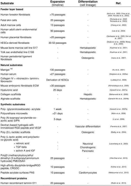
Table 1.2 Page 14 Summary of substrates used for propagation and/or differentiation of hESCs
Table 1.3 Page 23 List of advantages and disadvantages of different culture systems for stem cell bioprocessing
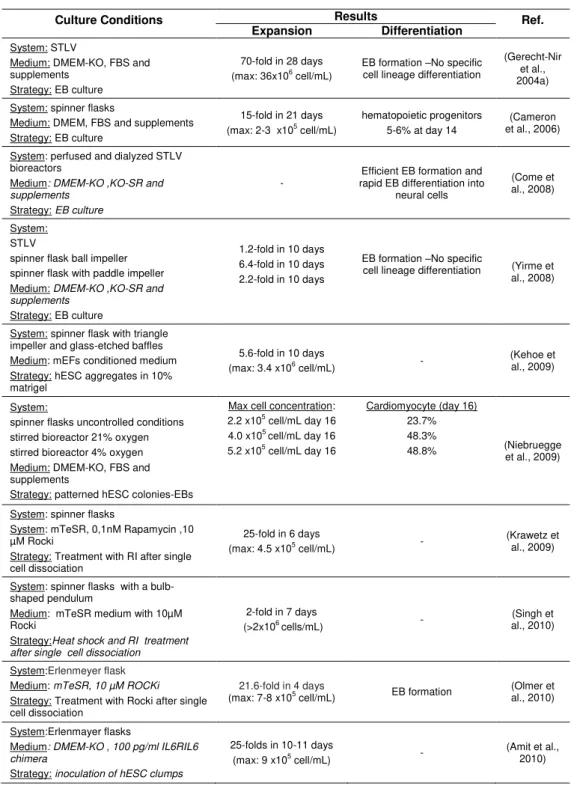
Table 1.4 Page 26 Summary of the studies involving the cultivation of hESCs as aggregates
Table 1.5 Page 28 Summary of the studies involving the cultivation of hESCs immobilized in microcarriers
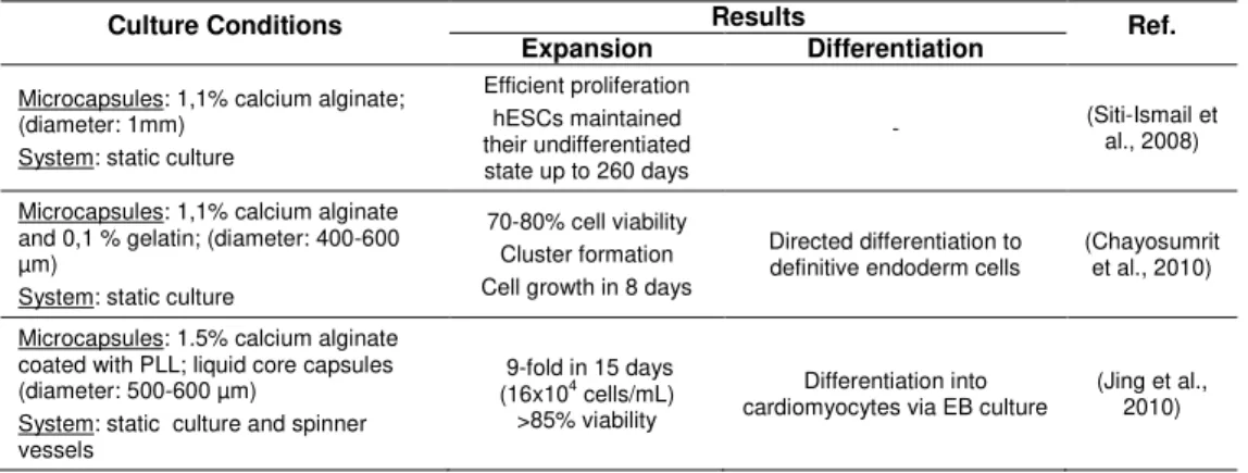
Table 1.6 Page 30 Summary of the studies involving the cultivation of microencapsulated hESCs
Table 1.7 Page 32 Culture systems for stem cell expansion and differentiation
Table 2.1 Page 65 Growth rate, doubling time and fold increase values of rPSCs
cultured in cytodex 1 and cytodex 3 microcarriers, using spinner vessels and a 250 mL bioreactor
Table 2.2 Page 67 Metabolic characterization of rPSCs cultured in cytodex 1 and cytodex 3 microcarriers, using spinner vessels and a 250 mL bioreactor
Table 3.1 Page 83 Comparison of neuronal differentiation protocols in static and stirred culture conditions for NT2 cells
Table 3.2 Page 96 Comparison of static and stirred culture conditions for NT2 neuronal differentiation
Table 4.1 Page 120 Growth kinetics of NT2 cell expansion as 3D-aggregates using different culture strategies
Table 4.2 Page 126 Characterization of NT2 neurospheres cultured in a fully controlled bioreactor
Table 5.1 Page 147 Operating parameters and growth kinetics characterization of hESCs expansion using different culture strategies
Table 6.1 Page 180 Expansion and cryopreservation of encapsulated and non-encapsulated hESC cultures
Chapter 1:
Introduction ………..……. 3
Chapter 2:
Expansion of Adult Pancreatic Stem Cells in
Stirred Tank
Bioreactors ………..……..
51
Chapter 3:
Novel Strategy for Neuronal Differentiation of
Human Stem Cells ……….………..……..
77
Chapter 4:
Integrating Stem Cell Expansion and Neuronal
Differentiation
……….………..………….
103
Chapter 5:
Improving Expansion of Pluripotent Human
Embryonic Stem Cells in Perfused Bioreactors through
Oxygen Control
……….………..………….
.
135
Chapter 6:
Microencapsulation Technology: a Powerful
Tool to Integrate Expansion and Cryopreservation of
Pluripotent Human Embryonic Stem Cells
……..………….
.
157
C
HAPTER
1
I
NTRODUCTION
This chapter was based on the following manuscript:
Serra, M., Brito, C. and Alves, P.M., 2010. Bioengineering strategies for stem cell expansion
TABLEOFCONTENTS
1. Introduction ... 3 2. Transferring stem cells to the clinic: what is needed? ... 6
2.1. Purity ... 6 2.2. Quality ... 6 2.3. Quantity ... 7
3. Stem cell bioprocessing ... 7
3.1. Stem cell sources... 8
3.2. Environmental factors that determine stem cell fate decisions ... 12 3.2.1. Extracellular matrix ... 13 3.2.2. Soluble factors ... 15
3.2.3. Cell-cell interactions ... 16
3.2.4. Physical forces ... 18
3.2.5. Physiochemical environment ... 18
3.3. Moving stem cells from 2-D monolayers to 3-D culturing approaches ... 21 3.3.1. Cell aggregates ... 24 3.3.2. Cell immobilized in microcarriers ... 25
3.3.3. Encapsulated cells ... 29
3.4. Bioreactors for stem cell cultivation... 31 3.4.1. Microfluidic culture systems ... 34
3.4.2 Rotary cell culture systems ... 34 3.4.3. Stirred culture vessels ... 35
1.
I
NTRODUCTIONHuman embryonic stem cells (hESCs) constitute an exciting emerging field. The inherent capacity of these cells to grow indefinitely (self-renewal) and their ability to differentiate into all mature cells of the human body (pluripotency), have made them an extremely attractive tool for regenerative medicine and tissue engineering (Nirmalanandhan and Sittampalam, 2009). Indeed, for many years, they are considered the great promise for treating degenerative disorders such as Parkinson diseases, type I diabetes and heart failure, hoped to provide a new source of neurons, insulin producing cells or cardiomyocytes to replace the degenerating tissues and/or impaired cells.
The first clinical trial with hESCs was approved by the US Food and Drug Administration (FDA) in January 2009 but only initiated in October 2010. The goal of this first study is to assess the safety and tolerability of oligodendrocyte progenitor cells derived from hESCs (GRNOPC1, Geron Comp.) in patients with neurologically complete spinal cord injuries, i.e. patients with complete loss of locomotor and sensory activity below the site of injury. The second endpoint is efficacy; it will use similar testing for evidence of any return of sensory function or lower extremity locomotion for one year after injection of GRNOPC1. This clinical trial holds high expectations as in previous experiments with rats these cells revealed to be safe and efficient in restoring some function (Zhang et al., 2006).
Recently, another hESC clinical trial was approved (November 2010) by the US FDA. Advanced Cell Technology, Inc. announced a Phase I/II multicenter clinical trial using retinal cells derived from hESCs to treat
patients with Stargardt’s Macular Dystrophy (SMD), one of the most
Today an increasing number of biotechnology industries have focused their interest on the development of both adult and embryonic stem cells as therapies; some examples are listed in Table 1.1.
In addition to clinical applications, hESCs have enormous prospective for the development of novel technologies in drug screening (Davila et al.,
2004; Ebert and Svendsen, 2010; Jensen et al., 2009). In fact, given the
high costs spent by pharmaceutical R&D to bring a new drug to market, there is ongoing effort to introduce new cell models which are practicable (robust, reproducible, etc.) and have improved throughput and predictivity. hESCs and their derivatives have all the potential to be used here as “bio
-tools”, also contributing to the reduction of animal experimentation (Davila et al., 2004; Jensen et al., 2009). For example, pure cultures of hepatocytes, cardiomyocytes and neuronal cells derived from hESCs would provide robust cell-based in vitro assays for toxicity measurements and for
drugs being development for cardiovascular or neurodegenerative disorders, respectively.
hESCs are also valuable models for scientific research. They can lead to a better understanding of the basic biology of the human body, embryonic development, pathogenesis of congenital defects and cancer formation (Bongso et al., 2008). In fact, it is possible to derive disease-specific hESCs from embryos with diagnosed mutations by preimplantation genetic diagnosis (Galat et al., 2010). As an example, hESC lines derived from embryos with Fanconi anemia-A mutation and fragile X mutation have already been established (Galat et al., 2010). These hESC lines will provide
in vitro models for study the phenotype of these mutations, allowing the
Table 1.1. Summary of some biotechnology industries that have focused their research on the development of both adult and embryonic stem cells as therapies. The main technologies developed and ongoing actions are presented.
Company Technology Indications Action and Clinical Status
Aastrom Biosciences Inc
(Minnesota,USA)
Autologous therapy (patient´s own cells) Tissue repair cells
Limb ischemia, bone,
cardiac regeneration Phase II, III, I clinical trial
Advanced Cell Technology Inc
(California, USA)
Multiple technologies
based on ESC Heart failure, macular degeneration , vascular ischemia
Received U.S. FDA approval for use retinal stem cells to treat Stargardt's Macular Dystrophy Phase I/II
Aldagen Inc
(North Carolina, USA)
ALD-201 Heart failure Phase I clinical trial
AmStem, Histostem
(California, USA)
Human umbilical cord blood
stem cells Buerger´s disease Hair loss treatment Phase I and II clinical trial completed Received Korea FDS approval Phase II and III clinical trials
Athersys (ATHX)
(Ohio, USA)
MultiStem Myocardial infarction, bone marrow transplantation
Phase I clinical trial
Celgene Corp
(New Jersey, USA)
Blood cancer treatments Phase I clinical trials
Cytori Therapeutics Inc (California, USA)
Adipose tissue derived
stem cells Tissue regeneration after breast surgery, cardiac ischemia, hear attack
In planning, pilot study
Its StemSource product line is sold globally for cell banking and research applications.
Geron Corp.
(California, USA)
hESCs derived
oligodendrocytes Spinal cord injury Received U.S. FDA approval Phase I
Neuralstem Inc
(Maryland, USA)
Neural Stem cells CNS injury (chronic spinal cord injury, amyotrophic lateral sclerosis)
Recently filed a new drug application with the FDA to begin a Phase I safety clinical trial for chronic spinal cord injury
NovoCell
(California, USA)
Stem cell engineering, cell
encapsulation Type I diabetes Proof of concept phase I/II
Osiris Therapeutics Inc. (Maryland, USA)
Autologous therapy (patient´s own cells taken from the bone marrow)
Crohn's disease Phase III clinical trial
ReNeuron Group Plc
(Guildford,UK)
REN009 stem cell therapy peripheral arterial
disease in diabetes Starting trials in 2011.
StemCells Inc.
(California, USA)
Human neural stem cells
(HuCNS-SC® product) Batten disease Starting human trials in 2011
Technology
Apceth
(München,Germany)
Development of GMP-grade protocols for mesenchymal stem cell production and gene transfer for cancer therapy applications
International Stem Cell Corporation
(California, USA)
Establishment of human stem cells via parthenogenesis (hpSC lines) to provide potential products as alternatives to ESCs. ISCO plans to create a bank of these hpSC lines
(UniStemCell™)
Lonza Bioscience
(Basel, Switzerland)
Establishment of Poietics® Human Adipose-Derived Stem Cells for use in adult stem cell research
Thermogenesis Corp
(California, USA)
2.
T
RANSFERRING STEM CELLS TO THE CLINIC:
WHAT IS NEEDED?
The successful translation of stem cells to the clinical and/or industrial fields will require contributions from fundamental research (from the
developmental biology to the “omics” technologies and advances in
immunology) and from existing industrial practice (biologics), especially on automation, quality assurance and regulation.
Attention is shifting also to the development of bioprocesses to produce hESCs or their derivatives in high purity, consistent quality and relevant quantity.
2.1. Purity
The tumorigenic potential of pluripotent stem cells is one of the important hurdles in the safety utilization of these cells. At present, protocols for the directed differentiation of stem cells are generally inefficient, resulting in low differentiated cell yields and contamination by other cell types. Of greater concern is the persistence of undifferentiated stem cells and the possibility of these cells form malignant tumors when transplanted in the host (Fujikawa et al., 2005). Therefore the use of efficient methods for differentiation and selection of pure populations of specialized cells will be essential before these cells being used clinically (Brignier and Gewirtz, 2010).
2.2. Quality
culture. Undifferentiated stem cells have to maintain their pluripotency and genetic and epigenetic stability after expansion while stem cell derivatives must express markers of the specific cell lineage and be fully functional after differentiation.
2.3. Quantity
Another important challenge is to achieve sufficient numbers of stem cell for an effective therapy. In general, these numbers fall in the range of millions to a few billion. For example, in Geron´s first clinical trial, patients will be injected in the spinal cord with small doses of GRNOPC1 (2x10⁶
cells, www.geron.com), but for the replacement of damage cardiac tissue after myocardial infarction 1-2x10⁹ cardiomyocytes are required (Jing
et al.,
2008). To achieve these high cell numbers robust, affordable and scalable bioprocesses need to be developed.
3.
S
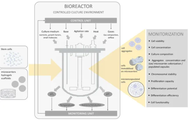
TEM CELL BIOPROCESSINGThe successful production of stem cell-based products relies on robust bioprocesses that should be designed following pertinent principles (Figure 1.1, Placzek et al., 2009).
Herein, the cell source and the signals that govern stem cell fate decisions are essential bioprocessing components. Next, the integration of a controlled culturing strategy for 3-D cell organization via cell self-assembly, cell immobilization to biomaterials/supports with a bioreactor-based system where the necessary conditions for cells to guide their fate are “perfectly tuned”, is a key factor to move stem cells from lab scale to clinical trials and large scale industrial applications. In this chapter, the importance of these process components on the design of stem cell bioprocesses will be presented, highlighting the main requirements needed to fulfil the end
product’s purity, quality and quantity.
3.1. Stem cell sources
There are several classes of stem cells including embryonic and adult stem cells, and the new type of induced stem cells, each one presenting its own benefits, limitations and challenges in bioprocess development (Figure 1.2). All of them share as common features the ability to proliferate indefinitely (unlimited self-renewal capacity) and vary in their differentiation potential.
hESCs are isolated from the inner cell mass (ICM) of blastocysts at day five of embryonic development. The first reports of hESCs were published in 1984 (SB Fishel et al., 1984) and 1994 (Bongso et al., 1994), but it was only in 1998 that Thomson and co-workers described the isolation of hESCs and the establishment of the firsts permanent and characterized hESC lines for research (Thomson et al., 1998). Today, more than 1000 hESC lines are reported in the literature (Löser et al., 2010). Some of these cell lines are well characterized and organized in international stem cell banks, for example, hESCreg (www.hescreg.eu), UK stem cell bank
(www.ukstemcellbank.org.uk), and National stem cell bank
Substantial efforts have been made towards the identification of phenotypic/genomic markers to characterize, validate and distinguish hESCs from other cell types. hESC lines can be identify by the presence of surface marker antigens (Tra series, SSEA series, GCT series, HLA, and CD markers) and transcriptional factors (Oct4, Nanog), by the chromossomal stability with serial culture, alkaline phosphatase positiveness and high telomerase activity (Allegrucci and Young, 2007; Bongso et al., 2008). As described above, these cells present a high proliferation capacity and are pluripotent, i.e., they possess the potential to differentiate into all cell types that compose an adult body, derived from the three germ layers (e.g. cardiomyocytes, neurons, pancreatic islets, hepatocytes, chondrocytes,) (Hay et al., 2007; Kroon et al., 2008; Mummery et al., 2003; Toh et al., 2009; Zhang et al., 2001). However, hESCs are still difficult to control with respect to their stem cell fate, and elicit ethical considerations, requiring the manipulation of human embryos. For clinical applications, these cells still present limitations related with immune rejection and the possibility of teratoma formation. On the other hand, adult stem cells (ASCs) do not present immunogenic complications on implantation since they can be isolated directly from the patient. ASCs exist in specific niches in the different organs (e.g. bone marrow, peripheral blood, pancreas, lung, brain, liver) (Lanza et al., 2004) contributing to the regeneration/repair of the tissue/organ where they reside. Depending on the source, ASCs can be isolated relatively easy, however they present as major limitations the difficulty in obtaining pure populations, the limited expansion capacity and the restricted differentiation potential, as they are often committed to their original cell lineage (multipotent cells).
state of pluripotency using defined reprogramming strategies including overexpression of a core transcription factors known to be required for maintenance of ESC pluripotence and proliferation (Oct4, Sox2, and either c-Myc and Klf4 or Nanog and Lin28) (Takahashi et al., 2007; Takahashi and Yamanaka, 2006; Yu et al., 2007). The creation of these induced pluripotent stem cells (iPSCs) elicited an explosion of scientific curiosity and industrial interest. This is mainly because iPSCs are similar to ESCs (namely cell morphology, cell-surface markers, self-renewal ability, potential to differentiate in vitro and in vivo into cells derived from all three
germ layers) (Takahashi et al., 2007; Takahashi and Yamanaka, 2006) and thereby could potentially replace ESCs for clinical applications, circumventing the ethical concerns regarding the use of embryos. Additionally, iPSCs present the benefit of being patient-derived cells, avoiding immune rejection in cell therapy applications. iPSC research is expanding rapidly, including modeling complex diseases in vitro and
3.2. Environmental factors that determine stem cell fate decisions
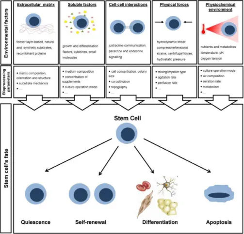
Stem cells develop their behaviour from cues that lie in the extracellular environment. These cues operate on different temporal and spatial scales, driving specific cellular behaviours and ultimately promoting/controlling cells’ self-renewal, differentiation or apoptosis (Figure 1.3).
Substantial efforts have been made to identify such stimuli. The
extracellular matrix (ECM), soluble factors, cell-cell interactions, physical forces and physiochemical factors have been suggested as the most relevant cues governing stem cell fate.
3.2.1. Extracellular matrix
Extracellular matrix (ECM) is a key component of the stem cell niche in vivo
and can influence stem cell fate via mediating cell attachment and migration, presenting chemical and physical cues, as well as binding soluble factors. In a natural setting, this environment encloses a complex and dynamic network of proteins, polysaccharides, proteoglycans and water that provide structural and organizational guides for tissue development. The activation of these signalling pathways through the adhesion of specific components of the ECM to cells via integrins/cadherins/cell surface receptors is not trivial as it is highly dependent on the composition, orientation and structure of the ECM (Lukashev and Werb, 1998).
Table 1.2. Summary of substrates used for propagation and/or differentiation of hESCs. (Adapted from (Abraham et al., 2009))
Substrate Expansion (timeline) Differentiation (cell lineage) Ref.
Feeder layer based
Human foreskin fibroblasts >70 passages - (Amit et al., 2003; Choo et al., 2004; Hovatta et al., 2003) Fetal skin cells 20 passages - (Richards et al., 2002; Richards et al., 2003) Adult marrow cells 13 passages - (Cheng et al., 2003)
Human adult uterin endonmetrial
cells 90 passages - (Lee et al., 2004)
Human placental fibroblasts >25 passages - (Genbacev et al., 2005; Kim et al., 2007) hESCs-derived fibroblasts 30-52 passages - (Stojkovic et al., 2005b; Wang et al., 2005) Mouse bone marrow cell line S17 - Hematopoietic (Kaufman et al., 2001)
Yolk sac endothelial line C166 - Hematopoietic (Kaufman et al., 2001)
Human periodontal ligament
fibroblasts - Osteogenic (Inanc et al., 2007)
Natural substrates
Matrigel TM 130 passages - (Xu et al., 2001)
Human serum >27 passages - (Stojkovic et al., 2005a)
Collagen IV + vitronectin+ laminin+
fibronectin Derivation of hESCs (Ludwig et al., 2006) Mouse embryonic fibroblasts ECM >30 passages - (Klimanskaya et al., 2005)
Hyaluronic acid 20 days - (Gerecht et al., 2007a)
Collagen scaffolds - Hepatic (Baharvand et al., 2006)
Alginate scaffolds - Hematopoietic (Gerecht-Nir et al., 2004b)
Synthetic substrates
Poly- (glycerolcosebacate)- acrylate 1 week - (Gerecht et al., 2007b)
Polyurethane microwells >21 days - (Mohr et al., 2006)
Poly (N-isopropyl
acrylamide-co-acrilic acid) SIPN 5 days - (Li et al., 2006) Dextran-based hydrogels with
immobilized RGD peptide and VEGF - Vascular differentiation (Ferreira et al., 2007) Poly (D,L-lactide) scaffolds - Osteogenic (Bielby et al., 2004)
Poly (L-lactic acide) and poly(lactic-co-glycolic acid)
+ retinoic acid + TGF-beta + activin A and IGF
- - - Neuronal Chondrogenic Pancreatic
(Levenberg et al., 2003)
Poly[2-(methacryloyloxy)ethyl dimethyl-(3-sulfopropyl)ammonium
hydroxide] PMEDSAH 25 passages -
(Villa-Diaz et al., 2010)
High-affinity disulphide-bridgedRGD
peptide, CRGDC 10 passages - (Kolhar et al., 2010) Peptide-acrylate surfaces PAS 10 passages Cardiomyocytes (Melkoumian et al., 2010)
Recombinant proteins
From clinical and industrial perspectives, the use of synthetic matrices may offer greater advantages in terms of reproducibility, quality control and costs (Kolhar et al., 2010; Melkoumian et al., 2010; Villa-Diaz et al., 2010). These matrices have wide diversity in properties that may be obtained and tailored with respect to mechanics, chemistry and degradation according to the case study. However, potential limitations to the use of synthetic materials include toxicity and limited repertoire of cellular interactions, unless they are modified with adhesion peptides or designed to release biological molecules.
3.2.2. Soluble factors
The outcome of stem cell culture depends also on the presence/concentration of growth/differentiation factors which provide survival, proliferation, differentiation signals to the cells. These regulatory molecules can be either added to the culture or secreted by the cells. Upon diffusion through the medium, these factors are sequestered by the ECM and bind to the cell surface receptors thus activating cellular functions. In alternative, to achieve a better control of the cellular microenvironment and ultimately enhance stem cell proliferation and/or differentiation, they can be immobilized on the surface of biomaterials (Ferreira et al., 2007).
Substantial efforts have been made to identify the factors regulating stem cell proliferation and/or differentiation. As an example, the basic fibroblast growth factor (bFGF) and several members of the transforming growth
factor beta (TGFβ) superfamily of ligands have been reported as vital
and differentiation factors in scalable culture systems is their high costs and low stability in medium. The engineering of more stable molecules or the development of appropriated perfusion systems would be potential strategies to reduce the concentration of these compounds without compromising the culture outcome.
In parallel, attempts have been made to minimize the use of these factors, for example, by including natural and/or synthetic small molecules that can be isolated/synthesized economically. Small molecules have been shown to target specific signal transduction pathways (e.g. Wnt, Hedgehog, retinoid, NF-κB), which either alone or in concert dictate the fate of stem cells, including the maintenance of undifferentiated phenotype (Sato et al., 2004) and pluripotency (Miyabayashi et al., 2007), improve cell viabilities (Watanabe et al., 2009) and promote differentiation of stem cells to cardiac (Tseng et al., 2006), hematopoietic (Naito et al., 2006), neuronal (Ding et al., 2003), and bone (Wu et al., 2004) cell phenotypes. With the advent of high-throughput screening technologies, small molecule libraries have been analyzed to identify molecular interactions leading to particular stem cell responses (revised in Ding and Schultz, 2004; McNeish, 2007).
3.2.3. Cell-cell interactions
Cell-to-cell communication, either in vivo or in vitro, can be established via
effectiveness; 2) be taken by the cells very quickly, leaving few to travel further, thus creating a heterogeneous environment where cells are exposed to different concentration gradients; 3) have their movement hindered by the ECM. These different cell-cell interactions drive a set of stem cell responses, from the induction of programs of differentiation (Tsai and McKay, 2000) to promote proliferation and self-renewal properties (Purpura et al., 2004). In particular, hESCs are standard cultivated as flat colonies in static adherent conditions. Typically, these colonies are maintained at an appropriate size to assure controlled self-renewal. It is well established that individual cells or small clumps do not grow efficiently while large colonies exhibit substantial levels of spontaneous differentiation (Azarin and Palecek, 2010; Bauwens et al., 2008).
3.2.4. Physical forces
A number of in vivo and in vitro studies have demonstrated that physical
forces (e.g. hydrodynamic/hydrostatic, mechanical and electrical) play a key role in the development of tissues and organs during embryogenesis as well as their remodelling and growth in postnatal life. Moreover, it has been found that stem cells are sensitive to fluid flow-induced shear stress (Glossop and Cartmell, 2009), compressive and tensional strains (Haudenschild et al., 2009), cyclical stretching (Shimizu et al., 2008) and hydrostatic pressures (Liu et al., 2009). In particular, Sargent et al
demonstrated that manipulation of hydrodynamic environments modulates the kinetic profile of gene expression and relative percentages of ESC differentiation (Sargent et al., 2010). In another study, Veraitch et al
reported that excessive centrifugal forces up to 1000 g cause shifts in phenotype and proliferation during expansion and differentiation of ESCs (Veraitch et al., 2008).
However, relatively few information is known about the impact of these physical forces on physiological mechanisms. Recently, Stolberg et al
proposed potential mechanisms for shear stress signalling that may play a role in endothelial differentiation trough the VEGF signalling pathway (Stolberg and McCloskey, 2009). Since scalable culture systems often employ perfusion or mixing that can apply mechanical forces to the cells, this kind of information will be extremely important to design efficient bioreactor-based strategies. The effect of shear-protection additives on hESC proliferation, viability and pluripotency will be important information for the establishment of scalable bioprocesses.
3.2.5. Physiochemical environment
and metabolites affects cell growth, viability and differentiation. In order to mimic the in vivo physiological environment and further improve culture
performance, different operation modes can be adopted, including fed-batch and perfusion. The fed-fed-batch strategy is often considered the most suitable for tuning cell metabolism; by providing nutrients in a rational manner, their uptake and consumption are energetically more efficient leading to reduced accumulation of metabolites in culture supernatant (Xie and Wang, 1994). However, as described above, growth factors play a crucial role in regulation of stem cell behavior. Thus perfusion mode has been preferentially adopted in the majority of stem cell bioprocesses aimed to control culture outcome, since it assures the continuous renewal of nutrients and other factors as well as the continuous removal of metabolic byproducts (Bauwens et al., 2005). Within this context, more knowledge regarding the in vivo stem cells microenvironment is needed, i.e. the
concentration gradients existed in stem cell niches in order to understand
their impact on stem cells’ fate decisions.
Although typically cultivated inside of incubators operated at standard conditions of temperature (37ºC), dissolved oxygen tension (20%) and pH (7.4), stem cell expansion and differentiation potential can be enhanced at different conditions. Up to now few studies have been conducted on the effect of temperature and pH in stem cell culture. For instance, it has been shown that mesenchymal stem cell differentiation is enhanced at lower temperatures (32ºC) than in 37ºC conditions (Stolzing and Scutt, 2006)
while high temperatures (39ºC) demonstrated to enhance
exposure of ESC cultures for 1-3 h to ambient conditions during passaging procedures (which resulted in a rapid drop in temperature and rise in pH) inhibits cell proliferation and reduces the expression levels of Oct-4 (Veraitch et al., 2008).
Stem cell niches are often located in regions of low oxygen tension (pO2)
and low pO2 typically decreases the rate of stem cell differentiation and
enhances stem cell proliferative potential (King and Miller, 2007; Millman et al., 2009). Regarding hESC culture, there is emerging evidence that reducing oxygen concentration towards physiological levels, i.e. low levels of oxygen (hypoxia – 1.5-8%) is beneficial for in vitro maintenance of their
pluripotent status: stem cells self-renewal is supported, spontaneous (uncontrolled) differentiation is reduced and karyotypic integrity is maintained (Ezashi et al., 2005; Prasad et al., 2009), contrasting to normoxia conditions (20% oxygen). The hypothesis is that these hypoxic environments protect the ESCs from oxygen toxicity while inducing the up-regulation of an array of genes orchestrating the earliest steps of embryonic development. Nonetheless, further investigation is required to support such assumption.
Based on these findings and in an attempt to unlock the full potential of stem cells, bioprocess engineers are focused on recreating in vitro the
dynamic environments experienced by cells in vivo. However, the design of
such complex microenvironments is not trivial. The degree of complexity involving the incorporation of various ECM proteins, soluble factors and cell populations into different physical stimuli and physiochemical conditions,
3.3. Moving stem cells from 2-D monolayers to 3-D culturing approaches
Stem cells are traditionally cultured in 2-D systems (e.g. Petri dishes, culture flasks and well plates). In particular, hESCs are propagated as colonies on top of a feeder layer of inactivated fibroblasts (Figure 1.4).
Over the last years, constant inadequacy of conventional 2-D culture systems in resembling the in vivo developmental microenvironment has
been observed in both basic biology and tissue engineering studies. In fact, tissue-specific architecture, mechanical and biochemical cues, cell-cell and cell-matrix communications are lost under such simplified and highly biased conditions. In addition, the inherent uncontrollability, heterogeneity and low production yields associated with these systems have made them unattractive and unsuitable for clinical and industrial applications.
Moving stem cells from 2-D cell monolayers to 3-D culturing strategies is
imperative to enhance cells’ performance and fully exploit cells’ potential.
The general recognition that spatial arrangement and directional cues have an important role in stem cells behaviour contributed for the acceptance of 3-D cultures as the most suitable system to mimic stem cells’ native
microenvironment. By providing a cellular context closer to what actually occurs in native microenvironment, these strategies can significantly
improve cell’s viability, identity and function (Cukierman et al., 2002; Lund et al., 2009; Pampaloni et al., 2007). In summary, engineered 3-D microstructures have the potential to provide a higher degree of efficiency, robustness, consistency and predictability to the cultures.
This section will address current 3-D culture strategies that could be used to generate large numbers of pluripotent hESCs and/or their derivatives with potential application in regenerative medicine and drug discovery. It is important to highlight that, an optimal hESC based bioprocess capable of embracing all the applications of these cells does not exist so far. Nonetheless, the knowledge gained during the last decades with murine ESCs (mESCs) and other stem cell model systems (e.g. human teratocarcinoma stem cells), in which the quantitative characterization of expansion and differentiation processes is included, have been providing important insights for the development of robust hESC production platforms.
Table 1.3. List of advantages and disadvantages of different culture systems for stem cell bioprocessing.
Culture Strategy Advantages Disadvantages
2-D Culture Static Cultures
easy visualization and cell morphology monitorization
easy handling
affordable system
ideal for small scale studies
low reproducibility
low scalability
difficult to control specific culture parameters and diffusion gradients
low cell production yields
limitation in resembling in vitro tissues
Cell aggregates easy handling
scalable system
high reproducibility
3- D cell-cell contact is preserved
can mimic stem cells’ native
microenvironment
high differentiation efficiency
high cell production yields
difficult to control culture outcome due to the occurrence of
uncontrolled/spontaneous differentiation (EB formation)
aggregate size (important to avoid diffusion gradients inside the aggregate structure that lead to necrotic centres and/or spontaneous differentiation)
single cell harvesting (difficult to dissociate aggregates without compromising cell viability)
cell damage due to physical forces (hydrodynamic shear, perfusion flow)
Microcarriers Non-porous
easy handling
scalable system
high reproducibility
easy visualization and cell morphology monitorization
No limitations in mass and gas diffusion
high surface to volume ratio (able to support high cell densities, reduce the process cost)
high cell production yields
microcarrier agglomeration (important to avoid diffusion gradients inside the cell-microcarrier aggregate structure that lead to necrotic centres and/or spontaneous differentiation)
cell-bead separation step required
cell damage due to physical forces (hydrodynamic shear, perfusion flow)
costs associated to material (microcarrier)
Porous easy handling
scalable system
high reproducibility
high surface to volume ratio (able to support high cell densities, reduce the process cost)
high cell production yields
protection from physical forces (hydrodynamic shear, perfusion flow)
difficulty in culture visualization and cell morphology monitorization
Limitations in mass and gas diffusion inside the pores that lead to necrotic centres and/or spontaneous differentiation
cell harvesting limitation (except for biodegradable supports)
cell-bead separation step required
costs associated to material (microcarrier)
Cell
Microencapsulation
easy handling
scalable system
high reproducibility
high surface to volume ratio (able to support high cell densities, reduce the process cost)
high cell production yields
protection from physical forces (hydrodynamic shear, perfusion flow)
3- D cell-cell and cell-matrix contacts are preserved, mimicking stem cells’
native microenvironment
biomaterial can be engineered to improve cell culture performance
process can be integrated in transplantation studies
difficulty in culture visualization and cell morphology monitorization
Limitations in mass and gas diffusion inside the pores that lead to necrotic centres and/or spontaneous differentiation
cell harvesting (decapsulation protocol could compromise cell viability)
3.3.1. Cell aggregates
By aggregation into spheroids, cells can re-establish mutual contacts and specific microenvironments that allow them to express a tissue-like structure. Within this context, the cultivation of stem cells as 3-D aggregates has been extremely explored during the last decades proving to be an efficient system for expansion/differentiation of progenitor cells, such as human neural precursor cells (Baghbaderani et al., 2008; Baghbaderani et al., 2010), pancreatic cells (Chawla et al., 2006) and hepatocyte progenitors (Gerlach et al., 2003; Miranda et al., 2009).
For ESCs, the 3-D aggregate culture strategy is usually associated with differentiation; the most robust method for generating differentiated cells from ESCs is through the formation of embryoid bodies (EBs), where ESC cultured in suspension self-aggregate and spontaneously differentiate into multiple tissues (Dang et al., 2004). EB differentiation has been shown to recapitulate aspects of early embryogenesis, including the formation of a complex 3-D arrangement where cell-cell and cell-matrix interactions are thought to support the development of three embryonic germ layers and their derivatives (Itskovitz-Eldor et al., 2000; Keller, 1995).
The main limitation of this system is, in fact, the inefficient control of stem cell expansion or in directing stem cell differentiation towards a specific lineage, thus resulting in a mixture of different cell types. This drawback demands the need of efficient integrative downstream approaches to further purify the culture outcome into a desired cell type population.
expansion of pluripotent stem cells. One year later, zur Nieden demonstrated that mESCs could be maintained for a total of 28 days in these culture systems by repeated aggregate dissociation (zur Nieden et al., 2007).
Besides propagation, many studies have been performed in directing differentiation of mESCs aggregates into a specific cell lineage (revised in (Jensen et al., 2009; King and Miller, 2007; Ulloa-Montoya et al., 2005).The knowledge gained with these model systems combined with developments in fundamental cell biology contributed to the design of controlled bioprocesses for hESCs. During the last 2 years, significant efforts have been made in 3-D aggregate culture systems for controlled expansion of undifferentiated hESCs and their directed differentiation into functional cell types (summarized in Table 1.4).
3.3.2. Cell immobilized in microcarriers