Bacillus pumilus
Reveals a Remarkably High Resistance
to Hydrogen Peroxide Provoked Oxidative Stress
Stefan Handtke1., Rebecca Schroeter2., Britta Ju¨rgen2
, Karen Methling3, Rabea Schlu¨ter1,
Dirk Albrecht , Sacha A. F. T. van Hijum , Johannes Bongaerts , Karl-Heinz Maurer , Michael Lalk ,1
Thomas Schweder2,7, Michael Hecker1,7, Birgit Voigt1,7*
1Institute for Microbiology, University of Greifswald, Greifswald, Germany,2Pharmaceutical Biotechnology, Institute of Pharmacy, University of Greifswald, Greifswald, Germany,3Institute of Biochemistry, University of Greifswald, Greifswald, Germany,4Centre for Molecular and Biomolecular Informatics (CMBI), Nijmegen Centre for Molecular Life Sciences, Radboud University Medical Center, Nijmegen, The Netherlands; and Division Processing and Safety, NIZO Food Research B.V., Ede, The Netherlands,5Department of Chemistry and Biotechnology, Aachen University of Applied Sciences, Ju¨lich, Germany,6AB Enzymes GmbH, Darmstadt, Germany,
7Institute of Marine Biotechnology, Greifswald, Germany
Abstract
Bacillus pumilusis characterized by a higher oxidative stress resistance than other comparable industrially relevant Bacilli such asB. subtilisorB. licheniformis. In this study the response ofB. pumilusto oxidative stress was investigated during a treatment with high concentrations of hydrogen peroxide at the proteome, transcriptome and metabolome level. Genes/ proteins belonging to regulons, which are known to have important functions in the oxidative stress response of other organisms, were found to be upregulated, such as the Fur, Spx, SOS or CtsR regulon. Strikingly, parts of the fundamental PerR regulon responding to peroxide stress inB. subtilisare not encoded in theB. pumilusgenome. Thus,B. pumilusmisses the catalase KatA, the DNA-protection protein MrgA or the alkyl hydroperoxide reductase AhpCF. Data of this study suggests that the catalase KatX2 takes over the function of the missing KatA in the oxidative stress response ofB. pumilus. The genome-wide expression analysis revealed an induction of bacillithiol (Cys-GlcN-malate, BSH) relevant genes. An analysis of the intracellular metabolites detected high intracellular levels of this protective metabolite, which indicates the importance of bacillithiol in the peroxide stress resistance ofB. pumilus.
Citation:Handtke S, Schroeter R, Ju¨rgen B, Methling K, Schlu¨ter R, et al. (2014)Bacillus umilusReveals a Remarkably High Resistance to Hydrogen Peroxide Provoked Oxidative Stress. PLoS ONE 9(1): e85625. doi:10.1371/journal.pone.0085625
Editor:Adam Driks, Loyola University Medical Center, United States of America
ReceivedSeptember 6, 2013;AcceptedDecember 5, 2013;PublishedJanuary 20, 2014
Copyright:ß2014 Handtke et al. This is an open-access article distributed under the terms of the Creative Commons Attribution License, which permits unrestricted use, distribution, and reproduction in any medium, provided the original author and source are credited.
Funding:This work was financially supported by funds of the Competence Network ‘‘Mipro - Microbes for production: A genomics-based approach to engineer novel industrial production strains’’ (0315594B) by the German Federal Ministry of Education and Research (BMBF, www.bmbf.de) and the project ‘‘Ausbau und Profilierung von COAST Fun-Gene’’ (UG11043, ESF/IV-BM-B35-0003/12) of the Bildungsministerium of Mecklenburg-Vorpommern (www.regierung-mv.de). The transcriptome analysis was financially supported by the Henkel KGaA (www.henkel.de). The funders had no role in study design, data collection and analysis, decision to publish, or preparation of the manuscript.
Competing Interests:The transcriptome analysis for this study was financially supported by Henkel KGaA. Dr. Sacha A.F.T. van Hijum is affiliated to NIZO Food Research B.V., and Dr. Karl-Heinz Maurer to AB Enzymes GmbH. Dr. Karl-Heinz Maurer was formerly employed at the company Henkel AG & Co. KGaA (Du¨sseldorf, Germany) which was involved as an associated partner in one of the projects. There are no patents, products in development or marketed products to declare. This does not alter the authors’ adherence to all the PLOS ONE policies on sharing data and materials, as detailed online in the guide for authors.
* E-mail: [email protected]
.These authors contributed equally to this work.
Introduction
Bacillus pumilusis a Gram-positive, rod-shaped and endospore-forming bacterium closely related to the industrially relevant bacteria Bacillus subtilis and Bacillus licheniformis. B. pumilus represents a potential alternative host for the industrial production of enzymes. For the evaluation and optimization of fermentation processes with this organism a comprehensive knowledge on its physiology and stress adaptation is required.
During fermentation processes a variety of stresses (e.g. salt, heat and oxidative stress) can impair the fitness of the production host and the quality of the fermentation product [1–3]. B. pumilus strains are highly resistant against UV radiation and hydrogen peroxide, which may explain the finding of viable spores of B. pumilusin hostile environments such as the interior of the Sonoran desert basalt and spacecrafts [4,5]. This natural potential and resistances of B. pumilus could be a major benefit for the
improvement of industrial production strains, since oxidative stress can occur in all phases of fermentation processes [1–3].
Reactive oxygen species (ROS) such as superoxide (O2?2),
hydrogen peroxide (H2O2) and hydroxyl radical (OH?) are
successive one-electron-reduction products of molecular oxygen and therefore occur in all aerobically living organisms [3,6,7]. Increased ROS production that exceeds the cell defense capacity leads to oxidative stress in the cell and to the oxidation of nucleic acids, proteins and lipids [2,3,8–10].
In B. subtilis, the cellular defense against oxidative stress is ensured by the detoxification of harmful agents, protection of macromolecules and the repair or removal of damaged molecules. The oxidative stress response of this organism is regulated by specific transcriptional regulators, such as PerR, SigB, LexA/ RecA, Spx and OhrR, as previously described in detail [11–13]. The oxidative stress response ofB. pumilusdiffers significantly from the response in B. subtilis, as major oxidative stress genes of B.
PLOS ONE | www.plosone.org 1 January 2014 | Volume 9 | Issue 1 | e85625
3 6
5 4
subtilisare missing in the genome ofB. pumilus, such as the catalase KatA or alkyl hydroperoxide reductase AhpCF. For some of these genes no homologs could be found in theB. pumilusgenome. This leads to the questions, which genes compensate the missing genes and are thus responsible for the oxidative stress resistance of B. pumilus. In this study we used a combination of proteomics, transcriptomics and metabolomics to investigate the individual peroxide stress response ofB. pumilus.
Materials and Methods
2.1 Strain, Media, Growth and Cell Sampling
Bacillus pumilusJo2 (DSM 14395) was used for all experiments described in this study. Cells were grown aerobically at 37uC and 180 rpm in minimal medium containing 15 mM (NH4)2SO4,
8 mM MgSO467 H2O, 27 mM KCl, 7 mM Na-citrate62 H2O,
50 mM Tris-HCl (pH 7.5) supplemented with 1.8 mM KH2PO4,
2 mM CaCl2, 1mM FeSO467 H2O, 10mM MnSO464 H2O,
4.5 mM glutamate, 0.2% w/v glucose and 0.04mM biotin.
Exponentially growing cells at an OD500 nmof 0.6 were exposed to a final concentration of 2 mM hydrogen peroxide. Proteome samples were taken from unstressed cultures before and 10 as well as 30 minutes after exposure to hydrogen peroxide. Samples were pulse-labeled with L-[35S]-methionine for 5 min, as described by Hoiet al.[14]. Samples for preparative gels were prepared from unlabeled cells 30 and 60 min after exposure to H2O2 [14]. Preparative gels were used only for spot identification via mass spectrometry.
Samples for RNA extraction were taken before (control) and 3 and 8 min after addition of H2O2. Cell samples for RNA extraction were mixed with 0.5 volumes of ice-cold killing buffer (20 mM Tris-HCl pH 7.5, 5 mM MgCl, 20 mM NaN3), and immediately harvested at 100006g for 5 min at 4uC.
2.2. Scanning Electron Microscopy
For the scanning electron microscopy, the cells were separated from the culture medium by filtration through a 0.2mm pore size polycarbonate filter. The filter were placed in fixation solution (1%
glutaraldehyde, 4% paraformaldehyde, 50 mM NaN3 in 5 mM
HEPES [pH 7.4]) for 1 h at room temperature and 4uC overnight. After fixation, the samples were treated with 2% tannic acid for 1 h, 1% osmium tetroxide for 2 h, 1% thiocarbohydrazide for 30 min, 1% osmium tetroxide overnight, and 2% uranyl acetate for 30 min with washing steps in between. The samples were dehydrated in a graded series of aqueous ethanol solutions (10– 100%) and then critical point-dried. Finally, filter were mounted on aluminum stubs, sputtered with gold/palladium and examined in a scanning electron microscope EVO LS10 (Carl Zeiss microscopy GmbH, Oberkochen, Germany).
2.3 Transmission Electron Microscopy
Cells were fixed in 1% glutaraldehyde, 4% paraformaldehyde,
50 mM NaN3in 5 mM HEPES for 1 h at room temperature and
then at 4uC overnight. Subsequent to embedding the cells in low gelling agarose, cells were postfixed in 2% osmium tetroxide for 2 h at 4uC. After dehydration in graded series of ethanol (20– 100%) for 10 min each step with 0.5% uranyl acetate in 70% ethanol for 30 min (at 4uC) in between, the material was embedded in Epon. Sections were cut on an ultramicrotome (Reichert Ultracut, Leica UK Ltd, Milton Keynes, UK), stained with uranyl acetate and lead citrate and analyzed with a transmission electron microscope LEO 906 (Carl Zeiss microscopy GmbH, Oberkochen, Germany).
2.4 2D-Gel Electrophoresis
Cytosolic protein extracts were loaded onto IPG-strips in the pH-range 4–7 (GE Healthcare Bio-Sciences AB, Finland) using 100mg protein for labeled samples and 500mg for preparative gels. 2D-PAGE was performed as described by Bu¨ttneret al.[15]. Autoradiography of radioactively labeled gels was performed as previously described [14]. Preparative gels were stained with Coomassie Brilliant Blue as described by Voigtet al.[16]. Proteins were excised from preparative gels, digested and the peptide solution spotted onto MALDI targets using the Ettan Spot Handling Workstation (GE Healthcare, UK). Identification was performed using MALDI-TOF-MS/MS (Proteome Analyzer
5800 MDS Sciex, USA) and an in-house B. pumilus Jo2 (DSM
14395) database as described by Wolf et al. [17]. Protein
quantification was done with the Delta2D proteome software (Decodon, Germany).
2.5 Microarray Experiment
Total RNA of B. pumilus was prepared by the acid phenol
method [18] with the modifications described elsewhere [19]. The isolated RNA was treated with DNase (RNase-free DNase Set, Quiagen, Germany) and subsequently concentrated and cleaned (RNA cleanup and concentration Kit, Norgen Biotek, Canada). Quantity of RNA was determined on a microscale spectropho-tometer (Nanodrop ND-1000, Peqlab Biotechnologie GmbH, Germany) and RNA integrity was analyzed using a capillary electrophoresis system (Bioanalyzer 2100, Agilent Technologies, USA). Synthesis and purification of fluorescently labeled cDNA was carried out according to Schroeter et al. [20] with minor modifications described below. After the labeling and clean-up step [20], 600 ng of respective Cy3- and Cy5 -labeled cDNA were admixed (ad. 44ml), denaturated and mixed with 11ml
pre-warmed blocking agent and 60ml hybridization buffer (both Gene expression hybridization kit, Agilent Technologies, USA). 100ml of the emerging cDNA mixture, respectively, were used for any hybridization. Custom-madeB. pumilusJo2 4644 K gene expres-sion microarrays were obtained from Agilent Technologies (https://earray.chem.agilent.com/earray/), containing 60-mer Oligonucleotide probes (SurePrint technology, Agilent Technolo-gies). Probe design was performed on the chromosome sequence of B. pumilusJo2 (Sequence Intellectual Property of Henkel KGaA). In addition to the annotated open reading frames (ORFs), ORFs were predicted using (i) Glimmer 3.0 [21], (ii) ZCURVE [22], (iii) Genemark HMM [23], and (iv) Prodigal [24]. Predicted ORFs were added to the design provided that: (i) they were non-overlapping with existing ORFs; or (ii) they were in the reverse complementary strand of existing ORFs. On the annotated and predicted ORFs, up to 5 probes were designed. Altogether, a total of 41377 probes were designed by means of OligoWiz 2.1.3 [25] using default parameters for prokaryotic long-mers. The arrays were hybridized and washed according to the manufacturer’s instructions (Two-Color Microarray-Based Gene Expression Analysis Protocol, Agilent Technologies, USA), followed by a last wash step with acetonitrile (Carl Roth GmbH+Co. KG, Germany) for 30 sec. Microarrays were scanned using the Agilent scanner Type G2565CA with high resolution upgrade G2539A and the software Scan Control 8.4.1 (Agilent Technologies, USA). Data were extracted from scanned images using Agilent’s Feature Extraction Software (version 10.5.1.1; Agilent Technologies, USA) using default settings. A common reference type of design was employed, and data from three biological replicate hybridizations for each point in time were used for data analysis. Spot signals were normalized using Lowess as described earlier [26]. Next, for each ORF a signal was determined by taking the median signal of Bacillus pumilusResistance to Hydrogen Peroxide
the up to 5 probes per ORF. Differential regulation was determined from the biological triplicate measurements by false-discovery rate (FDR) from the Cyber-T p-values [27] by means of multiple testing correction [26]. Differential regulation was defined as a 2-fold or higher differential expression with a FDR cut-off value of 0.05 or lower.
2.6 Metabolomic Analysis of Thiols as their Monobromobimane-derivatives
Cells were grown in minimal medium as described above and exponentially grown cells from 10 ml culture medium were harvested before oxidative stress, 10, 30 and 60 min after addition of hydrogen peroxide. The isolation of LMW-thiols for HPLC analysis was performed as described previously [28]. In brief, after centrifugation the cells were washed with 50 mM Tris–HCl (pH 8.0) and resuspended in 50% acetonitrile containing 20 mM Tris–HCl (pH 8.0), 1 mM penicillamine as internal standard and 2 mM monobromobimane (mBBr). Control samples were resus-pended without penicillamine and 5 mM N-ethylmaleimide (NEM) was used prior to addition of mBBr. Thiols were extracted at 60uC and directly labeled with mBBr. Labeling reaction was stopped with aqueous methane sulfonic acid in a final concentra-tion of 5 mM. BSmB (monobromobimane-derivative of BSH) standards were synthesized as described previously [7,29]. For detection and quantification of LMW-thiols, ion pairing HPLC was performed as described before [30]. For absolute quantifica-tion the ratio peak area thiol/peak area internal standard was used and an eight-point calibration between 10 nM and 2000 nM was generated.
2.7 Prediction of the PerR Consensus Sequence
Prediction of the PerR consensus sequence was done with the
PRODORICH database (http://prodoric.tu-bs.de/vfp/index2.
php) release 8.9 [31] using the consensus sequence as described by Fuangthonget al.[32].
Results and Discussion
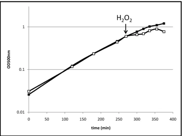
3.1 Effects of H2O2on Growth and Cell Morphology Exponentially growingB. pumiluscells were treated with 2 mM hydrogen peroxide. Thus, the concentration of H2O2 that was used to trigger the stress in this study was about 40-fold higher than those used for comparable analyses with B. subtilis or B. licheniformis[13,20]. The highest peroxide concentrations allowing growth for B. subtilis and B. licheniformis were 4 and 1 mM, respectively (Table S1).B. pumilusis still able to grow with 20 mM hydrogen peroxide. This indicates a striking resistance ofB. pumilus against peroxide stress. Compared to unstressed cells, growth was significantly impaired for a short time (approximately 15 min) after the H2O2treatment (Figure 1). However, after that time, cells continued to grow for about one hour. An electron microscopy analysis indicated that after exposure to H2O2most of the cells are morphologically intact, but some of the cells exhibited major damage of their envelope (Figure 2D). Furthermore, scanning electron microscopy revealed some atypically long cells (up to
approximately 10–20% two hours after H2O2 treatment,
Figure 2B, 2E) indicating an impact of hydrogen peroxide on processes involved in cell division.
3.2 Global Expression Profile
All values presented for up- and downregulation of genes or proteins are fold change values. The analysis of the soluble intracellular proteome of B. pumilus revealed 54 significantly upregulated and 111 downregulated proteins 10 min after H2O2
treatment (with a threshold of 2-fold, Table 1, Table S2, Figure 3). For the visualization of the fast and early response on proteome level, a labeling with35S-methionine was necessary. 30 minutes after initiating the stress, 73 proteins were up- and 59 proteins downregulated (Table 1, Table S2 and S3, Figure 4). Transcrip-tome analysis revealed an at least 2-fold increased transcription of 181 genes three minutes after treatment with H2O2; 76 of them were more than 3-fold upregulated. Eight minutes after treatment, the transcription of 558 genes appeared at least 2-fold increased (307 genes with an at least 3-fold increased transcription). Three minutes after the stress, 266 genes were transcribed with an at least 3-fold lower rate than under control conditions, for 296 genes this decreased transcription rate was shown eight minutes after treatment. To indicate quality of the transcriptome results, raw data for individual probes for selected genes (which were not found to be induced in the proteome analysis) are presented in Table S4. These data show similar basal values and changes following addition of hydrogen peroxide for all five probes corresponding to a gene.
To compare the physiological changes in H2O2 treated B. pumiluscells with the oxidative stress responses of other organisms, the upregulated genes and proteins were assigned to putative regulons known from related organisms like B. subtilis and B. licheniformis [13,20]. 139 of the upregulated genes and proteins could be assigned to these putative regulons (Table S2). The thus classified genes and proteins identified in this study are summa-rized and discussed below.
3.3 PerR Regulon
The PerR regulon is known to be highly induced by oxidative stress caused by hydrogen peroxide and paraquat [13]. As shown previously forB. licheniformis, theB. pumilusgenome encodes a PerR regulator protein with a high level of identity (93%) to the PerR-protein known fromB. subtilis[20]. Transcription of theperRgene was significantly increased immediately after stress (Table 1). This indicates a regulation mechanism of PerR in H2O2 treated B. pumiluscells that is similar to the de-repression model reported for B. subtilis[33].
In our study genes assigned to a putative PerR regulon, including those encoding the regulator proteins Fur and SpxA as well as the zinc-uptake protein ZosA, the heme biosynthesis complex HemABCD2LX and the general stress protein YjbC were significantly induced at transcriptional level (Table 1).
Strikingly, some of the PerR-regulated genes exhibiting the highest induction inB. subtiliscells subjected to hydrogen peroxide, are absent from the genome of theB. pumilusstrain used in our study, as well as from a previously publishedB. pumilus genome [34]. This applies e.g. for the genes encoding the catalase KatA and the DNA-protection protein MrgA. Furthermore,B. pumilus lacks not only the genesahpCandahpF, encoding subunits of the alkyl hydroperoxide reductase, but there are no genes annotated with this function in the genome.
Instead of KatA, a gene annotated as catalase KatX2 (53% sequence similarity toB. subtilisKatX) was significantly induced in B. pumiluscells at transcriptional and translational level (up to 10 and 20-fold, respectively, Table 1). Thereby, KatX2 was one of the proteins with the highest induction rates detected.B. subtilis andB. licheniformissubjected to hydrogen peroxide exhibit a more than 100-fold induction of KatA [13,20]. KatX2 comprises about 0.38% of the cytoplasmic protein present in the gel before addition of hydrogen peroxide. The values forB. subtilisandB. licheniformis are 0.13% in both strains (personal communication C. Scharf, B. Voigt). After addition of hydrogen peroxide KatX2 comprises about 3.8% of the cytoplasmic protein. This is comparable to the Bacillus pumilusResistance to Hydrogen Peroxide
Figure 1. Growth ofB. pumilus.Growth ofB. pumilusunder control conditions (filled squares) and stressed with 2 mM H2O2at OD500 nm0.6 (empty squares).
doi:10.1371/journal.pone.0085625.g001
Figure 2. Electron microscopy micrographs.Scanning (A,B,E) and transmission (C,D) electron microscopy micrographs ofB. pumilus cells under control conditions (A,C), 30 min (B,D) and 120 min after treatment with 2 mM H2O2(E).
doi:10.1371/journal.pone.0085625.g002
Bacillus pumilusResistance to Hydrogen Peroxide
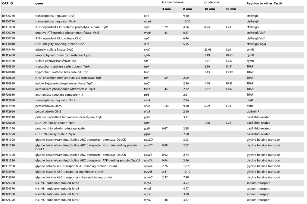
Table 1.Selected induced genes and proteins.
ORF ID gene transcriptome proteome Regulon in otherBacilli
3 min 8 min 10 min 30 min
BPJ13600 zinc-transporting ATPase ZosA zosA 12.74 28.72 perR
BPJ25410 glutamyl-tRNA reductase HemA hemA 3.44 3.99 perR
BPJ25390 porphobilinogen deaminase HemC hemC 2.68 3.90 perR
BPJ25370 delta-aminolevulinic acid dehydratase HemB hemB 2.52 3.72 perR
BPJ25400 putative cytochrome C biogenesis protein HemX hemX 2.86 4.25 perR
BPJ25380 uroporphyrinogen III synthase HemD hemD2 2.68 4.23 perR
BPJ25360 glutamate-1-semialdehyde 2,1-aminomutase HemL hemL 2.75 3.56 perR
BPJ21690 Fur family ferric uptake regulation protein Fur fur 1.92 3.62 perR
BPJ11620 transcriptional regulator Spx spxA 4.14 3.31 perR/spx/sigB
BPJ11610 putative N-acetyltransferase YjbC yjbC 2.41 4.41 perR/spx/sigB/sigM/sigW/sigX
BPJ09760 catalase KatX2 katX2 6.96 10.69 15.18 21.09 sigB/sigF
BPJ34450 putative ABC transporter permease YwjA ywjA 1.57 4.47 fur
BPJ30810 hydroxamate siderophore ABC transporter ATP-binding protein FhuC
fhuC1 1.51 2.46 fur
BPJ30830 hydroxamate siderophore ABC transporter permease FhuB
fhuB1 1.52 4.01 fur
BPJ30820 hydroxamate siderophore ABC transporter permease FhuG
fhuG1 1.53 3.20 fur
BPJ08440 ABC transport system permease bpj08440 4.11 7.49 fur
BPJ08430 putative iron complex transport system substrate binding protein
bpj08430 4.54 7.43 fur
BPJ08420 putative HTH-type transcriptional regulator bpj08420 3.58 5.59 fur
BPJ08580 putative nitroreductase YfhC yfhC 2.67 5.00 1.10 fur
BPJ08410 ferredoxin–NADP reductase 2 bpj08410 3.90 3.83 fur
BPJ37570 AraC family transcriptional regulator/putative FeuA-like substrate-binding domain ybbB
ybbB 4.93 12.84 fur
BPJ37580 iron complex ABC transporter substrate-binding protein FeuA
feuA 3.21 10.04 fur, btr, citB
BPJ37590 putative bacillibactin esterase YbbA ybbA 5.24 18.42 fur/btr/citB
BPJ07970 C56 family peptidase YfkM yfkM 2.94 7.61 7.39 3.09 fur/sigB
RBPU30260 FeS cluster assembly protein SufB sufB 1.87 2.10 1.73 Fe/S cluster biogenesis
RBPU30280 cysteine desulfurase SufS sufS 21.77 2.69 Fe/S cluster biogenesis
RBPU30290 FeS cluster assembly permease SufD sufD 1.73 Fe/S cluster biogenesis
RBPU30300 FeS cluster assembly ATPase SufC sufC 2.52 2.13 Fe/S cluster biogenesis
BPJ11040 diaminobutyrate–2-oxoglutarate aminotransferase RhbA rhbA 21.11 11.10 siderophore synthesis
BPJ11080 rhizobactin siderophore biosynthesis protein RhbE rhbE 21.18 5.28 siderophore synthesis
Bacillus
pumilus
Resistance
to
Hydrogen
Peroxide
PLOS
ONE
|
www.ploson
e.org
5
January
2014
|
Volume
9
|
Issue
1
|
Table 1.Cont.
ORF ID gene transcriptome proteome Regulon in otherBacilli
3 min 8 min 10 min 30 min
BPJ11090 rhizobactin siderophore biosynthesis protein RhbF rhbF 21.72 3.02 siderophore synthesis
BPJ35800 iron complex ABC transporter ATP-binding protein FhuC fhuC2 3.88 7.85 iron uptake
BPJ35810 iron complex ABC transporter permease FhuB fhuB2 3.32 7.15 iron uptake
BPJ35770 putative iron complex ABC transporter permease FhuG fhuG2 2.31 4.39 iron uptake
BPJ35780 putative iron complex ABC transporter substrate-binding protein FhuD fhuD 2.72 5.57 iron uptake
BPJ35830 putative iron transport-associated protein/putative siderophore bpj35830 3.65 5.84 iron uptake
BPJ35840 putative heme uptake protein IsdC bpj35840 4.91 7.62 iron uptake
BPJ35850 putative iron transport-associated protein bpj35850 3.89 6.47 iron uptake
BPJ28430 DinB-like domain-containing protein YuaE yuaE 2.25 2.87 spx
BPJ31980 thioredoxin-disulfide reductase TrxB trxB 3.97 3.93 3.59 spx
BPJ29110 putative NADH-dependent butanol dehydrogenase YugJ yugJ 2.32 1.08 4.60 spx
BPJ19830 methionine sulfoxide reductase MsrA msrA 1.46 2.24 spx
BPJ19820 peptide-methionine sulfoxide reductase MsrB msrB 1.48 2.27 spx
BPJ25870 thioredoxin TrxA trxA 1.40 2.58 spx/ctsR/sigB
BPJ35200 NADPH-dependent nitro/flavin reductase NfrA nfrA 2.50 2.47 5.21 spx/sigD/spo0A
BPJ24450 cystathionine gamma-lyase MccB mccB 21.58 7.58 spx/cymR
BPJ17710 putative cell division suppressor protein YneA yneA 2.24 44.25 lexA/SOS
BPJ10180 39-59exoribonuclease YhaM yhaM 0.71 2.81 lexA/SOS
BPJ21860 DNA polymerase 4 polY1 10.68 lexA/SOS
BPJ32300 excinuclease ABC subunit B uvrB 7.22 2.52 4.29 lexA/SOS
BPJ32290 excinuclease ABC subunit A uvrA 1.49 6.75 lexA/SOS
BPJ25860 excinuclease ABC subunit UvrC uvrC 3.65 lexA/SOS
BPJ17700 repressor LexA lexA 1.55 5.66 lexA/SOS
BPJ17730 DUF896 family protein YnzC ynzC 0.65 8.85 lexA/SOS
BPJ12460 phage-like PBSX protein XkdA xkdA 3.10 17.84 lexA/SOS
BPJ17720 resolvase-like protein YneB yneB 1.38 17.03 lexA/SOS
BPJ10160 putative exonuclease YhaO yhaO 8.76 lexA/SOS
BPJ16880 recombinase RecA recA 1.63 7.22 4.94 9.58 lexA/SOS/comK
BPJ35170 minor extracellular serine protease Vpr vpr 1.58 2.23 lexA/SOS/phoP
BPJ21470 hypothetical protein YpuD ypuD 1.93 7.12 lexA/SOS/sigB/sigM
BPJ10170 putative ATPase YhaN yhaN 8.73 lexA/SOS
BPJ13450 ATP-dependent Clp protease ATP-binding subunit ClpE clpE 2.78 45.41 ctsR
BPJ25460 ATP-dependent protease ATP-binding subunit ClpX clpX 2.67 ctsR
BPJ00800 DNA repair protein RadA radA 10.02 ctsR/sigB
Bacillus
pumilus
Resistance
to
Hydrogen
Peroxide
PLOS
ONE
|
www.ploson
e.org
6
January
2014
|
Volume
9
|
Issue
1
|
Table 1.Cont.
ORF ID gene transcriptome proteome Regulon in otherBacilli
3 min 8 min 10 min 30 min
BPJ00760 transcriptional regulator CtsR ctsR 9.40 ctsR/sigB
BPJ00770 transcriptional regulator McsA mcsA 10.26 ctsR/sigB
BPJ31850 ATP-dependent Clp protease proteolytic subunit ClpP clpP 1.79 4.26 8.74 1.73 ctsR/sigB
BPJ00780 putative ATP:guanido phosphotransferase McsB mcsB 1.43 8.87 ctsR/sigB/sigF
BPJ00790 ATP-dependent Clp protease ClpC clpC 6.44 ctsR/sigB/sigF
BPJ00810 DNA integrity scanning protein DisA disA 5.15 ctsR/sigB/sigM
BPJ15470 adenylyl-sulfate kinase CysC cysC 23.93 1.60 cymR
BPJ15480 uroporphyrin-3 C-methyltransferase CysG cysG 1.49 10.53 cymR
BPJ15460 sulfate adenylyltransferase Sat sat 1.37 13.07 cymR
BPJ20800 tryptophan synthase alpha subunit TrpA trpA 2.16 13.51 TRAP
BPJ20810 tryptophan synthase beta subunit TrpB trpB 1.13 13.99 TRAP
BPJ20820 N-(59-phosphoribosyl)anthranilate isomerase TrpF trpF 1.59 2.09 TRAP
BPJ20830 indole-3-glycerol-phosphate synthase TrpC trpC 2.36 1.49 10.53 TRAP
BPJ20840 anthranilate phosphoribosyltransferase TrpD trpD 1.44 2.72 1.37 13.07 TRAP
BPJ20850 anthranilate synthase component 1 trpE 2.61 TRAP
BPJ12980 transcriptional regulator OhrR ohrR 2.54 ohrR
BPJ12970 peroxiredoxin OhrA ohrA 10.66 9.88 6.29 1.30 ohrR
BPJ12990 peroxiredoxin OhrB ohrB 2.10 sigB/ohrR
BPJ19510 putative bacillithiol biosynthesis deacetylase YojG yojG 3.31 bacillithiol-related
BPJ20020 DUF1094 family protein YphP yphP 1.78 2.22 bacillithiol-related
BPJ21140 putative thioredoxin reductase YpdA ypdA 0.67 2.58 bacillithiol-related
BPJ22220 DUF1094 family protein YqiW yqiW 2.58 bacillithiol-related
BPJ31300 glycine betaine/carnitine/choline ABC transporter permease OpuCD opuCD 3.03 glycine betaine transport
BPJ31310 glycine betaine/carnitine/choline ABC transporter substrate-binding protein OpuCC
opuCC 0.88 2.62 glycine betaine transport
BPJ31320 glycine betaine/carnitine/choline ABC transporter permease OpuCB opuCB 0.92 2.74 glycine betaine transport
BPJ31330 glycine betaine/carnitine/choline ABC transporter ATP-binding protein OpuCA opuCA 0.94 2.46 glycine betaine transport
BPJ02950 glycine betaine ABC transporter ATP-binding protein OpuAA opuAA 2.76 10.75 glycine betaine transport
BPJ02960 glycine betaine ABC transporter membrane protein opuAB 2.41 10.19 glycine betaine transport
BPJ02970 glycine betaine ABC transporter substrate-binding protein opuAC 2.37 7.48 glycine betaine transport
BPJ29360 Na+/H+antiporter subunit MrpA mrpA 4.57 sodium transport
BPJ29370 Na+/H+antiporter subunit MrpB mrpB 4.77 sodium transport
BPJ29380 Na+/H+antiporter subunit MrpC mrpC 3.60 sodium transport
BPJ29390 Na+/H+antiporter subunit MrpD mrpD 1.48 3.87 sodium transport
Bacillus
pumilus
Resistance
to
Hydrogen
Peroxide
PLOS
ONE
|
www.ploson
e.org
7
January
2014
|
Volume
9
|
Issue
1
|
value of 3.6% forB. licheniformis(personal communication B. Voigt)
but higher than the value for B. subtilis (1.2%, personal
communication C. Scharf). These values indicate that in B. pumilusthere is a higher synthesis of KatX2 already in unstressed cells compared toB. subtilisandB. licheniformisKatA explaining the lower induction rate. InB. subtilis, KatX is the major spore catalase and under control of SigB and SigF [35,36]. We detected aB. subtilis PerR consensus sequence [32] containing 2 mismatches about 90 bases in front of the start codon of KatX2 indicating a possible involvement of PerR in its regulation.
3.4 Fur Regulon and Fe-metabolism
The PerR-regulatedfurgeneof B. pumilus, shows 95% similarity to thefur gene known fromB. subtilis and was induced 3.6-fold after stress [32]. The regulator protein Fur ofB. subtiliscontrols the expression of genes responsible for iron uptake [37]. Immediately after exposure to H2O2, cytosolic iron concentration is consider-ably reduced to prevent the formation of OHN by the Fenton
reaction [13]. Upregulation of the Fur-controlled genes may be a reaction of the cells to optimize iron uptake in order to face the resulting iron limitation. Alternatively it might be that Fur is H2O2 sensitive as it is inE. coli[38].
Nine genes of a putative Fur regulon showed a significantly increased expression in B. pumilus cells after H2O2 treatment, including the ABC transporter systemfhuB1C1G1(Table 1). The fhuCgene was induced by H2O2inB. subtilisandB. licheniformis, too [13,20]. Further Fur regulon member genes known to be induced by H2O2inB. subtilisshowing an induction in our study wereykuN, ykuP (flavodoxins) and the hypothetical protein ykuO. With an about 30-fold higher mRNA level 8 minutes after treatment, these were among the highest upregulated genes in this putative regulon. The putative nitroreductase YfhC, also induced in H2O2stressed B. subtiliscells, was the only member of the putative Fur regulon we observed to be upregulated at translational level.
The gene ywjA, encoding another ABC transporter of yet unknown function, the peptidase encoding gene yfkM and the bacillibactin esterase encoding gene ybbAwere upregulated, too. These genes are Fur-regulated in B. subtilis, but they were not upregulated by H2O2in this organism [13,39]. InB. subtilisandB. licheniformis, the siderophore biosynthesis complex encoded by dhbACEBFwas strongly upregulated by H2O2. In our study, these genes showed no significant changes in their expression level.
Other genes that exhibited higher transcription rates after H2O2 treatment were the iron ABC transporter protein encoding gene feuAand its upstream-located regulator ybbB (renamed btr inB. subtilis) [40]. Unlike B. subtilis, the B. pumilus genome encodes a second Fhu-related iron uptake system. Our study showed an induction of the genes encoding FhuC2-FhuB2-BPJ35820 as well as fhuG2 and fhuD immediately after subjecting the cells to the stress. Two further putative iron transporter systems,bpj35830 -bpj35840-bpj35850 and bpj08420-bpj08430-bpj08440, were in-duced, too. The proteins encoded by the latter genes showed no significant homology to any protein known from relatedBacillus species.
Furthermore, the proteomic approach revealed a strong induction of the siderophore synthesis proteins RhbA, RhbE and RhbF, encoded by the rhbABCDEF-operon (Table 1). A rather slight induction at the translational level was shown for the iron/ sulfur cluster biogenesis proteins SufB, SufS, SufD and SufC as previously shown forB. licheniformis[20]. ThesufUgene was found to be only slightly upregulated at the mRNA level.
Table 1. Cont. ORF ID gene transcriptome proteome Regulon in other Bacilli 3 m in 8 m in 10 min 3 0 m in BPJ29400 N a + /H + antiporter subunit M rpE mrpE 1.51 3 .20 sodium transport BPJ29410 N a + /H + antiporter subunit M rpF mrpF 1.72 3 .38 sodium transport BPJ29420 N a + /H + antiporter subunit M rpG mrpG 1.96 2 .60 sodium transport Selected genes and proteins that are induced in H2 O2 treated B. pumilus cells. Genes and proteins are listed, which could b e assigned to putative regulons known from other Bacilli .C omplete lists of upregulated as well as downregulated genes/proteins is given in supporting information Tables S2 and S3. For transcriptome, selected g enes are shown for 3 and 8 minutes after stress compared to the control conditions (0 min). For a complete list of induced and repressed genes see Table S3. Differential regulation w as determined from the biological triplicate measurements by false-discovery rate (FDR) from the Cyber-T p-values [27] by means of multiple testing correction [2 6]. Differential regulation was defined as a two-fold or higher differential expression w ith a FDR cut-off value o f 0 .05 o r lower. Protein quantification w as performed by the D elta 2D software (Decodon) from 3 biological replic ates with a FDR cut-off value of 0.05 or lower. doi:10.1371/journal.pone.0085 625.t001
Bacillus pumilusResistance to Hydrogen Peroxide
3.5 Spx Regulon and Bacillithiol
Another regulator protein assigned to the putative PerR regulon is SpxA, controlling the expression of the Spx regulon inB. subtilis
[41,42]. This gene exhibited an about 4-fold increased transcrip-tion rate in H2O2stressedB. pumiluscells. Some of the genes and proteins attributed to a putative Spx regulon in B. pumilus Figure 3. Cytosolic proteome 10 min after H2O2treatment.The cytosolic proteome ofB. pumiluscells 10 min after H2O2treatment. Cell samples were labeled with L–[35S]-methionine during the exponential growth phase (OD500 nm0.6), and 10 min after H2O2addition. Proteins were separated in a pH gradient 4 (right) –7 (left).
doi:10.1371/journal.pone.0085625.g003
Bacillus pumilusResistance to Hydrogen Peroxide
appeared to have rather moderately increased expression rates or were not induced after H2O2treatment.
In our study we detected six genes of a putative Spx regulon to be induced following H2O2 treatment (Table 1). The proteins
encoded by three of them, nitro/flavinreductase NfrA, putative NADPH-dependent butanol dehydrogenase YugJ and thiore-doxin-disulfide reductase TrxB, were induced in H2O2 treated cells, too. Upregulation ofmsrAB(methionine sulfoxide reductase Figure 4. Cytosolic proteome 30 min after H2O2treatment.The cytosolic proteome ofB. pumiluscells 30 min after H2O2treatment. Cell samples were labeled with L–[35S]-methionine during the exponential growth phase (OD500 nm0.6), and 30 min after H2O2addition. Proteins were separated in a pH gradient 4 (right) –7 (left).
doi:10.1371/journal.pone.0085625.g004
Bacillus pumilusResistance to Hydrogen Peroxide
operon) andtrxA(thioredoxin) was detected at transcriptional level only. The proteins TrxA and TrxB are described to act in direct detoxification of hydrogen peroxide [43–45]. Cystathionine gamma-lyase MccB and DinB-like domain-containing protein YuaE showed an induction only at proteome level.
The Spx-regulated srf operon, mediating competence and
metabolic functions in B. subtilis, is absent in the B. pumilus genome as shown before forB. licheniformis[42,46,47].
We noticed an increased transcription ofypdAandyqiWas well as an induction of theyphPgene product (Table 1). These genes co-occur with bacillithiol (Cys-GlcN-malate, BSH) synthesis genes [48]. However, only one gene encoding a protein involved in bacillithiol synthesis, yojG was transcribed at a slightly elevated level (Table S2). Bacillithiol is one of the major thiols inB. subtilis and known to be involved in resistance against organic peroxide stress and disulfide stress [7,49,50]. For further investigation, we analyzed the cytosolic metabolome of H2O2treatedB. pumiluscells concerning the concentration of thiol compounds. Our analysis revealed a bacillithiol level of 2.6 nmol per mg cell dry weight already under control conditions. Similar BSH concentrations have been detected inB. subtilis(0.6–2.2 nmol per mg) [7,48,51]. Ten minutes after H2O2treatment, the cytosolic concentration of bacillithiol increased to 5 nmol per mg cell dry weight (Figure 5). The increase continued up to a concentration of about 6.2 nmol per mg cell dry weight 60 minutes after stress. Since only one bacillithiol synthesis gene (yojG, renamedbshB2 inB. subtilis) was slightly upregulated, increase of bacillithiol concentration in the cells might be regulated allosterically, for example, by an oxidation of the BSH pool leading to a relief of feedback inhibition. [52,53].
3.6 SOS Regulon
H2O2 treatment leads to the formation of OHN by Fenton
reaction, which exhibits a high DNA-damaging potential. Lowering the concentration of iron in the cells reduces this threat. As a result,B. subtilisandB. licheniformiscells subjected to oxidative stress caused by H2O2, induced the SOS regulon, regulated by the
proteins RecA and LexA, responsible for repair of DNA [13,20,54,55].
The proteomic analysis displayed the induction of two proteins, excinuclease subunit UvrB and the recombinase RecA, assigned to a putative SOS regulon in B. pumilusfollowing H2O2 treatment (Table 1). The transcriptomic approach added further 13 upregulated genes belonging to this putative regulon; among them the excinuclease subunits encoding genesuvrAanduvrC. The operonyneABynzC, induced by H2O2and involved in suppression of cell division inB. subtilis, was also strongly induced in our study [13,56]. This might be an explanation for the formation of atypically long cells as described above. Showing an about 44-fold increased transcription rate,yneAbelongs to the strongest induced genes observed in our study. Furthermore, the putative DNA double-strand break repair cluster yhaONM exhibited a signifi-cantly higher transcription rate following H2O2addition [57].
3.7 CtsR Regulon
The CtsR regulon, mediating repair and/or degradation of misfolded and damaged proteins, was induced by several oxidative stressors inB. subtilisandB. licheniformis[13,20,58]. In our study, we detected an upregulation of nine genes assigned to a putative CtsR regulon inB. pumilusindicating a significant impact of H2O2 on protein quality (Table 1). The operon ctsR-mcsAB-clpC was transcribed with significantly higher intensity after the addition of H2O2as well as the genesclpE,clpXandclpP, encoding members of the proteolytic complex. Only ClpP was observed to be induced at the protein level. Furthermore, the DNA repair protein
encoding gene radA and the DNA integrity scanning protein
encoding genedisAshowed higher transcription rates compared to control conditions.
3.8 SigB Regulon
Besides the induction of the above described putative regulons more or less directly associated to oxidative stress, H2O2 treated cells exhibited an upregulation of 47 genes known to be under control of the general stress sigma factor SigB inB. subtilis(Table 1) Figure 5. Concentration of thiol compounds inB. pumiluscells.Cytosolic concentration of bacillithiol (BSH), CoA and cysteine (Cys) per mg cell dry weight (CDW) during the exponential growth phase (OD500 nm0.6 at 0 min) and 10, 30 and 60 min after H2O2treatment.
doi:10.1371/journal.pone.0085625.g005
Bacillus pumilusResistance to Hydrogen Peroxide
[59,60]. A part of a putative SigB-regulon inB. pumilusdetected to be upregulated in our study was thesigBgene itself with its signal cascade genesrsbRSTUVWandrsbXindicating an activation of the putative regulon via the general stress response cascade known fromB. subtilis[61].
Another of these putative SigB-dependent genes, encoding the putative universal stress protein NhaX, showed the highest induction rate detected in this study (more than 60-fold). Further strongly upregulated genes are the regulator protein encoding genemgsRandydaG(general stress protein), both also detected to be induced in H2O2 stressed B. licheniformis cells [20]. The upregulated genes mgsR and ydaG encode proteins with still unknown functions. Six of the upregulated putative SigB-dependent genes could be also detected to be induced in the proteomic approach. The putative general stress protein YtxH is among the strongest induced proteins (about 14-fold). The putative iron storage/DNA protecting protein Dps, providing peroxide resistance inB. anthracis, was induced in H2O2treatedB. pumilus cells, too [62].
3.9 CymR Regulon
The results of our study showed an upregulation of several proteins belonging to a putative CymR regulon. InB. subtilis, it is described to be involved in regulation of the sulfur metabolism [63]. An induction of genes belonging to this regulon has been shown in cells afflicted with oxidative stress caused by paraquat, but not stress caused by H2O2[13]. Our proteome study showed a strong induction of three putatively CymR-regulated proteins. The adenylyl-sulfate kinase (CysC) was with an induction of about 24-fold the strongest induced protein. An upregulation of the sulfate adenylyltransferase (Sat) catalyzing sulfate assimilation to 39 -phospho-adenylylsulfate was also detected (Table 1). Further proteins involved in cysteine biosynthesis were not significantly upregulated. The third upregulated protein is the uroporphyrin-3 C-methyltransferase (CysG). This enzyme catalyzes a reaction in a branch in the heme pathway producing precorrin2. An induction of the enzymes that continue the pathway from precorrin2 to siroheme could not be detected.
3.10 OtherB. pumilusUpregulated Genes/proteins
The OhrR-regulated peroxiredoxin-encoding gene ohrA is
reported to be involved in organic peroxide resistance inB. subtilis [64]. Following H2O2 treatment, there was no induction of this gene observed inB. subtilisandB. licheniformis[13,20]. In our study, we observed a strongly induced expression of this gene at transcriptional and translational level indicating an involvement of this peroxiredoxin in the H2O2resistance ofB. pumilus(Table 1). Transcription of the other organic peroxide resistance peroxir-edoxin (ohrB) as well as their regulator geneohrRwas also slightly induced in hydrogen peroxide treatedB. pumiluscells.
H2O2treatment induced some additional regulator genes. One of them isfadR, encoding a regulator protein mediating fatty acid degradation inB. subtilis[65]. Two genes putatively controlled by FadR, etfAB - encoding the electron transfer flavoprotein alpha and beta subunit, were also induced (Table S2). Another regulator, AbrB1, controlling the expression of genes induced by transition from exponential to stationary growth in B. subtilis [66], was induced at transcriptional and translational level. Similar results, but with significantly higher induction rates in the proteomic approach, were observed for the AbrB1-regulated peroxiredoxin YkuU and thiol-disulfide oxidoreductase YkuV. Furthermore, several putative regulator genes with still unknown targets were observed to be upregulated.Bpj13620,bpj17020andydcIshowed the highest changes in their expression rates. Genes encoding a
sensor kinase and a response regulator forming the two-component system YhcYZ were significantly induced directly after H2O2treatment. Its function is also unknown.
Several genes and proteins involved in transport processes were detected to be upregulated following H2O2 stress (Table 1, S2). H2O2 treatment caused an upregulation of the sodium uptake systemnatABand themrpABCDEFGcluster. This operon encodes a sodium excretion system that is considered to be the major sodium excretion system in bacteria and acts in pH homeostasis and multiple resistances inB. subtilis[67,68].
Strikingly, transcription of the glycine betaine uptake system consisting ofopuAA-AB-ACandopuCA-CB-CC-CDwas observed to be significantly induced after treatment, indicating that H2O2 impacts osmotic homeostasis inB. pumiluscells [69]. Furthermore, it is worth to mention that H2O2induced expression of a putative
TRAP regulon in B. pumilus cells. An upregulation of the
tryptophan-synthesis operon trpABFCDE as well as histidinol-phosphate aminotransferase HisC was observed in our analysis. However, neither addition of tryptophan nor addition of glycine betaine before peroxide treatment brought forth better growth or survival of stressedB. pumiluscells (data not shown).
3.11 Downregulated Genes/proteins
As shown for many other organisms, the adaptation mechanism ofB. pumiluscells to oxidative stress includes also a downregulation of vegetative cellular functions. Most of the down-regulated genes encode proteins involved in main metabolic pathways. As shown for B. subtilis and B. licheniformis, expression of the purine and pyrimidine synthesis genes was downregulated as well as genes involved in synthesis of arginine (Table S3) [13,20]. Contrary toB. subtilisandB. licheniformis, a repression of histidine synthesis genes was not observed. Instead, isoleucine and leucine synthesis genes were expressed in lower amounts following H2O2treatment. This repression might due to the iron sparing response described by Gaballa et al. [70]. Repression of enzymes involved in branched chain amino acid synthesis has been found during iron starvation inB. subtilis[37]. Furthermore, we observed a reduced expression of most of the aminoacyl-tRNA-synthetases, with the exception of tryptophanyl-tRNA-synthetasetrpS, which matched the upregula-tion of the tryptophan operon.
Strikingly, a stringent response, i.e. a downregulation of ribosomal proteins or elongation factors likefusA, tsf ortufA, as described for other organisms (B. subtilis, B. licheniformis, E. coli) could not be detected inB. pumilus[13,20,71].
Conclusion
The combination of proteomics and transcriptomics revealed a specific adaptation ofB. pumiluscells caused by the oxidative stress trigger H2O2. Although many of the induced genes and proteins could be assigned to well-known oxidative stress regulons like PerR, CtsR and Fur, there are particular mechanisms detectable which seem to be involved in the remarkable oxidative stress resistance ofB. pumilus. The concentration of H2O2that was used to trigger the stress in our study was about 40-fold higher than those used for comparable analysis ofB. subtilisorB. licheniformis. Our study could enlighten several points at which the peroxide stress response ofB. pumiluscells is different from its Gram-positive relatives. It is suggested that the catalase KatA is replaced by the catalase KatX2. Furthermore, our study revealed an induction of genes that are highly correlated to bacillithiol synthesis indicating an involvement of bacillithiol in the peroxide stress response ofB. pumilus. Metabolome analysis demonstrated a basal level of this protective metabolite but also an increase of the cytosolic Bacillus pumilusResistance to Hydrogen Peroxide
bacillithiol concentration during peroxide stress. Furthermore, a considerable set of H2O2 induced unique proteins with so far unknown function could be identified in this study. These proteins are worth to address in follow up studies to elucidate their specific role in the oxidative stress adaptation of this organism. Finally, sinceB. pumilusis an organism of industrial interest, understanding its oxidative stress response and defining marker genes for the analysis of fermentation processes is important to prevent possible negative influences on the process and the product quality.
Supporting Information
Table S1 Determination of minimal inhibition
concen-tration of hydrogen peroxide.
(XLSX)
Table S2 Genes and proteins that are upregulated after
addition of hydrogen peroxide.
(XLSX)
Table S3 Genes and proteins that are downregulated
after addition of hydrogen peroxide.
(XLSX)
Table S4 Individual signal on the array for five probes
for selected genes.
(XLSX)
Acknowledgments
We thank Annette Meuche for excellent technical assistance and Chris Hamilton (University of East Anglia) for providing the bacillithiol standard.
Author Contributions
Conceived and designed the experiments: SH R. Schroeter R. Schlu¨ter JB KHM ML TS MH BV. Performed the experiments: SH R. Schroeter KM R. Schlu¨ter DA. Analyzed the data: SH R. Schroeter BJ SvH KM. Wrote the paper: SH R. Schroeter KM R. Schlu¨ter BV TS MH.
References
1. Schweder T, Hecker M (2004) Monitoring of stress responses. Adv Biochem Eng Biotechnol 89: 47–71.
2. Stadtman ER, Levine RL (2003) Free radical-mediated oxidation of free amino acids and amino acid residues in proteins. Amino Acids 25: 207–218. 3. Farr SB, Kogoma T (1991) Oxidative stress responses inEscherichia coliand
Salmonella typhimurium. Microbiol Rev 55: 561–585.
4. Benardini JN, Sawyer J, Venkateswaran K, Nicholson WL (2003) Spore UV and acceleration resistance of endolithicBacillus pumilusandBacillus subtilisisolates obtained from Sonoran desert basalt: implications for lithopanspermia. Astrobiology 3: 709–717.
5. Kempf MJ, Chen F, Kern R, Venkateswaran K (2005) Recurrent isolation of hydrogen peroxide-resistant spores ofBacillus pumilusfrom a spacecraft assembly facility. Astrobiology 5: 391–405.
6. Imlay JA, Fridovich I (1991) Assay of metabolic superoxide production in
Escherichia coli. J Biol Chem 266: 6957–6965.
7. Newton GL, Rawat M, La Clair JJ, Jothivasan VK, Budiarto T, et al. (2009) Bacillithiol is an antioxidant thiol produced in Bacilli. Nat Chem Biol 5: 625– 627.
8. Blokhina O, Virolainen E, Fagerstedt KV (2003) Antioxidants, oxidative damage and oxygen deprivation stress: a review. Ann Bot 91 Spec No: 179–194. 9. Imlay JA (2003) Pathways of oxidative damage. Annu Rev Microbiol 57: 395–
418.
10. Aruoma OI, Halliwell B, Gajewski E, Dizdaroglu M (1991) Copper-ion-dependent damage to the bases in DNA in the presence of hydrogen peroxide. Biochem J 273 (Pt 3): 601–604.
11. Zuber P (2009) Management of oxidative stress inBacillus. Annu Rev Microbiol 63: 575–597.
12. Imlay JA (2008) Cellular defenses against superoxide and hydrogen peroxide. Annu Rev Biochem 77: 755–776.
13. Mostertz J, Scharf C, Hecker M, Homuth G (2004) Transcriptome and proteome analysis ofBacillus subtilisgene expression in response to superoxide and peroxide stress. Microbiology 150: 497–512.
14. Hoi le T, Voigt B, Ju¨rgen B, Ehrenreich A, Gottschalk G, et al. (2006) The phosphate-starvation response ofBacillus licheniformis. Proteomics 6: 3582–3601. 15. Bu¨ttner K, Bernhardt J, Scharf C, Schmid R, Ma¨der U, et al. (2001) A comprehensive two-dimensional map of cytosolic proteins ofBacillus subtilis. Electrophoresis 22: 2908–2935.
16. Voigt B, Schweder T, Becher D, Ehrenreich A, Gottschalk G, et al. (2004) A proteomic view of cell physiology ofBacillus licheniformis. Proteomics 4: 1465– 1490.
17. Wolf C, Hochgra¨fe F, Kusch H, Albrecht D, Hecker M, et al. (2008) Proteomic analysis of antioxidant strategies ofStaphylococcus aureus: diverse responses to different oxidants. Proteomics 8: 3139–3153.
18. Vo¨lker U, Engelmann S, Maul B, Riethdorf S, Vo¨lker A, et al. (1994) Analysis of the induction of general stress proteins ofBacillus subtilis. Microbiology 140 (Pt 4): 741–752.
19. Homuth G, Masuda S, Mogk A, Kobayashi Y, Schumann W (1997) The dnaK operon ofBacillus subtilisis heptacistronic. J Bacteriol 179: 1153–1164. 20. Schroeter R, Voigt B, Ju¨rgen B, Methling K, Po¨ther DC, et al. (2011) The
peroxide stress response ofBacillus licheniformis. Proteomics 11: 2851–2866. 21. Delcher AL, Harmon D, Kasif S, White O, Salzberg SL (1999) Improved
microbial gene identification with GLIMMER. Nucleic Acids Res 27: 4636– 4641.
22. Guo FB, Zhang CT (2006) ZCURVE_V: a new self-training system for recognizing protein-coding genes in viral and phage genomes. BMC Bioinfor-matics 7: 9.
23. Borodovsky M MR, Besemer J., Lomsadze A. (2003) Current Protocols in Bioinformatics.
24. Hyatt D, Chen GL, Locascio PF, Land ML, Larimer FW, et al. (2010) Prodigal: prokaryotic gene recognition and translation initiation site identification. BMC Bioinformatics 11: 119.
25. Wernersson R, Juncker AS, Nielsen HB (2007) Probe selection for DNA microarrays using OligoWiz. Nat Protoc 2: 2677–2691.
26. van Hijum SA, de Jong A, Baerends RJ, Karsens HA, Kramer NE, et al. (2005) A generally applicable validation scheme for the assessment of factors involved in reproducibility and quality of DNA-microarray data. BMC Genomics 6: 77. 27. Baldi P, Long AD (2001) A Bayesian framework for the analysis of microarray
expression data: regularized t -test and statistical inferences of gene changes. Bioinformatics 17: 509–519.
28. Po¨ther DC, Liebeke M, Hochgra¨fe F, Antelmann H, Becher D, et al. (2009) Diamide triggers mainly S Thiolations in the cytoplasmic proteomes ofBacillus subtilisandStaphylococcus aureus. J Bacteriol 191: 7520–7530.
29. Sharma SV, Jothivasan VK, Newton GL, Upton H, Wakabayashi JI, et al. (2011) Chemical and Chemoenzymatic syntheses of bacillithiol: a unique low-molecular-weight thiol amongst low G+C Gram-positive bacteria. Angew Chem Int Ed Engl 50: 7101–7104.
30. Po¨ther DC, Gierok P, Harms M, Mostertz J, Hochgra¨fe F, et al. (2013) Distribution and infection-related functions of bacillithiol inStaphylococcus aureus. Int J Med Microbiol 303: 114–123.
31. Mu¨nch R, Hiller K, Barg H, Heldt D, Linz S, et al. (2003) PRODORIC: prokaryotic database of gene regulation. Nucleic Acids Res 31: 266–269. 32. Fuangthong M, Herbig AF, Bsat N, Helmann JD (2002) Regulation of the
Bacillus subtilis furandperRgenes by PerR: not all members of the PerR regulon are peroxide inducible. J Bacteriol 184: 3276–3286.
33. Lee JW, Helmann JD (2006) The PerR transcription factor senses H2O2by metal-catalysed histidine oxidation. Nature 440: 363–367.
34. Gioia J, Yerrapragada S, Qin X, Jiang H, Igboeli OC, et al. (2007) Paradoxical DNA repair and peroxide resistance gene conservation inBacillus pumilus SAFR-032. PLoS One 2: e928.
35. Bagyan I, Casillas-Martinez L, Setlow P (1998) The katX gene, which codes for the catalase in spores ofBacillus subtilis, is a forespore-specific gene controlled by sigmaF, and KatX is essential for hydrogen peroxide resistance of the germinating spore. J Bacteriol 180: 2057–2062.
36. Petersohn A, Engelmann S, Setlow P, Hecker M (1999) The katX gene of
Bacillus subtilisis under dual control of sigmaB and sigmaF. Mol Gen Genet 262: 173–179.
37. Baichoo N, Wang T, Ye R, Helmann JD (2002) Global analysis of theBacillus subtilisFur regulon and the iron starvation stimulon. Mol Microbiol 45: 1613– 1629.
38. Varghese S, Wu A, Park S, Imlay KR, Imlay JA (2007) Submicromolar hydrogen peroxide disrupts the ability of Fur protein to control free-iron levels in
Escherichia coli. Mol Microbiol 64: 822–830.
39. Kunst F, Ogasawara N, Moszer I, Albertini AM, Alloni G, et al. (1997) The complete genome sequence of the gram-positive bacterium Bacillus subtilis. Nature 390: 249–256.
40. Gaballa A, Helmann JD (2007) Substrate induction of siderophore transport in
Bacillus subtilismediated by a novel one-component regulator. Mol Microbiol 66: 164–173.
41. Choi SY, Reyes D, Leelakriangsak M, Zuber P (2006) The global regulator Spx functions in the control of organosulfur metabolism inBacillus subtilis. J Bacteriol 188: 5741–5751.
Bacillus pumilusResistance to Hydrogen Peroxide
42. Nakano S, Ku¨ster-Scho¨ck E, Grossman AD, Zuber P (2003) Spx-dependent global transcriptional control is induced by thiol-specific oxidative stress in
Bacillus subtilis. Proc Natl Acad Sci U S A 100: 13603–13608.
43. Spector A, Yan GZ, Huang RR, McDermott MJ, Gascoyne PR, et al. (1988) The effect of H2O2upon thioredoxin-enriched lens epithelial cells. J Biol Chem 263: 4984–4990.
44. Chae HZ, Chung SJ, Rhee SG (1994) Thioredoxin-dependent peroxide reductase from yeast. J Biol Chem 269: 27670–27678.
45. Fernando MR, Nanri H, Yoshitake S, Nagata-Kuno K, Minakami S (1992) Thioredoxin regenerates proteins inactivated by oxidative stress in endothelial cells. Eur J Biochem 209: 917–922.
46. Nakano S, Erwin KN, Ralle M, Zuber P (2005) Redox-sensitive transcriptional control by a thiol/disulphide switch in the global regulator, Spx. Mol Microbiol 55: 498–510.
47. Veith B, Herzberg C, Steckel S, Feesche J, Maurer KH, et al. (2004) The complete genome sequence ofBacillus licheniformisDSM13, an organism with great industrial potential. J Mol Microbiol Biotechnol 7: 204–211.
48. Gaballa A, Newton GL, Antelmann H, Parsonage D, Upton H, et al. (2010) Biosynthesis and functions of bacillithiol, a major low-molecular-weight thiol in Bacilli. Proc Natl Acad Sci U S A 107: 6482–6486.
49. Lee JW, Soonsanga S, Helmann JD (2007) A complex thiolate switch regulates theBacillus subtilisorganic peroxide sensor OhrR. Proc Natl Acad Sci U S A 104: 8743–8748.
50. Chi BK, Gronau K, Ma¨der U, Hessling B, Becher D, et al. (2011) S-bacillithiolation protects against hypochlorite stress inBacillus subtilisas revealed by transcriptomics and redox proteomics. Mol Cell Proteomics 10: M111 009506.
51. Chi BK, Roberts AA, Huyen TT, Ba¨sell K, Becher D, et al. (2013) S-bacillithiolation protects conserved and essential proteins against hypochlorite stress infirmicutesbacteria. Antioxid Redox Signal 18: 1273–1295.
52. Upton H, Newton GL, Gushiken M, Lo K, Holden D, et al. (2012) Characterization of BshA, bacillithiol glycosyltransferase from Staphylococcus aureusandBacillus subtilis. FEBS Lett 586: 1004–1008.
53. Gaballa A, Antelmann H, Hamilton CJ, Helmann JD (2013) Regulation of
Bacillus subtilisbacillithiol biosynthesis operons by Spx. Microbiology 159: 2025– 2035.
54. Miller MC, Resnick JB, Smith BT, Lovett CM, Jr. (1996) TheBacillus subtilis dinR
gene codes for the analogue of Escherichia coli LexA. Purification and characterization of the DinR protein. J Biol Chem 271: 33502–33508. 55. Love PE, Lyle MJ, Yasbin RE (1985) DNA-damage-inducible (din) loci are
transcriptionally activated in competentBacillus subtilis. Proc Natl Acad Sci U S A 82: 6201–6205.
56. Kawai Y, Moriya S, Ogasawara N (2003) Identification of a protein, YneA, responsible for cell division suppression during the SOS response inBacillus subtilis. Mol Microbiol 47: 1113–1122.
57. Krishnamurthy M, Tadesse S, Rothmaier K, Graumann PL (2010) A novel SMC-like protein, SbcE (YhaN), is involved in DNA double-strand break repair and competence inBacillus subtilis. Nucleic Acids Res 38: 455–466.
58. Leichert LI, Scharf C, Hecker M (2003) Global characterization of disulfide stress inBacillus subtilis. J Bacteriol 185: 1967–1975.
59. Petersohn A, Brigulla M, Haas S, Hoheisel JD, Vo¨lker U, et al. (2001) Global analysis of the general stress response ofBacillus subtilis. J Bacteriol 183: 5617– 5631.
60. Hecker M, Reder A, Fuchs S, Pagels M, Engelmann S (2009) Physiological proteomics and stress/starvation responses inBacillus subtilisandStaphylococcus aureus. Res Microbiol 160: 245–258.
61. Hecker M, Vo¨lker U (2001) General stress response ofBacillus subtilisand other bacteria. Adv Microb Physiol 44: 35–91.
62. Tu WY, Pohl S, Gizynski K, Harwood CR (2012) The iron-binding protein Dps2 confers peroxide stress resistance onBacillus anthracis. J Bacteriol 194: 925– 931.
63. Even S, Burguiere P, Auger S, Soutourina O, Danchin A, et al. (2006) Global control of cysteine metabolism by CymR in Bacillus subtilis. J Bacteriol 188: 2184–2197.
64. Fuangthong M, Atichartpongkul S, Mongkolsuk S, Helmann JD (2001) OhrR is a repressor of ohrA, a key organic hydroperoxide resistance determinant in
Bacillus subtilis. J Bacteriol 183: 4134–4141.
65. Matsuoka H, Hirooka K, Fujita Y (2007) Organization and function of the YsiA regulon ofBacillus subtilisinvolved in fatty acid degradation. J Biol Chem 282: 5180–5194.
66. Perego M, Spiegelman GB, Hoch JA (1988) Structure of the gene for the transition state regulator, abrB: regulator synthesis is controlled by thespo0A
sporulation gene inBacillus subtilis. Mol Microbiol 2: 689–699.
67. Ito M, Guffanti AA, Oudega B, Krulwich TA (1999) mrp, a multigene, multifunctional locus inBacillus subtiliswith roles in resistance to cholate and to Na+and in pH homeostasis. J Bacteriol 181: 2394–2402.
68. Kajiyama Y, Otagiri M, Sekiguchi J, Kosono S, Kudo T (2007) Complex formation by themrpABCDEFG gene products, which constitute a principal Na+/H+antiporter inBacillus subtilis. J Bacteriol 189: 7511–7514.
69. Kempf B, Bremer E (1995) OpuA, an osmotically regulated binding protein-dependent transport system for the osmoprotectant glycine betaine inBacillus subtilis. J Biol Chem 270: 16701–16713.
70. Gaballa A, Antelmann H, Aguilar C, Khakh SK, Song KB, et al. (2008) The
Bacillus subtilisiron-sparing response is mediated by a Fur-regulated small RNA and three small, basic proteins. Proc Natl Acad Sci U S A 105: 11927–11932. 71. VanBogelen RA, Kelley PM, Neidhardt FC (1987) Differential induction of heat shock, SOS, and oxidation stress regulons and accumulation of nucleotides in
Escherichia coli. J Bacteriol 169: 26–32.
Bacillus pumilusResistance to Hydrogen Peroxide