Mart a Coelho Abrant es
Dissert ation present ed t o obt ain t he Ph.D degree in Biology
Instituto de Tecnologia Química e Biológica | Universidade Nova de Lisboa
Oeiras,
July, 2012
Insert here an im age
wit h rounded corners
and Cu
2+in t he hum an pat hogen
Ent erococcus faecalis
Me
2+Me
2+Me
2+Me
2+Me
2+Me
2+Me
2+Me
Me
2+ 2+Me
2+Me
2+Me
2+Me
2+Me
2+Me
2+Me
2+Me
2+Me
2+Me
2 +Me
2+Me
2+Me
2+Me
2+Me
2 +Me
2+Me
2+Me
2+Me
2+Me
2+Me
2+Me
2+Me
2+Me
Me
2+ 2+Me
2+Me
2+Me
2+Me
2+Me
2+Me
2+Me
2+Me
2+Me
2+Me
2+
Me
2+Me
2+Me
2+Me
2+Me
2+Me
2+Me
2+
Me
Me
2+2+
Me
2+Me
Me
2+2+
Me
2+Me
2+Me
2+Me
2+Me
Me
2+2+
Me
2+Me
2+Me
2+Me
2+Me
2+Me
2+Me
2+Me
2+Me
2+Me
2+Me
2+
Me
Me
2+ 2+Me
2+
Me
2+Me
2+Me
2+Me
2+Me
2+Me
2+Me
2+Me
2+Me
2+Me
2+Me
2+Me
2+Me
2+Me
2+Me
2+Me
2+Me
2+Me
2+Me
2+Me
2+
Me
2+Me
2+Me
2+Me
2+Me
2+Me
2+Me
2+Me
2+Me
2+Me
Me
2+ 2+Me
2+Me
Me
2+Me
2+ 2+Me
Me
2+2+
Me
2+Me
2+Me
Me
2+ 2+Me
2+Me
2+Me
2+Me
2+Me
2+Me
2+Me
2+Me
2+Me
2+Me
2+Me
2+Me
2+Me
2+Me
2+Me
Me
2+Me
2+ 2+Me
2+Me
2+Me
2+Me
2 +Me
2+Me
2 +Me
2+Me
2+Me
2 +Me
2+Me
2+Me
2+Me
2+Me
2+Me
2+Me
2+Me
2+Me
2+
Me
2+Me
2+Me
2+Me
2+Me
2+Me
Me
2+ 2+Me
2 +Me
2+Me
2+Me
2+Me
2+Me
2+Me
2+Me
2+Me
2 +Me
2 +Me
2 +Me
2 +Me
2 +Me
2 +Me
2 +Me
2 +Me
2 +Me
2 +Me
2 +Me
2 +Me
2 +Me
2 +Me
2 +Me
2 +Me
2 +Me
2 +Me
2 +Me
2 +Me
2 +Me
2 +Me
2 +Me
2 +Me
2 +Me
2 +Me
2 +Me
2 +Me
2 +Me
2 +Me
2+
Me
2 +Me
2 +Me
2 +Me
2 +Me
2 +Me
2+
Me
2 +Me
2+
Me
2 +Me
2 +Me
2 +
Me
2 +Me
2 +Me
2 +Me
2+Me
2 +Me
2 +Me
2 +Me
2 +Me
2 +Me
2 +Me
2 +Me
2+Me
2+Me
2+Me
2+
Me
2+Me
2+Me
2+Me
2+Me
2+Me
2+Me
2+Me
2+Me
2+Me
2+Me
2+
Me
2+Me
2+Me
2+Me
2+Me
2+Me
2+Me
2+Me
2+Me
2+Me
2+Me
2+Me
2+Me
2+Me
2+Me
2+Me
2+Me
2+Me
2+Me
2+Me
2+Me
2+Me
2 +Me
2 +Me
2 +Me
2 +Me
2 +Me
2 +Me
2 +Me
2+
Me
2 +
Me
2 +Me
2 +Me
2 +Me
2 +Me
2 +Me
2 +Me
2 +Me
2 +Me
2 +Me
2 +Me
2 +Me
2+Me
2 +Me
2 +Me
2 +Me
2+Me
2 +Me
2 +Me
2 +Me
2 +Me
2 +Me
2+Me
2+Me
2 +Me
2Marta Maria Coelho dos Sant os Abrant es
Dissert at ion present ed t o obtain t he Ph.D degree in Biology
Instituto de Tecnologia Quím ica e Biológica | Universidade Nova de Lisboa
Oeiras, July, 2012
im port ance of Zn
, Mn
and Cu
in t he
hum an pat hogen Enterococcus faecalis
iii
Supervisors
Doctor Maria de Fátima Lopes
–
Auxiliary Investigator at Instituto deTecnologia Química e Biológica, Universidade Nova de Lisboa.
Professor Doctor Jan Kok
–
Full Professor at the Department ofMolecular Genetics, Centre for Life Sciences, University of Groningen.
Examiners
Professor Doctor Jorge Humberto Gomes Leitão – Auxiliary Professor at the Department of Bioengineering, Instituto Superior Técnico, Universidade Técnica de Lisboa.
Doctor Teresa Maria Leitão Semedo-Lemsaddek – Auxiliary Investigator at Centro de Investigação Interdisciplinar em Sanidade Animal, Faculdade de Medicina Veterinária, Universidade Técnica de Lisboa.
Professor Doctor Ana Rosa Leal Lino – Investigator at Centro de Química e Bioquímica, Faculdade de Ciências, Universidade de Lisboa.
v
“It is not the strongest
of the species that
survives, nor the most intelligent, but the one
most responsive to change”
vii
Acknowledgements
Welcome to what is generally the most read part of a thesis!
This was the most difficult part to write, as it takes me “on a trip down Memory Lane”…
My PhD was a very enriching personal and professional experience,
for which I have many people to thank for (and I hope not to forget anyone).
I have to start by acknowledging the Institutions that provided me the
conditions to perform the work here presented. To ITQB, for accepting me
as a student and for its excellent conditions to perform science; and to GBB
(Groningen Biomolecular Sciences and Biotechnology Institute) for
accepting me as a guest PhD student and giving me the opportunity to use
their excellent facilities, learn new techniques and work in a great new
environment, which all together had a major contribution to this thesis. I
also thank the financial support provided by Fundação para a Ciência e
Tecnologia.
To my supervisors, Fátima and Jan, I’m very grateful to have worked
and learnt from you. It was a great pleasure to be your student.
Fátima, we had a first “successful experience” when I was a Biochemistry student on my internship under your supervision. Luckily for
me, a few years later we decided to start another (hopefully) successful
journey. Thank you so much for having me as you PhD student (you never
make us feel like just students), and for all the knowledge and support; in
science and sometimes in private matters I was able to count on you. A
BIG BIG thank you! I also want to thank you for starting the collaboration
with the MolGen group, where I was able to perform part of my PhD work
viii
Jan, I first want to thank you for accepting me as your PhD student,
which gave me the chance to have the great experience of living in The
Netherlands. I really enjoyed living in your country, especially for the nice
people I had the chance to meet (not so much for the weather!). I also want
to thank your concern for me, to fit within the group, which luckily was never
a problem. Back in the lab, I learnt a lot from you, particularly during our
brainstorming meetings, from where I would usually leave with a headache
and much more knowledge and passion for science.
I also want to dedicate a special thanks to Oscar, who very willingly
initiated the collaboration with Fátima, thus opening the doors of MolGen to
Fátima and I. Thank you for your kindness, your warm welcomes and
concern for my work and general well-being at MolGen.
To all MolGeners, past and present, I want to thank for the nice moments we’ve spent and for making me feel so welcome in the group. Within the group, I have to start by thanking my fellow compatriot João that
helped literally since day one to fit in the group, with work or just anything,
whenever I needed. Thank you for having made things easier for me while I
was at MolGen. I have to thank the people in the Strep lab, namely
Jan-Willem, who would cheer up the lab with his singing, thank you for the
support; Tomas, thank you for the work discussions and the nice dinners;
Sulman, it was great to share the bench with you, you can always cheer
someone up! Thank you for all your help, support and fun times! Hope you
come back to Portugal soon. Speaking of trips to Portugal, Imke, miss our
fun times at MolGen, the nights out and the heavenly dinner at your home –
can´t wait to see you back in Portugal! Katrin and Ana, we had lots of fun,
miss you girls! Rutger, thanks for your kindness, help and support. I have to
also thank Evert-Jan and Sacha for the funniest lunches ever, and in
particular Evert-Jan for so promptly helping me, using DISCLOSE, which
ix my abstract and for the great Sinterklaas party at your home. Much more
people are part of the good memories from MolGen: Bogusia, Aleksandra, Auke, Tom, Martijn, Wout, Akos, Robyn, Tariq, Lieke… and the list goes on, thank you all!
Back in Portugal, I want to thank the members of my lab, mostly from the past: Tânia, we’ve met a “few” years ago, on my internship and since then you’ve helped me, I’ve learnt a lot from you, could always count on you and we had so much fun in and out of the lab! Thanks for everything!
Frederic could always bring fun and laughter to the lab! I really enjoyed and
miss our times together. Renata, thanks for the help and all the fun times,
specially outside the lab, such as in Amsterdam and thanks for receiving
and showing me Paris! Breaks like those can do tremendously good when you’re doing a PhD! Neuza, Teresa, Sofia and Marta, thank you for your help and support. Thanks to all Microbiology group, in particular to Teresa
Crespo, Victória San-Romão and Paula Alves.
To the friends I was lucky to gain at ITQB, making my workplace even more pleasant: Zé, my “Sporting partner”, you’ve helped me in so many occasions, particularly with your wise words, and of course with our “therapeutic” trips to Sporting matches! I can´t thank you enough… Sandra, you’re the calm friend one can always rely on. Thank you for all the help and fun times together! Sónia and Vânia, I’ve been able to count on you
too, thanks for everything. Marta, we go a long way back before we met
again at ITQB (a nice surprise!). I knew I could count on you before as I know I’ll be able to count on you in the future. Thanks for all the support.
x
supported me unconditionally. Your friendship is precious to me, thank you
for everything.
To my family, who has supported me always. To my father, who
always supported my choices and trusted me; in a way, this journey was
also his. To my brother that had to make sure I would be safe and well on
my first stay in Groningen and (in his words) carried my bags! To my oldest
niece Vera, my good times in The Netherlands were also my time away from her. Still to today she asks me if I will have to go back… Sooner than we think you will be able to read this and this thesis is part of my legacy to
you, my princess. To my stepmother Ana, my youngest niece Inês, Tatão,
my uncles, aunts and cousins, and other friends who closer or far have
been with me, helping, supporting and giving me joy throughout this journey…
xi
Abstract
Enterococcus faecalis is a commensal bacterium able to colonize
different sites in the human host, such as the gastrointestinal tract, the
genito-urinary tract and the oral cavity. It can also be found in numerous
other environments, including soil, sand, water, food products and plants.
These bacteria show a dual behavior: they can behave quite harmlessly as
commensals, but are able to become opportunistic pathogens and cause
serious infections, such as urinary tract infections and endocarditis, in
hospital settings. The question as to how these bacteria are able to change
from commensalism to pathogenicity has directed many recent studies to
focus on the environmental host conditions that may trigger this transition
as well as on the underlying molecular mechanisms.
Metals are very important elements in the host environment, as they
are key components of many proteins and are involved in numerous cell
processes in both the host and the invading pathogen. The maintenance of
metal homeostasis is fundamental to both to ensure that metabolism and
cell functions are functioning properly. Variations in this homeostasis must
be tightly regulated. In several Gram positive pathogens, metal
homeostasis and regulation has been linked to their pathogenicity. The lack
of knowledge on this subject in E. faecalis motivated the work presented in
this thesis.
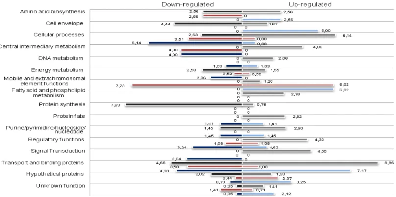
Our studies initially addressed the genome-wide transcriptional
responses of E. faecalis V583, a multi-resistant strain isolated from a
bloodstream infection, to zinc, manganese and copper ions (Chapter 2). A
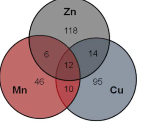
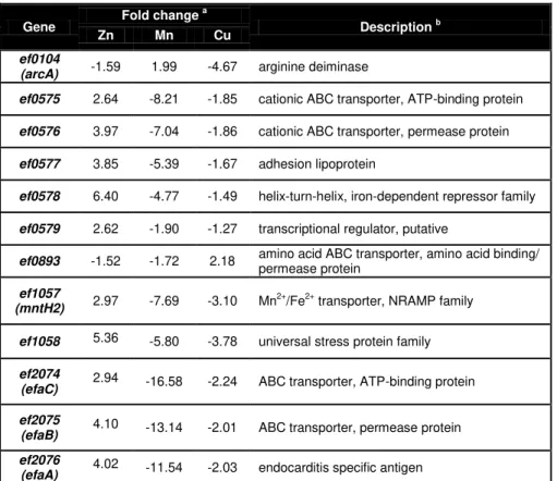
set of differentially expressed genes common to the three metal
xii
studied (Chapter 3). Some of the highest up-regulated genes in response to
zinc excess were examined in Chapter 4, focusing on their role in the ability
of E. faecalis to cause infections.
The transcriptomic experiments provided new information on the
responses of E. faecalis to zinc, manganese and copper excess and
revealed that the most relevant mechanisms involved were transport
systems (Chapter 2). Our findings provided the first evidence for the correct
annotation of many E. faecalis genes, as most of those transporters were
annotated as metal transporters. Furthermore, genes encoding other types
of transporters and proteins involved in energy, amino acid metabolism and
cellular processes were also shown to be relevant for metal homeostasis in
E. faecalis. Several genes related to the cell wall appeared to be
particularly important in the response to excess copper. Although the cop
operon for copper transport and regulation is described as the main player
in copper homeostasis in Enterococcus hirae, our results suggest that in E.
faecalis other transport genes may also be involved.
In a subsequent study, we focused on manganese transport systems
and their regulation (Chapter 3). The genes encoding these systems were
identified and were shown to be regulated by EfaR, a DtxR family regulator.
Furthermore, several transporter mutants were examined to evaluate the
role of these proteins, and that of their regulator, on cell processes relevant
for E. faecalis colonization and infection of the host. The studies revealed
that EfaR is the key player for manganese homeostasis in E. faecalis.
Moreover, EfaR was able to influence a number of cell processes important
for colonization and infection, namely biofilm formation, oxidative stress and
intramacrophage survival. Our observations provided a link between
xiii the study of genes found to be highly up-regulated under zinc excess. In
Chapter 4 we focused on the gene encoding a cation P-type ATPase,
denoted ZntAEf, and on ef0759, encoding a putative SapB protein.
Mutations were made in these genes in order to elucidate their role in E.
faecalis defense against host attack. The zinc-responsive ATPase ZntAEf
was shown to play a role in the response to zinc overload, lysozyme
treatment, oxidative stress, all used by the host to defend itself against
bacteria and contributing to intramacrophage survival. EF0759 also
influenced E. faecalis survival inside macrophages, although likely by a
different mechanism, as the corresponding mutant was not more
susceptible to the other three host weapons mentioned. As saliva contains
lysozyme and a relatively high concentration of zinc, ZntAEf may be relevant
for the success of E. faecalis as an orthodontia pathogen. This constitutes
the first link between zinc homeostasis and pathogenicity in this organism.
The work here presented reveals the importance of transition metals
and the mechanisms and regulation of their transport in processes involved
in E. faecalis colonization and infection of the host. Thus, it establishes a
novel link between metal homeostasis and pathogenicity of E. faecalis and
xv
Resumo
Enterococcus faecalis é uma bactéria comensal que coloniza
naturalmente mamíferos e insectos. No hospedeiro humano é capaz de
colonizar diferentes locais, tais como o tracto gastrointestinal, o tracto
génito-urinário e a cavidade oral; também pode estar presente noutros
ambientes, nomeadamente solo, areia, água, produtos alimentares e
plantas. Estas bactérias têm um comportamento dualístico: como
comensais, podem ser inofensivas, mas também se podem tornar
patogéneos oportunistas e causar infecções graves em ambiente
hospitalar, tais como infecções urinárias e endocardites. A questão de
como os enterococcus são capazes de passar de comensais a
patogénicos tem levado muitos estudos recentes a focarem-se nas
condições ambientais do hospedeiro que possam potenciar esta transição
e também nos mecanismos moleculares subjacentes.
Os metais são elementos importantes nos ambientes do hospedeiro
pois são componentes fundamentais de muitas proteínas e estão
envolvidos em inúmeros processos celulares tanto no hospedeiro como
nas bactérias. A homeostase da concentração de metais é fulcral em
ambos para garantir que o metabolismo e as funções celulares decorrem
de forma adequada. As variações nesta homeostase têm de ser reguladas
de forma rigorosa. Em vários patogéneos Gram positivos, a homeostase e
a regulação dos metais foram correlacionadas com a patogenicidade
dessas bactérias. A falta de conhecimento sobre esta temática
relativamente à bactéria E. faecalis motivou o trabalho apresentado nesta
tese.
Inicialmente, os nossos estudos foram dirigidos para o perfil
xvi
cobre, da estirpe E. faecalis V583, uma estirpe multi-resistente isolada de
uma infecção sanguínea (Capítulo 2). Um conjunto de genes que foram
diferencialmente expressos em comum aos três metais e relacionados com
o transporte de manganês foram objectos do estudo apresentado no
Capítulo 3. Alguns dos genes que foram mais sobrexpressos na presença
de excesso de zinco foram estudados no Capítulo 4, focando o papel
destes genes na capacidade de E. faecalis causar infecções.
As experiências de transcriptómica forneceram novas informações
evidenciando que os transportadores são os mecanismos mais relevantes
na resposta ao excesso de zinco, manganês e cobre. Os nossos
resultados concederam a primeira prova empírica da correcta anotação de
vários genes de E. faecalis, pois a maioria dos sistemas de transporte
revelados nestas experiências (Capítulo 2) estavam anotados como
associados ao transporte de metais. Adicionalmente, genes que codificam
outros tipos de transportadores e proteínas envolvidas no metabolismo em
geral e em processos celulares mostraram também ter um papel relevante
na homeostase de metais em E. faecalis. Vários genes relacionados com a
parede celular parecem ter um papel particularmente importante na
resposta ao excesso de cobre. Embora o operão cop, responsável pelo
transporte e regulação do cobre, esteja descrito como tendo o papel
principal na homeostase deste metal em Enterococcus hirae, os nossos
resultados sugerem que em E. faecalis há outros genes que codificam
transportadores que também podem estar envolvidos na homeostase do
cobre.
Os estudos seguintes focaram-se nos sistemas de transporte de
manganês e na sua regulação (Capítulo 3). Os genes que codificam estes
sistemas, identificados no Capítulo 2, revelaram ser regulados pela
proteína EfaR, pertencente à família de reguladores DtxR. Adicionalmente,
xvii processos celulares relevantes para a colonização e infecção do
hospedeiro. Os estudos mostraram que o regulador EfaR tem uma função
crucial na homeostase do manganês em E. faecalis. Os mesmos estudos
revelaram também que EfaR é capaz de influenciar um número de
processos celulares importantes para a colonização e infecção,
nomeadamente a formação de biofilmes, a resistência ao stress oxidativo e
a sobrevivência no interior de macrófagos. As nossas observações
demonstram a existência de uma ligação entre a homeostase do
manganês e a patogenicidade em E. faecalis.
A análise dos resultados das experiências de transcriptómica com
zinco impulsionou o estudo, apresentado no capítulo 4, de genes
consideravelmente sobrexpressos na presença de excesso de zinco,
nomeadamente um gene que codifica uma ATPase de tipo P, denominada
ZntAEf e o gene ef0759, codificando uma proteína SapB putativa. Foram
construídas estirpes com mutações nestes genes, com o intuito de
esclarecer as suas funções na resposta de E. faecalis às defesas do
hospedeiro. A ATPase ZntAEf, que responde à presença de zinco, mostrou
ter um papel relevante na resistência ao excesso de zinco, à lisozima e ao
stress oxidativo. Estes três elementos de defesa do hospedeiro são
particularmente importantes nos macrófagos, onde o transportador ZntAEf
demonstrou ser crucial para a sobrevivência de E. faecalis. A proteína
EF0759 demonstrou ter também um papel activo na sobrevivência de E.
faecalis dentro de macrófagos. No entanto, o mecanismo pelo qual
contribui para este fenótipo deverá ser diferente do mecanismo da proteína
ZntAEf, já que o respectivo mutante não se revelou mais susceptível às três
referidas defesas do hospedeiro, comparativamente com a estirpe
selvagem. Dado que a saliva contém lisozima e uma concentração
xviii
sucesso de E. faecalis como patogéneo periodontal. Este trabalho constitui
a primeira ligação entre a homeostase do zinco e a patogenicidade deste
microrganismo.
O trabalho apresentado nesta tese revela a importância dos metais de
transição e dos mecanismos de regulação dos seus transportadores, e
permite o reconhecimento da ligação entre homeostase de metais e a
patogenicidade de E. faecalis, sendo fornecidas novas pistas para futuras
estratégias de combate a este importante patogéneo.
xix
Abbreviations
∆ deletion
ABC ATP-binding cassette
ADI arginine deiminase
ATP Adenosine-5’-triphosphate
BHI Brain Heart Infusion
BLAST Basic Local Alignment Search Tool
bp Base pairs
CDF cation diffusion facilitators
cDNA complementary DNA
CFU colony forming unit
chelGM17 chelated GM17
cm chloramphenicol
DISCLOSE DISsection of Clusters Obtained by Series of transcriptome
data
DNA Deoxyribonucleic acid
ebm EfaR binding motif
ECM ExtraCellular Matrix
Ehk Enterococcal histidine kinase
Err Enterococcal response regulator
ery erythromycin
GEO Gene Expression Omnibus
GM17 M17 medium with 0.5% glucose
GRAS Generally Recognized As Safe
GSH Glutathione
xx
LAB Lactic Acid Bacteria
LB Luria-Bertani medium
LTA Lipotheichoic acids
MCO multi-copper oxidase
MCS multiple cloning site
moi multiplicity of infection
MOODS MOtif Ocurrence Detection Suite
NCBI National Center for Biotechnology Information
NRAMP Natural Resistance-Associated Macrophage Protein
OD Optical Density
PAI Pathogenicity Island
PCR Polymerase Chain Reaction
PTS Phosphotransferase system
r
resistance
RNA Ribonucleic acid
RND resistance nodulation family
ROS Reactive Oxygen Species
rpm rotations per min
SDS Sodium Dodecyl Sulfate
SI Survival Index
sqRT-PCR semi-quantitative Reverse Transcriptase PCR
TCS Two Component System
tet tetracycline
v/v volume/volume
YT Yeast Extract with Tryptone
xxi
Table of Contents
Acknowledgements VII
Abstract XI
Resumo XV
Abbreviations XIX
Table of Contents XXI
Chapter 1
General Introduction 1
Chapter 2
Impact of manganese, copper and zinc ions on the transcriptome of the nosocomial pathogen Enterococcus faecalis V583 53
Chapter 3
EfaR is a major regulator of Enterococcus faecalis manganese transporters and influences processes involved in host
colonization and infection 109
Chapter 4
ZntAEf mediates Enterococcus faecalis defense against
zinc overload, lysozyme and oxidative stress 143
Chapter 5
Chapter 1
3
Table of Contents
Enterococcus 5
Transition from commensalism to pathogenicity 7
Virulence 8
Environmental stresses 10
Metals 12
Metal sensor proteins 15
Metal transport 17
Uptake systems 17
Efflux systems 17
Zinc 18
Zinc regulation 20
Manganese 21
Manganese regulation 24
Copper 26
Copper regulation 28
Metal regulation in enterococci 29
Scope of this thesis 31
5
Enterococcus
Enterococci are commensal inhabitants of the gastrointestinal tract of
humans and other animals and can colonize the genitourinary tract and the
oral cavity [1]. They are also capable of surviving in the most diverse
environmental niches such as soil, sand, water, food products and plants
[2]. These bacteria are differentiated by their capacity to grow between
10°C and 45°C, in 6.5% NaCl and at pH 9.6, to survive upon heating at
60°C for 30 min, to hydrolyze esculine into esculitine [3] and to react with
the Lancefield group D antisera [1]. Enterococci produce L(+)-lactic acid
homofermentatively from glucose. They belong to the large group of the
lactic acid bacteria (LAB). Contrary to other LAB, enterococci are not
considered “Generally Recognized As Safe” (GRAS) and their detection in
water is regarded as an indicator of fecal contamination [4].
Presently forty-one species of Enterococcus are recognized
(http://old.dsmz.de/microorganisms/bacterial_nomenclature_info.php?genus=Enter
ococcus); Enterococcus faecalis and Enterococcus faecium are the two
most common species found in the human microbiota [3].
Enterococci are important in several areas of our daily lives. In the food
industry, they have a beneficial role due to their ability to degrade casein
and stimulate the growth of certain LAB, which is exploited for the ripening
of cheeses such as cheddar and mozzarella [5]; their capacity to grow
under harsh conditions makes them suited for fermentation of other
products as well, such as sausages [6-8]. The putative negative aspect of
enterococci in food is their ability to produce biogenic amines in cheese and
fermented sausages [9, 10]. Enterococci have also been successfully used
6
Although harmless in healthy individuals, enterococci have been
emerging as important nosocomial pathogens causing wound-,
bloodstream- and urinary tract infections and endocarditis mainly in
hospitalized patients with severe underlying diseases or with an impaired
immune system under prolonged antibiotic treatments. Enterococci rank
second as a cause of urinary tract infections in both the United States and
Europe [14, 15] and are the third leading cause of endocarditis after
streptococci and Staphylococcus aureus, being responsible for 5 to 20% of
all cases of endocarditis (predominantly E. faecalis) [16]. E. faecalis and E.
faecium are associated with approximately 60% and 40%, respectively, of
hospital-acquired infections caused by enterococci [17, 18]. The strong
association of enterococci with infections in hospital settings and the
difficulty in devising a successful medical treatment have been attributed to
their inherent capacity to withstand environmental stresses and their innate
and acquired resistance to many commonly used antibiotics [1, 19, 20]
making them well equipped to survive and colonize hospital environments
[21, 22]. Also, the pathogenicity of enterococci is potentially increased by
their highly efficient ability to transfer genetic material [23]. Vancomycin has
been used as the drug of last resort in the treatment of Gram positive
bacterial infections, including those caused by enterococci. Resistance to
vancomycin poses a serious problem in the treatment of enterococcal
infections [19], and favors propagation and persistence of enterococci in
hospitals; furthermore, there is an increased risk of horizontal transfer of
this resistance determinant to other medically relevant vancomycin-
susceptible species [19, 24, 25], as has already been observed for S.
aureus [26]. Vancomycin resistance genes have been found in both clinical
and dairy enterococcal isolates, which facilitates the propagation of these
7
E. faecalis V583 was the first reported vancomycin resistant clinical
isolate in the United States. It was obtained from a patient suffering from a
persistent bloodstream infection [28]. V583 is part of the high-risk clonal
complex 2, which comprises mostly isolates derived from hospital infections
worldwide [29, 30], and was the first E. faecalis strain to have its genome
sequence published [30]. Since then, 29 other enterococcal genomes were
sequenced [31, 32]. Comparative genome hybridization-based studies have
revealed considerable variation in the genomic content of different E.
faecalis and E. faecium strains. This variation is mainly a result of the
presence or absence of mobile genetic elements such as phages and
conjugative transposable elements and differences within the E. faecalis
and E. faecium pathogenicity islands [33, 34].
The mechanisms by which inoffensive commensal enterococci may
become major hospital-acquired pathogens are still not understood. The
identification of traits related to the pathogenicity of these bacteria will
contribute to the understanding of the dual nature of this organism.
Transition from commensalism to pathogenicity
The invasion of the bloodstream or other internal areas of the host is a
critical step in the transition of enterococci from commensals to pathogens.
During the process of tissue invasion, enterococci encounter an
environment vastly different from that at sites of colonization, where there is
limited nutrient availability, high redox potentials and host defenses are
present. Infecting enterococci likely express genes favoring growth under
8
Virulence
E. faecalis possess several virulence factors that help establishing
infection and persisting in the presence of host immune responses [35].
Several of these virulence factors have been characterized and are
presented in Table 1 (reviewed in [24, 36, 37]).
Table 1: E. faecalis genes encoding virulence factors and their putative
roles.
Gene Virulence factor Putative role Reference
agg Aggregation substance Adhesion [38]
efaA Endocarditis specific antigen Adhesion and infection [39]
ace Adhesin to collagen Adhesion to ECM [40]
esp Enterococcal surface protein Adhesion and infection [41]
cylA-M Cytolysin Tissue damage [42]
gelE Gelatinase Tissue damage [43]
epa Enterococcal polysaccharide
antigen Not determined [44]
cpsA-K Capsular polysaccharide
Resistance to host
defense [45]
sprE Serine protease Tissue damage [46]
hypR Hydrogen peroxide regulator Resistance to host
defense [47]
gls24 Glucose starvation protein Persistence in the host [48]
cylR1-R2 Two-component system cylA-M regulation [42]
fsrA-C Agr-like regulatory system gelE and sprE
regulation [46]
etaRS OmpR-like two-component
system Not determined [49]
perR Peroxide regulator Not determined [50]
9 Even though several factors may contribute to virulence of enterococci,
a widespread distribution of putative virulence determinants in enterococcal
isolates independent of their origin has been reported [51-53] and to date,
no single virulence factor has been found to be ubiquitous in clinical
isolates or has been demonstrated to be essential for enterococcal
infections [54]. Furthermore, the importance of these virulence factors does
not always seem to be supported by the findings of clinical studies [21]. In
addition, though the detection of virulence genes may point to a virulence
potential in food strains, foodborne enterococcal infections have never
been reported. Nonetheless, food isolates may contribute to the spread of
virulence genes by horizontal transfers rather than being a direct cause of
infection [36].
The ability to cause infection can also be associated with a strain’s competence to form biofilms [55], which would possibilitate the survival in
less favorable conditions such as antibiotic-rich environments and promote
persistance on medical devices. The capacity of enterococci to bind to
various medical devices such as ureteral stents [56], intravascular
catheters, biliary stents and silicone gastrostomy devices has been
associated with their ability to produce biofilms [57, 58]. Hence, this ability
enhances the capacity of enterococci to cause infections and contributes to
the emergence of enterococci in hospitals [20, 59].
For enterococci to become pathogens, they had to develop
mechanisms of adaptation that clearly include more than production of
biofilms or expression of virulence factors and enabled them to cope with
10
Environmental stresses
In order to survive and colonize the human gastrointestinal tract,
bacteria must overcome several biological barriers. Microorganisms that
survive the gastric acidity of the stomach are able to transit to the intestine,
where they encounter stresses associated with variations in pH, low oxygen
availability, elevated osmolarity, nutrient limitations and relatively high
concentrations of host-produced detergents [61-63]. Environmental stress
responses in E. faecalis have been well studied for more than a decade
and have shown their exceptional ability to survive and persist in a variety
of adverse environments. In these studies, several E. faecalis genes have
been identified to play important roles in the survival to environmental
stresses and thus enabling the infection process (Table 2).
The ability of most bacteria to monitor and adapt to changing
conditions is often mediated through two-component signal transduction
systems. Two-component systems (TCS) generally consist of a sensory
histidine kinase and a response regulator. The histidine kinase senses the
signal and transfers a phosphoryl group to the response regulator, which
can then regulate gene expression [64]. These systems are usually
involved in environmental stress responses and other cellular processes
important for survival and for infection of the host. It has been described
that the E. faecalis response regulators Err04, Err08 and Err18 are involved
in heat shock response. Moreover, TCS Err-Ehk05 has a role in the
regulation of the sagA gene, which is involved in various stress responses
(see Table 2; [65]) ; TCS Err-Ehk10 is involved in heat and acid pH
response (EtaRK; [49]), in bile salts and, to a lesser extent in hyperosmotic
stress response, and has a potential role in the expression of the heat
11 Table 2: Genes described to be involved in environmental stress
responses in E. faecalis.
Genes Related stress Reference
sagA Bile, NaCl, SDS, ethanol, oxidative, heat, alkaline, acid [66]
gsp62 Bile, SDS, acid, oxidative, heat, ethanol, tert-butyl
hydroperoxide, sodium chloride [67]
gsp65 SDS, acid, oxidative, heat, ethanol, sodium chloride [68]
gls24 Bile, carbohydrate and complete starvation, CdCl2 [69]
clpPBCEX Heat [70]
ctsR Heat (Clp cluster regulator) [71]
dnaK Bile, heat [63]
groEL Bile, heat [63]
sigV Glucose and complete starvation, heat, ethanol, acid [7]
rsiV Glucose and complete starvation, heat, ethanol, acid
(sigV regulator) [7]
relA Alarmone synthesis and degradation. Alarmone
accumulates with heat, alkaline and vancomycin stress [72]
hypR Oxidative stress [73]
sodA Oxidative stress [74]
katA Oxidative stress [75]
gor Oxidative stress [76]
npr Oxidative stress [77]
trxB Oxidative stress [77]
ahpF Oxidative stress [77]
ahpC Oxidative stress [77]
qacZ Biocide – benzalkonium chloride [78] SDS – Sodium dodecyl sulfate
As discussed so far, bacteria have to be able to obtain the appropriate
nutrients and adapt to the environmental conditions for survival and growth.
One important requirement is the maintenance of metal ion homeostasis,
particularly for the proper control of regulatory networks that govern gene
expression and for virulence. In pathogenic bacteria, the mechanisms for
metal ion homeostasis or, more specifically, metal ion transport may be the
key to major adaptations to intracellular survival, colonization and infection
12
In the next part of this Introduction chapter, the biological importance of
metals and the bacterial mechanisms for metal regulation will be discussed
with particular emphasis on zinc, manganese and copper ions, as these
metals were the focus of the work presented in this thesis.
Metals
Transition metals are essential for all organisms, from bacteria to man.
Approximately a quarter to a third of all proteins in any organism are
metalloproteins [80]. They play important roles in the regulation of gene
expression and in the activity of biomolecules. They are able to function as
catalysts for biochemical reactions, as stabilizers of protein structures and
bacterial cell walls, and can serve in maintaining osmotic balance [81, 82].
It is known that transition metals are also crucial for microbial invasion
and infection, as bacterial pathogens must acquire metal nutrients in order
to cause disease. The strict requirement for these elements during
pathogenesis is due to their involvement in numerous processes, ranging
from bacterial metabolism to accessory virulence factor function [83]. As
metals are required for essential cellular processes, in both the host and
the bacteria, vertebrates tend to frustrate this bacterial requirement by
sequestering these elements [84].
Metal concentrations in the host can vary tremendously (Fig. 1) and for
colonization and infection, bacteria need to have the proper mechanisms
13
Figure 1– Concentrations of zinc (Zn) [85-87], manganese (Mn) [85, 86] and copper (Cu) ions [85-87] in different parts of the human body (figure adapted from http://www.humanillnesses.com).
Metal ions are most often required in trace amounts. Suboptimal or
elevated intracellular metal concentrations have severe, pleiotropic effects
on many aspects of bacterial cellular metabolism. This obliges cells to be
capable of scavenging trace metal ions from their environment to meet
cellular requirements and, on the other hand, to have resistance
mechanisms for when metal concentrations exceed physiological needs
[88]. The existence of such resistance mechanisms is very important
because high metal concentrations can cause severe problems such as
damage to cell membranes, alteration of enzyme specificity, disruption of
cellular functions and damage to the structure of DNA. Toxicity occurs
through displacement of metals from their native binding sites or through
ligand interactions, which may lead to alterations in the conformational Blood serum:
Zn: 15.3 M Mn: 9 nM
Cu: 15.7 M
Gastric juice :
Zn: 0.13 M Cu: 1.2 M Saliva:
Zn: 133.3 M Mn: 35.8 M
Cu: 2.8 M
Lung tissue:
14
structure of nucleic acids and proteins, interference with oxidative
phosphorylation and osmotic balance [89, 90].
Tight regulation of metal transport systems is the purview of
metalloregulators that sense cytosolic metal levels and regulate the
transcription of genes that maintain metal homeostasis accordingly [91-93],
while metallochaperones storage metals or deliver them to proteins that
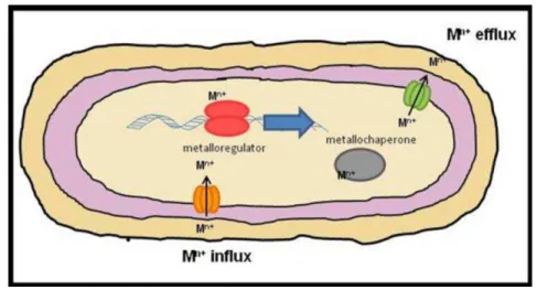
need metal ions for function (Fig. 2; [92, 94-96]).
Figure 2 - Cellular processes for metal homeostasis. Membrane protein transport systems are responsible for the uptake (orange) and efflux (green) of
metal ions according to cell’s needs; metalloregulators (red) regulate transcription of the referred metal transport system genes (blue arrow); metallochaperones (grey) help with proper metal trafficking and/or storage. Mn+; n-valent metal ion.
For maintaining metal homeostasis, specific metalloregulatory proteins
must be capable of discriminating the right ligand from a pool of transition
metals that often have similar sizes (ionic radii) and net charges (often 2+;
[88]). The fidelity of their discrimination controls the abundance of different
metals within cells with consequences for metal occupancy of other
15 substantially determined by the Irving– Williams series, for divalent metals (Mg2+ and Ca2+ (weakest binding) < Mn2+ < Fe2+ < Co2+ < Ni2+ < Cu2+ > Zn2+,
and Cu+ is also highly competitive) [83].
Metallochaperones have an important role in metal sequestration;
however in prokaryotes, metal ion homeostasis is mostly maintained by
metalloregulatory proteins. These proteins are able to bind metal ions
directly and repress, derepress or activate the transcription of operons that
encode metal-specific efflux pumps and/or membrane bound transporters,
metal reductases, soluble cytoplasmic or periplasmic metal transport
proteins, metal-sequestering proteins, as well as the metal-responsive
transcriptional regulator itself [97].
Metal sensor proteins
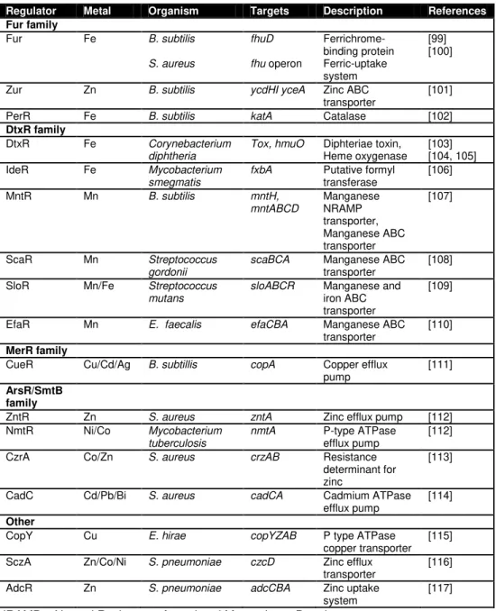
Recent structural studies have contributed to the differentiation of four
main distinct families of metal sensor proteins (Table 3). The Fur and DtxR
families regulate most of the genes encoding proteins involved in metal ion
uptake; in these cases, generally, the metal ion functions as a co-repressor
in turning off uptake-related genes under metal-replete conditions. The
MerR and ArsR/SmtB families regulate the expression of many genes
required for metal ion detoxification and efflux; for these regulators, metal
binding may lead to activation (MerR) or to derepression (ArsR/SmtB) of
the resistance operon [98].
As more and more studies on metal transport and regulation are being
reported, regulators from other families have been revealed as also playing
16
adaptation of bacterial genomes to changing conditions and different
environments.
Table 3: Examples of Gram positive metalloregulatory proteins.
Regulator Metal Organism Targets Description References Fur family
Fur Fe B. subtilis
S. aureus
fhuD
fhu operon
Ferrichrome-binding protein Ferric-uptake system [99] [100]
Zur Zn B. subtilis ycdHI yceA Zinc ABC
transporter
[101]
PerR Fe B. subtilis katA Catalase [102]
DtxR family
DtxR Fe Corynebacterium
diphtheria
Tox, hmuO Diphteriae toxin, Heme oxygenase
[103] [104, 105]
IdeR Fe Mycobacterium
smegmatis
fxbA Putative formyl transferase
[106]
MntR Mn B. subtilis mntH,
mntABCD Manganese NRAMP transporter, Manganese ABC transporter [107]
ScaR Mn Streptococcus
gordonii
scaBCA Manganese ABC transporter
[108]
SloR Mn/Fe Streptococcus mutans
sloABCR Manganese and iron ABC transporter
[109]
EfaR Mn E. faecalis efaCBA Manganese ABC
transporter
[110]
MerR family
CueR Cu/Cd/Ag B. subtillis copA Copper efflux pump
[111]
ArsR/SmtB family
ZntR Zn S. aureus zntA Zinc efflux pump [112]
NmtR Ni/Co Mycobacterium tuberculosis
nmtA P-type ATPase efflux pump
[112]
CzrA Co/Zn S. aureus crzAB Resistance determinant for zinc
[113]
CadC Cd/Pb/Bi S. aureus cadCA Cadmium ATPase efflux pump
[114]
Other
CopY Cu E. hirae copYZAB P type ATPase
copper transporter [115]
SczA Zn/Co/Ni S. pneumoniae czcD Zinc efflux transporter
[116]
AdcR Zn S. pneumoniae adcCBA Zinc uptake
system
17
Metal transport
Metal transport is basically performed by uptake and efflux systems,
which will be further detailed below.
Uptake Systems
Metal uptake systems mostly rely on ATP-binding cassette (ABC)
transporters. In Gram positive bacteria, the prototypical ABC transporter
consists of a lipoprotein, a hydrophobic membrane protein, and an ATPase,
with the two latter present as homo- or heterodimers. The lipoprotein is
tethered to the outer side of the cell membrane and functions as a ligand
binding protein [118]. Different ABC transporters translocate different
substrates, ranging from small ions to large polypeptides, and they
therefore play a wide variety of physiological roles. Mutations in ABC
transporter genes are the underlying cause of a number of human genetic
disorders. This fact contributes to their particular economic and medical
importance as they can pump cytotoxic molecules from cells, thus
conferring resistance to antibiotics, herbicides and chemotherapeutic drugs
[119]. Here ABC transporters will be referred to only when they play a role
in metal ion homeostasis (Table 3).
Efflux Systems
Microorganisms use efflux systems to export toxic metals from the
cytoplasm of their cells. The various metal efflux systems belong to known
protein families such as: the resistance nodulation (RND) family, which are
18
by a chemiosmotic gradient or a potassium gradient [120]; and P-type
ATPases that are driven by ATP hydrolysis [121].
The RND family of proteins is less common in bacteria. P-type
ATPases and CDF proteins can be found in eukaryotes and in bacteria.
The type ATPases are mostly metal cation transporters. Prokaryotic
P-type ATPases transport a range of divalent metal cations that include Cu2+,
Ag2+, Cd2+, Zn2+ and Mn2+ (Table 3; [122]).
The study of the roles of essential metal ions and the regulation of their
homeostasis has gained more and more relevance, mostly because of their
involvement in bacterial pathogenesis.
In this thesis we focused on zinc, manganese and copper, three
essential metal ions, that are crucial in many life processes across all
kingdoms and that have particularly relevant roles in bacterial colonization
and pathogenesis. The next sections will deal with each of these metals
separately, referring mainly to their biological roles, their benefits and their
associated diseases, and discussing the mechanisms involved in their
regulation and in homeostasis maintenance.
Zinc
Zinc is an essential element for all living organisms. It is the second
most abundant transition metal in seawater and in humans. This metal has
been suggested to interact with as many as 10% of host proteins [123].
Factors that contribute to zinc’s prominence among metal ions include its chemical characteristics as a relatively strong Lewis acid in enzymes and
the fact that it is the only essential transition metal that lacks biological
19 cofactor [125]. Hence, zinc serves as a cofactor in all six classes of
enzymes (oxidoreductases, transferases, hydrolases, lyases, isomerases
and ligases) as well as several classes of regulatory proteins [126, 127].
Zinc concentrations are highly variable in the human body, as shown
previously in Fig. 1. High concentrations of zinc are toxic as this metal ion
can interact with thiols and block essential reactions in the cell.
Zinc is known to be essential for all highly proliferating cells in the
human body, especially the immune system. In addition, its
immunosuppressive properties might be used for therapy in non-toxic
concentrations [128]. A role for zinc in neurodegenerative diseases such as
Alzheimer’s disease has also been suggested (Table 4; [129]).
Zinc may be used by the host as a weapon against bacterial infections.
In fact, zinc levels in the human body are increased during inflammation
[130-132]. Curiously, many of us probably do not acknowledge that zinc
toxicity is used every day to fight the development of dental plaque with
zinc-containing toothpaste [83].
Table 4: Impact and role of zinc in the human host.
Beneficial properties of zinc Reference
Immunosuppressant [128]
Antioxidant [133]
Cardiovascular protector [134]
Zinc depletion
Effects
Decreased immune function [128]
Symptoms (some examples)
Gastrointestinal problems, frequent infections, dermatitis, endocrine disorders, cancer, degenerative diseases
[128, 135]
Zinc toxicity
Effects
Damage immune cells, microbicidal [134, 136]
Symptoms (some examples)
Nausea, vomiting, renal failure [134]
Zinc unbalance
20
Bacteria are predicted to incorporate zinc into approximately 4–6% of all of their proteins [137]. Whereas the total cellular zinc concentration is in
the millimolar range, femtomolar concentrations of free Zn2+ trigger
transcription of genes involved in zinc uptake or efflux machinery. This
suggests an extraordinary intracellular zinc-binding capacity and shows that
cells exert tight control over cytosolic metal concentrations, even for
relatively low-toxicity metals such as zinc [138].
Zinc Regulation
Prokaryotes contain diverse mechanisms of zinc uptake and efflux and
carry different metalloregulators from distinct protein families to control
these systems (Table 3).
The Fur homologue Zur, a zinc uptake regulator, is a major regulator
responsible for guaranteeing the necessary uptake of zinc in several
bacteria, including B. subtillis [101] and S. aureus [139]. Fur is the
prototype for a large family of regulators that sense iron (Fur), zinc (Zur),
manganese (Mur) and nickel (Nur) sufficiency and peroxide stress (PerR)
[140, 141]. The emerging consensus is that most of these proteins are
likely to function as metal-dependent, DNA-binding repressors [142]. Zur
controls zinc transport by binding to a Zur box in the presence of Zn2+ and
thus repressing transcription of ABC transporter genes [143-145]. Although
Zur regulates potential virulence determinants such as metalloproteases in
S. suis, the effect of zur inactivation on virulence appears to be minimal
[139, 143].
The ArsR/ SmtB family of metalloregulators, which negatively
regulates genes involved in metal efflux, includes zinc-dependent
21 P-type ATPases and CDFs, in the absence of bound metal. Allosteric
binding of specific metals regulates binding to DNA, triggering derepression
[113, 141]. One of the members, S. aureus ZntR, regulates the expression
of its own gene and the adjacent gene, zntA. Together they form an operon
involved in the resistance to Zn2+ and Co2+ [112].
E. coli ZntR, on the other hand, is a member of the MerR family of
regulators. It binds to the zntA promoter region leading to repression of
zntA transcription. In the presence of metals, namely Zn2+, Cd2+ or Pb2+,
ZntR becomes an activator of zntA expression, so that ZntA efflux system
can expel the excess metals [146, 147].
Manganese
Manganese is the 12th most abundant element on the surface of the
earth and is naturally present in rocks, soil, water and food such as cereals,
fruits, vegetables and tea [148]. This metal is an essential micronutrient for
all forms of life [149]. Manganese is necessary in vertebrates for a
multitude of functions such as skeletal system development, energy
metabolism, activation of certain enzymes, nervous system function,
reproductive hormone function; it is also an antioxidant that protects cells
from damage by free radicals (Table 5; [148]). Unlike zinc, there is little
information regarding the effects of manganese deficiency on immune
development and function [149].
The importance of Mn2+ for cellular physiology of bacteria has only
recently been better revealed. By virtue of its redox activity and ability to
function as a Lewis acid catalyst, manganese participates in several vital
22
Table 5: Impact and role of manganese in the host.
Beneficial properties of manganese Reference
Antioxidant [148]
Manganese depletion
Neurological and behavioral effects [150-152]
Epilepsy, Down’s syndrome, osteoporosis [151]
Manganese toxicity
Manganism [153-155]
Parkinson’s disease [155, 156]
Mn2+ has a primary role in the protection against oxidative stress and a
variety of roles in lipid, protein and carbohydrate metabolism, biosynthesis,
and signal transduction [157]. Mn2+-dependent bacterial proteins include
phosphoglyceromutase, enolase, pyruvate kinase, type I protein
phosphatases, Mn2+-dependent superoxide dismutases and catalases [158,
159]. Mn2+ also influences spore composition, structure and germination in
some bacteria [160]. Mn2+ is particularly critical for the LAB, many of which
have been shown to require Mn2+, instead of Fe2+, for survival [161-163].
LAB lack catalase but contain superoxide dismutases that require Mn2+ as
a cofactor [161, 164]; Mn2+ itself can also act directly to detoxify superoxide
and hydrogen peroxide [161, 165, 166]. There are also descriptions of this
metal acting catalytically as an antioxidant in bacteria by associating with
anions including phosphate, and metabolic intermediates such as lactate or
malate [161, 165]. As an essential transition element, Mn2+ is extremely
limited within the human body, where it may be complexed with carrier
proteins. The concentration of this metal is 1000-fold higher in secretions
than in internal body sites, as shown in Fig. 1. The change in the external
Mn2+ concentration is one factor that signals the bacterium to change its
expression of virulence factors in response to its environment [166].
Emerging data have revealed that vertebrates resist bacterial
23 calprotectin, a well-known mammalian calcium-binding protein, chelates
manganese and zinc within abscesses [167]. The inhibition of bacterial
metal uptake represents a promising alternative area of research for the
design of new antimicrobials [167].
Vertebrates also seem to limit metal availability to intracellular
pathogens through the expression of the protein NRAMP-1 (Natural
Resistance-Associated Macrophage Protein 1). This kind of proteins is
expressed by many cell types including neutrophils and macrophages
[168]; they have been suggested to transport iron and manganese out of
the lysosome [168, 169]. Depletion of divalent metals, namely Mn2+, from
the bacteria-containing phagosome by NRAMP-1 might have a simple and
species-general bacteriostatic effect by removing an essential element from
the ecological niche of intracellular pathogens. This might enhance the
bactericidal activity of macrophages by rendering the pathogen more
sensitive to killing by oxygen radicals [170].
A hallmark of the susceptibility of a host cell to an invading pathogen is
the relative susceptibility of the host cell and the pathogen to reactive
oxygen and nitrogen species [158]. The use of Mn2+ complexes by bacteria
to provide protection against reactive oxygen species (ROS) might increase
their fitness. In fact, the involvement of complexes of Mn2+ with low
molecular-weight anions such as lactate, bicarbonate or malate in the
defense against ROS could form the basis of a widespread and so far
overlooked defense mechanism [165]. In addition, a recent study on
antibiotics has shown that exposure of Gram positive (E. faecalis and S.
aureus) and Gram negative (E. coli) bacteria to bactericidal antibiotics
induces the production of ROS by an oxidative damage cellular death
pathway involving the tricarboxylic acid cycle, a transient depletion of