557
online | memorias.ioc.fiocruz.br
Mem Inst Oswaldo Cruz, Rio de Janeiro, Vol. 106(5): 557-561, August 2011
Beside the considerable information available on myxozoan species collected from different geographical areas (Lom & Dyková 2006), little is known about those from South America and in particularly about those from the Brazilian Atlantic coasts, where a diverse as-semblage of several hundred species of fish live. Within the myxozoans the genus Myxidium is widely distribut-ed infecting several marine and freshwater fishes, some amphibians and reptiles (Lom & Dyková 2006).
Some Myxidium spp infecting the fish galbladders in the Atlantic ichthyofauna from different regions have been recorded (Lom & Dyková 1992, 2006, MacKenzie & Ka-lavati 1995, MacKenzie et al. 2005): Myxidium incurva-tum, collected from several species of the French coasts (Lom & Dyková 1992), Myxidium sphaericum in Belone belone and other hosts, Myxidium bergensis in Pollachius virens and other hosts, Myxidium oviforme in Pagon im-berbis from Mediterranean Sea and in other fish species in the Atlantic Ocean, Myxidium gadi in Pollachius
pol-lachius from French region (Mackenzie & Kalavati 1995), Myxidium gigantissimum in Alepocephalus australis off South African coast (Dubina & Isakov 1976), Myxidium baueri in Patagonotothen sima from Falkland Islands (Kovaleva & Gaevskaya 1982, Kalavati et al. 1996), My- xidium trachinarum in Echiichthys vipera from England (Canning et al. 1999) and Myxidium finnmarchicum in Merlangius merlangus from Norway (MacKenzie et al. 2010) (Table). A Brazilian myxosporidian check-list were published (Gioia & Cordeiro 1996) in which the great ma-jority of the results were based on light microscopy, how-ever, this record does not report any Myxidium sp. from the Brazilian coast. Until now, Dactylopterus volitans was not reported as a host of Myxidium sp.
This paper describes, based on light and ultrastruc-tural data, a new Myxidium species parasite of a marine fish collected in the Brazilian Atlantic coast.
MATERIALS AND METHODS
Small fragments of parasitized gallbladder walls and free spores were removed from the bile of the economi-cally important marine fish flying gurnard, D. volitans (Linnaeus, 1758) (Teleostei: Dactylopteridae) (Brazilian common name coió). Sixty four fishes (39 males and 25 females) with an average total length of ~25 cm (range 21-29 cm) (n = 64), collected in the Atlantic coast (22º58’S 43º00’W), near the city of Niterói, state of Rio de Janeiro (RJ), Brazil, were examined and the spores were measured Financial support: CNPq, CAPES
+Corresponding author: azevedoc@icbas.up.pt Received 29 December 2010
Accepted 6 June 2011
Myxidium volitans
sp. nov., a parasite of the gallbladder
of the fish,
Dactylopterus volitans
(Teleostei: Triglidae)
from the Brazilian Atlantic coast - Morphology and pathology
Carlos Azevedo1,2/+, Graça Casal1,3, Sérgio Carmona São Clemente4, Leila Maria Silva Lopes4, Patrícia Matos5, Abdel Azeem Abdel-Baki2, Elsa Oliveira1, Edilson Matos6
1Departamento de Biologia Celular, Instituto de Ciências Biomédicas e Laboratório de Patologia,
Centro Interdisciplinar de Investigação Marinha e Ambiental, Universidade do Porto, Largo Prof. Abel Salazar 2, P-4099-003 Porto, Portugal
2Zoology Department, College of Science, King Saud University, Riyadh, Saudi Arabia 3Departamento de Ciências, Instituto Superior de Ciências da Saúde–Norte, Gandra, Portugal
4Laboratório de Inspeção e Tecnologia dos Alimentos, Faculdade de Veterinária, Universidade Federal Fluminense, Niterói, RJ, Brasil 5Laboratório de Animais Aquáticos, Universidade Federal do Pará, Belém, PA, Brasil
6Laboratório de Pesquisa Carlos Azevedo, Universidade Federal Rural da Amazônia, Belém, PA, Brasil
Myxidium volitans sp. nov. (Myxozoa: Myxidiidae) parasitizing the hypertrophied green-brownish gallbladder of the teleost Dactylopterus volitans, collected in the Atlantic coast near Niterói, Brazil was described based on ul-trastructural studies. The spores were fusiform, sometimes slightly crescent-shaped on average 21.7 ± 0.3 µm (mean ± standard deviation) (n = 50) long and 5.6 ± 0.4 µm (n = 30) wide. The spore wall was thin and smooth, comprising two equally-sized valves joined by a hardly visible sutural ridge. Spores containing two pyriform polar capsules (PC) (5.0 ± 0.4 × 2.3 ± 0.3 µm) (n = 30) are situated in each extremity of the spore. The PC wall was composed of hyaline layer (0.20-0.29 µm thick) and by a thin external granular layer. Each PC contains a polar filament (PF) with irregular arrangements that was projected from its apical region to the bases of PC and coiled laterally from bases to the tip of PC. Some regular striations and S-like structures in the periphery of the PFs with four-five ir-regular sections were observed. Based on the spore morphology, ultrastructural differences and the specificity of the host we describe this parasite as a new myxosporidian, named M. volitans sp. nov.
using Nomarski differential interference contrast micros-copy equipped with a micrometric ocular and image soft-ware. Measurements were based on fresh spores and data are presented as the mean ± standard deviation (range).
For transmission electron microscopy (TEM), the free spores and small fragments of parasitized gallblad-ders were fixed in 5% glutaraldehyde in 0.2 M sodium cacodylate buffer (pH 7.4) for 24 h at 4ºC, washed over-night in the same buffer at 4ºC and post-fixed in 2% os-mium tetroxide buffered with the same buffer for 4 h at the same temperature. After dehydration in an ascending graded ethanol series and propylene oxide, the fragments of gallbladders and free spores were embedded in Epon. Semithin sections were stained with toluidine blue. Ul-trathin sections, cut with a diamond knife, were double-stained with aqueous uranyl acetate and lead citrate and observed in a JEOL l00CXII TEM operated at 60 Kv.
RESULTS
During a parasitological survey conducted to detect microparasites it was observed that some specimens of the marine fish D. volitans showed a hypertrophy of the gallbladder with a green-brownish colour compared with the light green colour of the non parasitized gallbladders. Several developmental life cycle stages and free spores were observed immersed in the bile and located within the epithelial cells of the gallbladder wall (Figs 1-5).
Based in the spore morphology, the parasite was iden-tified as belonging to genus Myxidium Bütschli, 1882 and
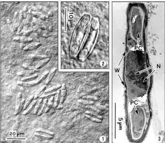
Figs 1-3: light and ultrastructural micrographs of a fish-infecting myxosporean Myxidium volitans sp. nov. found in the gallbladders of the marine fish Dactylopterus volitans collected in the Atlantic Brazilian coast. 1: several free spores observed by differential inter-ference contrast microscopy (DIC); 2: DIC image of two free spores, showing the wall, two evident polar capsules (PC) located at each extremity of the spore and the binucleated sporoplasm cell; 3: longitu-dinal section of a spore observed with low magnification showing the spore wall (W), two PCs each at the extremity of the spore and two nuclei (N) of the sporoplasm.
following Lom and Dyková (2006), we propose the es-tablishment of a new species classified as follows: Phy-lum Myxozoa Grassé, 1970, Class Myxosporea Bütschli, 1881, Order Bivalvulida Shulman, 1959, Family Myxidii-dae Thélohan, 1892, Genus Myxidium Bütschli, 1882.
Myxidium volitans sp. nov. (Figs 1-13)
Description - Developmental stages, including im-mature and im-mature spores, with morphological charac-ters of the genus Myxidium Bütschli, 1882 were observed free and immersed in the bile and in the epithelial cells of the gallbladder wall. The development was asynchro-nous with all the developmental stages and immature spores inter cellular in the epithelial cells of the gall-bladder wall (Figs 4-7). Some free mature spores were observed in the initial tract of the intestine. The spore bodies are fusiform, sometime slightly crescent-shaped with smooth surface and more or less rounded ends (Figs 1-3). The spore wall, is thin and smooth, comprised two equal-sized valves without ridges or projections (Figs 1-3). Sutural lines joining valves were hardly visible. Mature fresh spores have the following dimensions: 21.7 ± 0.3 µm (range 21.3-22.0) (n = 50) in length and 5.6 ± 0.4 µm (5.2-5.9) (n = 30) in width and contain two pyri-form polar capsules (PC) that are 5.0 ± 0.4 (4.6-5.5) µm long and 2.3 ± 0.3 (2.0-2.5) µm (n = 30) wide, these are situated in each extremity of the spore (Figs 1-3). The PC wall measured 0.20-0.29 µm (n = 30) thick (Figs 7-11). The PC wall consists of a continuous external dense layer and an internal hyaline thick material which contains an apical pore without a visible stopper (Figs 7-9). The PC contains a polar filament (PF) with irregular
559 Mem Inst Oswaldo Cruz, Rio de Janeiro, Vol. 106(5), August 2011
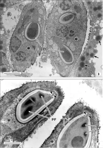
Figs 6, 7: ultrastructural aspects of the spores of Myxidium volitans sp. nov. infecting gallbladders of the marine fish Dactylopterus volitans
collected in the Atlantic Brazilian coast. 6: two immature spores (*) ob-served in the peripheral epithelial cell wall of the gallbladder showing the polar capsules (PC) and the sporoplasm nuclei (N). Several dense droplet structures possibly the bile component (arrows) are located near the spores; 7: extremities of two spores (*) showing the PC wall (W), the longitudinal section of the PCs, some polar filament sections (PF) projected across the PC wall (arrowheads) and the apical pole of the PC without evident stopper covering the pore (2 arrowheads).
Figs 8-11: ultrastructural aspects of different sections of the polar capsules (PC) and polar filaments (PF) obtained at different levels of
Myxidium volitans sp. nov. spores infecting gallbladders of the marine fish Dactylopterus volitans from the Atlantic Brazilian coast. In fa-vourable sections was possible to observe the longitudinal striation (P) at the periphery of the PF (arrows) and some S-like structures (arrow-heads) located within the PC matrix. The PC wall (W) composed of two layers: the external one is granulo-fibrillar and dense, while the internal is composed of light hyaline material. Internally the matrix contains some transverse section of the PF. A dense granulo-fibrillar mass (*) appeared frequently in the matrix of the PC.
ments that is projected laterally from its apical region to the bases of PC and coiled from the bases to the tip of PC (Figs 7-11). Some regular and parallel longitudinal striations were observed at the periphery of the PF. At the matrix of the PC there are 1-2 S-like structures that are adherent to the periphery of the PF (Figs 8-11). Sche-matic drawings of the spore and PF based on ultrathin serial sections are shown in Figs 12, 13.
Type host - D. volitans (Linnaeus, 1758) (Teleostei: Dactilopteridae) (Brazilian common name coió).
Site of infection - Different developmental stages and immature spores in the epithelial cells of the gallbladder wall and later developmental stages, immature and ma-ture spores immersed in the bile.
Type locality - Atlantic coast near Niterói (22º58’S 43º00’W).
Type data and depository - A glass slide with several semithin sections of the mature spores and some other developmental stages of the hapantotypes was deposited in the Myxozoa Type Slide Collection at the Instituto Nacional de Pesquisa da Amazônia - INPA, Amazonas, Manaus, Brazil, under acquisition 004/11.
Prevalence of infection - Twenty two out 64 (40.7%) of D. volitans specimens [15/39 (38.5%) for females, 7/25 (28%) for males].
Pathology signs - The signs of the infection were mac-roscopically observed by a well evidenced gallbladder hypertrophy and by the appearance of a green-brownish colour of the infected gallbladders in contrast to the light green colour in the non-infected specimens. Some spores were observed in the initial tracts of the intestine.
M o rp h o lo g y o f M . v o lit a n s s p. n ov
. • C
arl os A ze ve do e t a l. 0 D IS CUS SI O N In th e p re se n t p ap e r w e u se th e lig h t m ic ro sc o p y a n d ltr a st ru ct u ra l d at a to d e sc ri b e th e sp or e s o f th e m y x o -or ea n is o lat ed fr om th e g al lb la d d e rs o f th e m a ri ne fi sh vo lit a n s w h ic h h av e w id el y g eo g ra p h ic d is tr ib u tio n th e A tla n tic O ce a n (E sc h me y e r & D em o st e r 1 9 9 0 ) . e p re se n t st u d y rev ea ls a st ru ct u ra l or g a n iz at io n a n d ila rit ie s to g e n u s M y xid iu m T h él o h a n , 18 9 2 (f a m ily y x id iid a e) (L om & D y k o v á 1 9 9 2 , 2 0 0 6 , M a cK e n z ie K al av at i 1 9 9 5 ). T h is g e n u s is ch a ra ct e ri ze d b y h av in g n g at ed sp or e s, th at a re sl ig h tly c re sc e n t-sh ap ed , w ith o P C in o p p o si te e n d s, d is ch a rg e te rm in al ly a n d a lo n -itu d in al c u rv e d s u tu ra l l ine ( L
om & D
y k o v á 2 0 0 6 ). C o n si d e ri n g th e se p a rat io n o f m a ri ne a n d fr e sh w at e r y x o sp or ea n sp e ci e s in to tw o m aj or b ra n ch e s (or cl a d e s) re su lts o f th e re ce n t m o le cu la r d at a u si n g sm al l s u b u n it . 1 2 : se m is ch e m at ic d ra w in g o f th e sp o re o f M y x id iu m v o lit a n s s p . v. p a ra sit e fr o m g a llb la d d e rs o f th e m a ri n e fi sh D a c ty lo p te ru s v o li-s c o lle c te d in th e A tla n tic B ra z ili a n c o a st sh o w in g th e m o rp h o lo g y th e d if fe re n t st ru c tu re s a n d th e re o f th e m a ri n e fi sh D . v o lit a n s m t h e B ra z ili a n c o a st . TABLE
Comparative measurements (in µm) and other characters of the spores of different species of Myxidium spp infecting the gallbladders in the fish collected from Atlantic Ocean
Myxidium spp Hosts (countries)
SpL (µm) SpW (µm) PCL (µm) PCW (µm) PFc (n) References
M. incurvatum Thélohan, 1892 Several species(France) 8-20 4.0-8.2 3.0-5.5 2.4-2.7 5-7 Lom and Dyková (2006)
M. sphaericum Thélohan, 1895 Belone belonea,b (England) 15-20 5-10 3-5 2.5-5.0 3-4 MacKenzie and Kavalati (1995)
M. bergensis Auerbach, 1910 Pollachius virensa,b (Norway) 17.5-20.0 6.2-7.5 5.4 - 5-6 MacKenzie and Kavalati (1995)
M. oviforme Parisi, 1912 Apogon imberbisa,b (Italy) 11.75-16.25 7.50-9.50 4.25-5.0 - 4-5 MacKenzie and Kavalati (1995)
M. gadi Georgévitch, 1916 Pollachius pollachius (France) 12-15 5.0-7.5 2.5-4.5 2.5-4.0 5-6 MacKenzie and Kavalati (1995)
M. gigantissimum Dubina & Isakov, 1976 Alepocephalus australisa(South Africa) 97.5 8.5 ~32 - 13-14 Dubina and Isakov (1976)
M. baueri Kovaleva & Gaevskaya, 1982 Merluccius australis (Falkland Islands)b 19-24 9-11 4.5-5.5 3-4 - Kovaleva and Gaevskaya (1982)
M. trachinorum Canning et al., 1999 Echiichthys vipera (England) 17.2 8.8 6.8 3.7 Up to 82c Canning et al. (1999)
M. finnmarchicum MacKenzie et al., 2010 Merlangius merlangus (Norway) 17.6-22.4 6.4-9.6 4.8-6.4 3.2-4.8 4-5 MacKenzie et al. (2010)
M. volitans sp. nov. Dactylopterus volitans (Brazil) 21.7
(21.5-22.0) 5.6 (5.3-5.8) 5.0 (4.7-5.3) 2.3 (2.0-2.5) 2-3 Rarely 4 Present study
a: type species; b: several species; c: extruded polar filament (mean in µm); PCL: polar capsule length; PCW: polar capsule width; PFc: polar capsule coils; SpL: spore length; SpW: spore width.
561 Mem Inst Oswaldo Cruz, Rio de Janeiro, Vol. 106(5), August 2011
DNA sequences (Kent et al. 2000, 2001, Fiala 2006), we consider for this discussion only Myxidium spp hosted in the marine Atlantic fishes. On the other hand, taking in consideration the role of tissues and organs prefer-ences in differentiating between closely related species for identification of species (Lom & Dyková 2006), we establish spore comparison only infecting fish gallblad-ders from hosts collected from Atlantic areas.
So, comparing the characteristics of M. volitans sp. nov. (morphology and dimensions of the spore and PCs, number of coils and arrangements of the PFs, and host species), none of these characters were simultaneously equals when compared with those reported for Myxi-dium spp infecting fish collected in the different geo-graphical areas of the Atlantic Ocean (Table). The spores of M. volitans sp. nov. are substantially smaller (21.7 ± 0.3 µm long) than M. gigantissimum (97.5 µm long) and larger than M. oviform spores (11.75-16.25 µm) and than M. trachinarum spores (17.2 µm).
All other spores of the different Myxidium spp re-ported in the Table have similar ranges and mean of di-mensions, however, with different PC arrangement and polar capsule coils (PFc) number.
While the spores of M. volitans sp. nov. have PF coiled 2-3 times, in all other species the PFc are coiled more times, excepted the PC of M. sphaericum that have a similar number of PFc (3-4). These two species have, however, different morphology of spores: M. volitans have fusiform slightly crescent-shaped with more or less round end, while M. sphaericum have fusiform spores with broad at the centre and a relatively blunt extremity in valvular view (MacKenzie & Kalavati 1995).
Moreover, the ultrastructural analyses showed the PC and the arrangement of the PC tapering from its tip to the bases and an irregularly fold from the bases to tip of the PF, and this structural organization is different of those of the previously described species. Additionally the presence of the longitudinal striation on the periphery of the PC and the presence of the some S-like structures in the PC matrix were never reported in the previously described species. And finally, the species here described differs from the previously reported Myxidium spp (Table) in terms of its host specificity (Lom & Dyková 1992).
The confusion in the literature regarding the validity and host specificity among some of these Myxidium spp recorded in the Table, have been reported some years ago. Considering the similar morphology of the spores of M. sphaericum and M. bergensis, it was suggested that two of these species may be conspecific (Noble 1957, Moser et al. 1989). Recently was referred and discussed by MacKenzie et al. (2010) the existence of a great con-fusion and doubt regarding the validity and host speci-ficities of M. incurvatum, M. phaericum, M. bergensis and M. gadi which need detailed investigation on the morphological and molecular data.
ACkNOwLEDGEMENTS
To the technical assistance of Joana and JoãoCarvalheiro.
REFERENCES
Canning EU, Curry A, Anderson CL, Okamura B 1999. Ultrastruc-ture of Myxidium trachinorum sp. nov. from the gallbladder of the lesser weever fish Echiichthys vipera.Parasitol Res 85: 910-919.
Dubina VP, Isakov LS 1976. New species of myxosporeans from gal-New species of myxosporeans from gal-bladder of deep-sea fish. Parazitologiia 10: 556-560.
Eschmeyer WN, Demoster LJ 1990. Dactylopteridae. In JC Quero, JC Hureau, C Karrer A Post, L Saldanha (eds.), Check-list of the fishes of the eastern tropical Atlantic (CLOFETA), vol. 2, JNICT, Lisboa, p. 690-691.
Fiala I 2006. The phylogeny of Myxosporea (Myxozoa) based on small subunit ribosomal RNA gene analysis. Int J Parasitol26: 1521-1534.
Gioia I, Cordeiro NS 1996. Brazilian myxosporidians’ check-list (Myxozoa). Acta Protozool 35:137-149.
Kalavati C, Longshow M, MacKenzie K 1996. Two species of myxo-zoan parasites (Myxosporea: Bivalvulida), including a new genus
Patagonotothen sima (Richardson, 1845) (Pisces: Teleostei) in the southwest Atlantic. Syst Parasitol 34: 67-70.
Kent ML, Andree KB, Bartholomew JL, El-Matbouli M, Desser SS, Devlin RH, Feist SW, Hedrick RP, Hoffmann RW, Khattra J, Hallet SL, Lester RJG, Longshow M, Palenzuela O, Siddall ME, Xiao C 2001. Recent advances in our knowledge of the Myxozoa.
J Eukaryot Microbiol 48:395-413.
Kent ML, Khattra L, Hedrick RP, Devlin RH 2000. Tetracapsula renicola n. sp. (Myxozoa: Saccosporidae); the PKX myxozoan - the cause of proliferative kidney disease of salmonid fishes. J Parasitol86: 103-111.
Kovaleva AA, Gaevskaya AV 1982. New species of myxosporeans from fishes of the Falkland-Patagonian region. Parazitologiia 16: 353-359.
Lom J, Dyková I 1992. Myxosporidia (Phylum Myxozoa). Protozoan parasites of fishes. Developments in aquaculture and fisheries science, vol. 26, Elsevier Science, Amsterdam, p. 159-235.
Lom J, Dyková I 2006. Myxozoan genera: definition and notes on taxonomy, life-cycle terminology and pathogenic species. Folia Parasitol 53: 1-36.
MacKenzie K, Collins C, Kalavati C, Hemmingsen W 2010. Myxi-dium finnmarchicum n. sp. (Myxosporea: Myxidiidae) from the gall bladder of whiting Merlangius merlangus (L.) (Pisces: Tel-eostei) in North Norway. Zootaxa2673: 56-64.
MacKenzie K, Kalavati C 1995. Species in the genus Myxidium Bütsch-li, 1882 (Myxosporea: Bivalvulida) parasitizing the gall bladders of gadid fish in the northeast Atlantic. J Nat Hist29: 851-863.
MacKenzie K, Kalavati C, Gaard M, Hemmingsen W 2005. Myxospo-rean gall bladder parasites of gadid fishes in the north Atlantic: their geographical distributions and an assessment of their economic im-portance in fisheries and mariculture. Fish Res 76: 454-465.
Moser M, Kent ML, Dennis D 1989. Gall bladder Myxosporea in coral reef fishes from Heron Island, Australia. Aust J Zool 37: 1-13.