Effects of bromocriptine on serum
prolactin levels, pituitary weight and
immunoreactive prolactin cells in
estradiol-treated ovariectomized rats:
an experimental model of
estrogen-dependent hyperprolactinemia
1Departamento de Fisiologia, 2Unidade de Endocrinologia Ginecológica, Hospital de
Clínicas de Porto Alegre, Universidade Federal do Rio Grande do Sul, and
3Departamento de Patologia, Fundação Faculdade Federal de Ciências Médicas de
Porto Alegre, 90050-170 Porto Alegre, RS, Brasil M.F. Ribeiro1,P.M. Spritzer1,2,
L.M. Barbosa-Coutinho3,
M.C. Oliveira3,
M.A. Pavanato1, I.S.B. Silva1
and F.M. Reis1,2
Abstract
The present study was designed to assess the effects of bromocriptine, a dopamine agonist, on pituitary wet weight, number of immunoreac-tive prolactin cells and serum prolactin concentrations in estradiol-treated rats. Ovariectomized Wistar rats were injected subcutaneously with sunflower oil vehicle or estradiol valerate (50 or 300 µg rat-1
week-1) for 2, 4 or 10 weeks. Bromocriptine (0.2 or 0.6 mg rat-1 day-1)
was injected daily during the last 5 or 12 days of estrogen treatment. Data were compared with those obtained for intact control rats. Administration of both doses of estrogen increased serum prolactin levels. No difference in the number of prolactin cells in rats treated with 50 µg estradiol valerate was observed compared to intact adult animals. In contrast, rats treated with 300 µg estradiol valerate showed a significant increase in the number of prolactin cells (P<0.05). Therefore, the increase in serum prolactin levels observed in rats treated with 50 µg estradiol valerate, in the absence of morphological changes in the pituitary cells, suggests a “functional” estrogen-in-duced hyperprolactinemia. Bromocriptine decreased prolactin levels in all estrogen-treated rats. The administration of this drug to rats previously treated with 300 µg estradiol valerate also resulted in a significant decrease in pituitary weight and number of prolactin cells when compared to the group treated with estradiol alone. The general antiprolactinemic and antiproliferative pituitary effects of bromocrip-tine treatment reported here validate the experimental model of estro-gen-induced hyperprolactinemic rats.
Correspondence
P.M. Spritzer
Departamento de Fisiologia Universidade Federal do Rio Grande do Sul Rua Sarmento Leite, 500 90050-170 Porto Alegre, RS Brasil
Fax: 55 (051) 226-7191
Presented at the XI Annual Meeting of the Federação de Sociedades de Biologia Experimental, Caxambu, MG, Brasil, August 21-24, 1996.
Research supported by FINEP (No. 66.91.0509.00) and CNPq (No. 520107/94-2).
Received April 19, 1996 Accepted November 4, 1996
Key words
•Prolactin levels
•Pituitary growth
•Immunoreactive prolactin cells
•Bromocriptine
Estrogen is known to have a stimulatory role in prolactin synthesis and release (1,2). This steroid also induces pituitary tumors, depending on the experimental model and dose (3,4). Dopamine, acting via its specific receptor in the anterior pituitary, tonically inhibits pituitary prolactin secretion and lactotroph proliferation (5-7). On the other hand, estradiol appears to be a potent antidopaminergic agent in vivo. (8)
Bromo-criptine (2-bromo-α-ergocryptine, a dopa-mine agonist) has been used to exadopa-mine the neuroendocrine mechanism of dopamine that controls prolactin secretion in vivo. In
addi-tion, dopamine agonist therapy for pituitary prolactinomas results in the reduction of prolactin secretion and tumor regression (9,10).
There are only a few animal models avail-able which are sensitive to dopamine ago-nists, such as the SMtTW tumor, a spontane-ous prolactin-secreting transplantable tumor (4), and some studies using estrogen-induced hyperprolactinemia and pituitary enlargement (3,11-15). These studies did not report which doses and duration of in vivo estrogen ad-ministration are required to promote prolac-tin hypersecretion without pituitary enlarge-ment or if bromocriptine effects on prolactin levels and lactotroph proliferation may vary with different schedules of estrogen treat-ment.
The present study was designed to assess the effects of bromocriptine on pituitary wet weight, number of prolactin cells and serum prolactin levels in ovariectomized animals subacutely or chronically stimulated with estrogen, in order to validate an experimen-tal model for the study of the interaction between estrogen and dopamine in control-ling prolactin secretion and lactotroph pro-liferation in vivo. Data were compared with
those obtained for intact or ovariectomized control groups.
Ninety-four female Wistar rats, 3 months old, were maintained under conditions of controlled light and temperature with free access to water and standard rat chow. Rats were bilaterally ovariectomized under light ether anesthesia except for nine intact fe-male rats which were used as a control group. All procedures used on the rats were per-formed according to the NIH Guide to the Care and Use of Laboratory Animals. The rats were injected subcutaneously with sun-flower oil vehicle or estradiol valerate (Berlimed-Schering), 50 or 300 µg rat-1 week-1
for 2, 4 or 10 weeks. Bromocriptine (Sandoz) was injected daily (0.2 or 0.6 mg rat-1 day-1)
during the last 5 or 12 days of estrogen treatment.
Twenty-four hours after the last hormone or vehicle injection, rats were decapitated, trunk blood samples were collected, and se-rum was harvested and stored at -20oC until
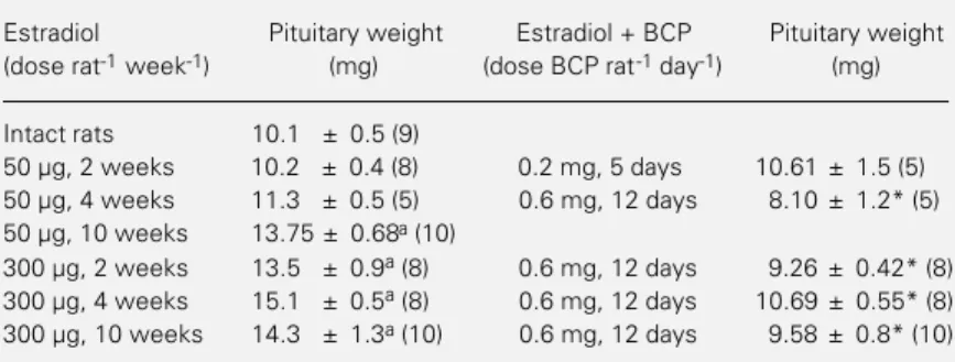
assayed for prolactin by a double-antibody radioimmunoassay. Pituitary glands were re-moved immediately after decapitation and wet weights were determined with an elec-tronic balance. Pituitary glands were placed in formalin and processed for prolactin im-munohistochemistry as previously described (13,16). Briefly, the antiserum used was pro-vided by the National Institute of Arthritis, Diabetes, and Digestive and Kidney Dis-eases (NIADDK). Rat prolactin anti-body produced in rabbits was used at 1:2000 dilution. After inhibiting endogenous per-oxidase with 1% methanol-H2O2, sections Table 1 - Effect of the administration of bromocriptine, a dopamine agonist, on pituitary
wet weights of intact or ovariectomized rats treated with estradiol valerate.
Results are reported as mean ± SEM. The number of animals is given in parentheses. BCP, Bromocriptine. aP<0.05 versusintact rats (Duncans test); *P<0.05versus estra-diol (t-test).
Estradiol Pituitary weight Estradiol + BCP Pituitary weight
(dose rat-1 week-1) (mg) (dose BCP rat-1 day-1) (mg)
Intact rats 10.1 ± 0.5 (9)
50 µg, 2 weeks 10.2 ± 0.4 (8) 0.2 mg, 5 days 10.61 ± 1.5 (5)
50 µg, 4 weeks 11.3 ± 0.5 (5) 0.6 mg, 12 days 8.10 ± 1.2* (5)
50 µg, 10 weeks 13.75 ± 0.68a (10)
were treated with normal goat serum for 30 min to reduce nonspecific binding. The pri-mary antiserum was applied for overnight incubation, treated with biotin anti-rabbit IgG for 30 min and finally incubated with the avidin-biotin peroxidase complex (Vector, Burlingame, CA) for 60 min. Diaminobenzi-dine (DAB) (Serva, Heidelberg) was used for 5 min as the chromogen. Controls con-sisted of 2 sections of normal rat pituitary. One section was processed exactly as done for the experimental sections (positive con-trol) and the other, in which the primary antibody was omitted, was used as the nega-tive control. Rinsing with phosphate buff-ered saline was performed after each step.
Two hundred nucleated cells were counted at X400, using a 1-mm2 grid in the
microscope eyepiece, and the number of prolactin-containing cells was recorded. Cells were independently counted by 3 observers. The data were tabulated, the average count for each anterior pituitary was calculated and the result was reported as percent of cells containing prolactin in relation to total cellularity.
Serum prolactin content was measured by a double-antibody radioimmunoassay us-ing materials kindly provided by the NIADDK. Prolactin was radioiodinated by the chloramine T method. NIADDK rat pro-lactin RP-3 was used as the standard. Assay sensitivity was 2 ng/ml and the intra- and interassay coefficients of variation were 8.6% and 12%, respectively.
Statistical analysis was performed by ei-ther the Student t-test or by analysis of
vari-ance, followed by Duncan’s multiple range test for the comparisons of multiple means. A P value <0.05 was considered to be statis-tically significant.
Table 1 shows the pituitary weights of intact and ovariectomized rats injected with estradiol valerate (50 or 300 µg/week) alone or in combination with bromocriptine. No difference in mean pituitary weight was ob-served in rats treated with estradiol (50 µg)
for 2 or 4 weeks compared to intact adult animals. Only rats treated for 10 weeks showed a significant increase in pituitary weight (P<0.05). In contrast, rats treated with 300 µg estradiol valerate showed a significant increase in pituitary weight as early as at 2 weeks of treatment (P<0.05). Bromocriptine, at the higher dose, signifi-cantly decreased the pituitary weights of all groups studied (P<0.05) when compared to the respective control rats treated with the same dose of estradiol valerate alone.
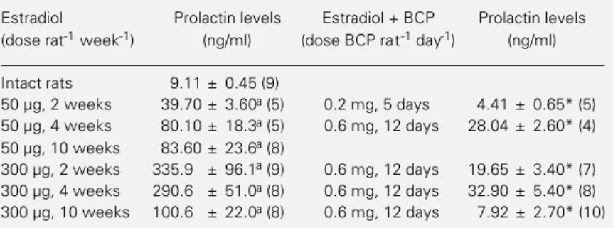
Table 2 illustrates the serum prolactin levels of intact rats and rats treated with estradiol alone or in combination with bromocriptine. Estrogen treatment (50 or 300 µg/week) for 2, 4 or 10 weeks significantly increased serum prolactin levels. The addi-tion of bromocriptine to the treatment with estradiol valerate resulted in a significant decrease in serum prolactin levels when com-pared to the respective control group treated with estrogen alone.
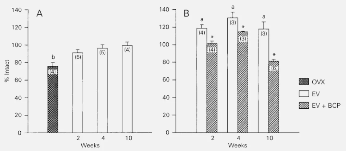
Figure 1A shows the percentage of im-munoreactive prolactin cells in ovariecto-mized rats subacutely or chronically treated with estradiol valerate (50 µg) or vehicle. When these groups were compared to intact control rats, only ovariectomized rats with-out hormonal treatment presented a signifi-cantly lower number of prolactin cells. No difference in the percentage of lactotrophs
Table 2 - Serum prolactin levels of intact or ovariectomized rats treated with estradiol valerate alone or in combination with bromocriptine.
Results are reported as mean ± SEM. The number of animals is given in parentheses. BCP, Bromocriptine. aP<0.05versus intact rats (Duncans test); *P<0.05 versus estradiol (t-test).
Estradiol Prolactin levels Estradiol + BCP Prolactin levels
(dose rat-1 week-1) (ng/ml) (dose BCP rat-1 day-1) (ng/ml)
Intact rats 9.11 ± 0.45 (9)
50 µg, 2 weeks 39.70 ± 3.60a (5) 0.2 mg, 5 days 4.41 ± 0.65* (5) 50 µg, 4 weeks 80.10 ± 18.3a (5) 0.6 mg, 12 days 28.04 ± 2.60* (4) 50 µg, 10 weeks 83.60 ± 23.6a (8)
was observed in rats treated with estradiol valerate (50 µg) for 2, 4 or 10 weeks. In contrast, all rats treated with 300 µg estradiol valerate for 2, 4 or 10 weeks presented a significant increase in the number of prolactin cells when compared to the intact control group (Figure 1B). As shown in Figure 1B, subse-quent administration of bromocriptine to the rats treated with the highest dose of estrogen resulted in a significant reduction in the percentage of immunoreactive prolactin cells. Estrogen administration to ovariecto-mized rats resulted in pituitary gland en-largement and in an increase in immunore-active prolactin cells only after long-term treatment with 50 µg/week or at the higher dose (300 µg), after all periods of time stud-ied. Therefore, the increase in serum prolac-tin levels before morphological changes are observed in rats subacutely treated with the dose of 50 µg estradiol valerate suggests a “functional” estrogen-induced hyperprolac-tinemia. This state of “functional hyperpro-lactinemia”, which is similar to human idio-pathic hyperprolactinemia (17), is a suitable model for the study of the neuroendocrine mechanisms of prolactin secretion, with the serum prolactin levels being taken as an example in the present study.
Thus, our findings obtained under differ-ent experimdiffer-ental conditions of estrogen ad-ministration to ovariectomized rats indicate two states of estrogen-dependent
hyperpro-lactinemia: a functional hyperprolactinemia and a hyperprolactinemia associated with pituitary enlargement and lactotroph prolif-eration.
The present data show that bromocriptine decreased serum prolactin levels under all experimental conditions, even when no pitu-itary enlargement was observed. Using in-creasing doses of bromocriptine in estrogen-treated rats, we have recently reported that the effect of bromocriptine in reducing the number of immunoreactive lactotrophs was observed only after long-term estrogen treat-ment including bromocriptine administra-tion for 12 days (16). No difference was observed when bromocriptine was adminis-tered for the last 5 days of short-term estro-gen treatment, suggesting that the duration of bromocriptine treatment required to de-tect a change in the number of lactotrophs by immunohistochemistry should be longer than 5 days (16). In the present study, we demon-strated a decline in lactotroph proliferation after only two weeks of estrogen treatment when bromocriptine was administered for 12 days. Our experiments do not address the question of the mechanisms underlying the different responses of prolactin cell prolif-eration to bromocriptine. However, it will be interesting to determine whether other sched-ules of concomitant administration of estro-gen and bromocriptine as well as the asso-ciation with other steroidal hormones have
% Intact
140
120
100
80
60
40
20
0
OVX
EV
EV + BCP
140
120
100
80
60
40
20
0
2 4 10 2 4 10
Weeks Weeks
A B
b
(4)
(5) (5)
(4)
(4)
(4) (3)
(3) (3)
(6)
* *
*
a a
a Figure 1 - Effects of estradiol
the same effects.
In conclusion, the present report describes a useful experimental model in which hy-potheses concerning the mechanisms of ac-tion of estradiol in the inducac-tion of prolactin hypersecretion, pituitary enlargement and lactotroph proliferation can be tested. The general antiprolactinemic and antiprolifera-tive pituitary effects of bromocriptine treat-ment reported here validate this model of estrogen-induced hyperprolactinemic rats. Further studies are needed to define the
ex-act role of dopamine and dopamine agonists at the molecular level in hyperprolactinemic states and their connection with steroidal hormones.
Acknowledgments
The authors would like to thank the staff of the Laboratory of Neuroendocrinology, Faculty of Medicine of Ribeirão Preto, Uni-versity of São Paulo, for providing goat anti-rabbit gammaglobulin.
References
1. Voogt JL, Chen CL & Meites J (1970). Serum and pituitary prolactin levels be-fore, during and after puberty in female rats. American Journal of Physiology, 218: 396-399.
2. Chen CL & Meites J (1970). Effects of estrogen and progesterone on serum and pituitary prolactin levels in ovariectomized rats. Endocrinology, 86: 503-505. 3. Lloyd HM, Meares JD & Jacobi J (1975).
Effects of oestrogen and bromocriptine
on in vivo secretion and mitosis in
prolac-tin cells. Nature, 255: 497-498.
4. Schussler N, Farnoud R, Rauch C, Roche M, Berthet M, Thomas F, Peillon F & Bayet MC (1994). Effect of the slow-re-lease formulation of somatuline (BIM 23014) on estrogen-induced hyperprolac-tinemia and lactotroph hyperplasia in the female rat. Neuropeptides, 26: 399-404. 5. Foord SM, Peters JR, Dieguez C, Scanlon
MF & Hall R (1983). Dopamine receptors on intact anterior pituitary cells in culture: functional association with the inhibition of prolactin and thyrotropin. Endocrinol-ogy, 112: 1567-1577.
6. Trouillas J, Chevallier P, Claustrat B, Hooghe-Peters E, Dubray C, Rousset B & Girod C (1994). Inhibitory effects of the dopamine agonists quinagolide (CV 205-502) and bromocriptine on prolactin se-cretion and growth of SMtTW pituitary tumours in the rat. Endocrinology, 134: 401-410.
7. Friedman E, Adams EF, Hoog A, Gejman PV, Carson E, Larsson C, De Marco L, Werner S, Fahlbush R & Nordenskjold M (1994). Normal structural dopamine type 2 receptor gene in prolactin-secreting and other pituitary tumors. Journal of Clinical
Endocrinology and Metabolism, 78:
568-574.
8. Ferland L, Labrie F, Euvrard C & Raynaud J-P (1979). Antidopaminergic activity of estrogens on prolactin release at the pitu-itary level in vivo. Molecular and Cellular
Endocrinology, 14: 199-204.
9. Chiodini PG, Liuzzi A, Cozzi R, Verde G, Oppizzi G, Dallabonzana D, Spelta B, Silvestrini F, Borghi G, Luccarelli G, Rainer E & Horowski R (1981). Size reduction of macroprolactinomas by bromocriptine or lisuride treatment. Journal of Clinical
En-docrinology and Metabolism, 53: 737-743.
10. Tindall GT, Kovacs K, Horvath E & Thorner MO (1982). Human prolactin-producing adenomas and bromocriptine: A histo-logic, immunocytochemical, ultrastruc-tural, and morphometric study. Journal of
Clinical Endocrinology and Metabolism,
55: 1178-1183.
11. Lyle SF, Wright K & Collins DC (1984). Comparative effects of tamoxifen and bromocriptine on prolactin and pituitary weight in estradiol-treated male rats. Can-cer, 53: 1473-1477.
12. Casanueva F, Cocchi D, Locatelli V, Flauto C, Zambotti F, Bestetti G, Rossi GL & Müller E (1982). Defective central nervous system dopaminergic function in rats with estrogen-induced pituitary tumours, as as-sessed by plasma prolactin concentra-tions. Endocrinology, 110: 590-599.
13. Oliveira MC, Spritzer PM, Poy M, Coronel AX, Dahlem N, Moraes JT & Barbosa -Coutinho LM (1993). Progestin effects on prolactin secretion and on immunoreac-tive prolactin cells in estradiol-treated ovariectomized rats. Hormone and
Meta-bolic Research, 25: 600-602.
14. Phelps CJ & Himer WC (1988). Effects of bromocriptine on prolactin cellular hyper-trophy, proliferation and secretory activity in diethylstilbestrol-induced pituitary tu-mors. Molecular and Cellular Endocrinol-ogy, 58: 137-148.
15. McComb DJ, Hellmann P, Thorner MO, Scott D, Evans WS & Kovacs K (1986). Morphologic effects of bromocriptine on spontaneously occurring pituitary prolac-tin-cell hyperplasia in old Long-Evans rats.
American Journal of Pathology, 122: 7-16.
16. Spritzer PM, Ribeiro MF, Oliveira MC, Barbosa-Coutinho LM, Silva ISB, Dahlem N, Cericatto R & Pavanato MA (1996). Effects of tamoxifen on prolactin levels, pituitary immunoreactive prolactin cells and uterine growth in estradiol-treated ovariectomized rats. Hormone and
Meta-bolic Research, 28: 171-176.