Cop
yright
© ABE&M t
odos os dir
eit
os r
eser
vados
.
Changes in prolactin secretion
in the short- and long-term
after adrenalectomy
Efeito da evolução temporal da adrenalectomia na secreção de prolactina
Maria Thereza C. Laguna-Abreu1, Carla Germano2, Ayrton C. Moreira2, José Antunes-Rodrigues3, Lucila Elias3, Margaret Castro2
ABSTRACT
Objective: To evaluate the modulation of the hypothalamus-pituitary-adrenal axis (HPA) on
prolactin secretion in rats after adrenalectomy (ADX). Materials and methods: Plasma corti-costerone, ACTH, and prolactin concentrations were measured by radioimmunoassay in rats after bilateral ADX in the short- (3 hours and 1day) and long-term (3, 7, and 14 days). Results:
Animals that underwent ADX showed undetectable corticosterone levels and a triphasic ACTH response with a transient increase (3h), a decrease (1d), and further increase in the long-term after ADX. Sham animals showed a marked increase in corticosterone and ACTH levels three hours after surgery, with a decrease to basal levels thereafter. Plasma prolactin levels were not changed after ADX. Conclusion: There are different points of equilibrium in the HPA axis after the glucocorticoid negative feedback is removed. Prolactin plasma secretion is not altered in the short or long- term after ADX, suggesting that the peptidergic neurons essential for prolactin release are not activated after ADX. Arq Bras Endocrinol Metab. 2012;56(4):244-9
Keywords
Adrenalectomy; prolactin; corticosterone; adrenocorticotropic hormone
RESUMO
Objetivo: Avaliar a modulação do eixo hipotálamo-hipóise-adrenal (HHA) sobre a secreção de
prolactina após adrenalectomia (ADX). Materiais e métodos: Quantiicamos por RIE corticoste-rona, ACTH e prolactina plasmáticos em ratos após curtos (3 horas e 1 dia) e longos (3, 7 e 14 dias) períodos de ADX bilateral. Resultados: Animais ADX mostraram níveis indetectáveis de corticosterona. As concentrações plasmáticas de ACTH apresentaram resposta trifásica: aumento transitório (3h), diminuição (1d) e novo aumento após longos períodos de ADX. Animais Sham mostraram aumento de corticosterona/ACTH após três horas de cirurgia, diminuindo posterior-mente aos níveis basais. As concentrações plasmáticas de prolactina não se alteraram após ADX.
Conclusão: Existem diferentes pontos de equilíbrio do eixo HHA após a remoção da
retroali-mentação negativa exercida pelos glicocorticoides. A secreção de prolactina não se alterou após curtos/longos períodos de ADX, sugerindo que os neurônios peptidérgicos essenciais para a li-beração de prolactina não estão ativados durante os diferentes períodos de ADX. Arq Bras Endocrinol Metab. 2012;56(4):244-9
Descritores
Adrenalectomia; prolactina; corticosterona; hormônio adrenocorticotróico
1 Laboratory of Molecular Biology and Biopathology, Universidade de Uberaba (Uniube), Uberaba, MG, Brazil 2 Division of Endocrinology, Department of Internal Medicine, School of Medicine of Ribeirao Preto, Universidade de Sao Paulo (FMRP-USP), Ribeirao Preto, SP, Brazil 3 Department of Physiology, FMRP-USP, Ribeirao Preto, SP, Brazil
Correspondence to:
Margaret de Castro Av. Bandeirantes, 3900
14049-900 – Ribeirão Preto, SP, Brazil [email protected]
Received on Dec/5/2011 Accepted on Feb/6/2012
INTRODUCTION
P
rolactin is a polypeptide hormone synthesized and secreted from specialized cells of the anterior pitui tary gland, called lactotrophs. In humans, an alternativeCop
yright
© ABE&M t
odos os dir
eit
os r
eser
vados
.
of the immune system activity, and in the maintenance of the internal environment by regulation of osmotic balan ce in some species (1,2). More recently, circulat ing and locallyproduced PRL has also been implicated as risk factors for cancer development (3,4).
Although hypothalamic dopamine provides inhi bitory control over prolactin synthesis and secretion, other factors produced in the brain, pituitary gland, and peripheral organs have also been shown to inhibit or stimulate prolactin secretion. In addition, prolactin itself can act on the hypothalamus to regulate its own secretion by means of a feedback mechanism (1,2). Prolactinreleasing stimuli not only include nursing, but also light, noise, smells, and stress (2,5). Prolactin secretory response differs depending on the stress sti mulus, such as ether (6,7), restraint (6,812), thermal stress (13), hemorrhage (9), social conlict (14), and even academic stress in humans (15).
Prolactin immunoreactivity is found in numerous hypothalamic areas in a variety of mammals. In the rat hypothalamus, prolactin immunoreactivity is detectable in the dorsomedial, ventromedial, supraoptic, and pa raventricular (16,17) nuclei. Vasopressin and oxytocin are synthesized in the magnocellular neurons located mainly in the posterior division of the paraventricular and supraoptic nuclei, and may be involved in the re gulation of prolactin secretion (18,19). Previous stu dies have clearly indicated that disturbance in water and electrolyte regulation alters adenohypophysial prolactin secretion (20). We have previously studied the effect of acute extracellular volume expansion by intravenous injection of isotonic or hypertonic saline on the secre tion of prolactin, corticosterone, vasopressin, oxytocin, and atrial natriuretic peptide. We demonstrated that the increment in prolactin and oxytocin was blocked by the inhibition of the hypothalamuspituitaryadrenal (HPA) axis by dexamethasone (21). Therefore, pro lactin and oxytocin responses induced by isotonic or hypertonic extracellular volume expansion are likely to be modulated by the HPA axis.
HPA axis is regulated by circadian rhythm, stress ful stimuli, and glucocorticoid negative feedback. Hy pothalamicpituitary activity is dramatically enhanced after adrenalectomy (ADX), which causes changes in pituitary corticotrophs, and might play a role in prolac tin response after stress (2224). Therefore, in the pre sent study, we evaluated the modulation of HPA axis on plasma prolactin secretion in rats in the short and longterm after ADX.
MATERIALS AND METHODS
Animals and experimental design
Adult male Wistar rats weighing 200 g were housed in individual cages (30 x 19 x 13 cm), in a room provided with temperature (23 ± 1ºC) and light control (12h light – 12h dark cycle) with free access to pelleted food and tap water. All experimental protocols were performed according to the guidelines of the Ethics Committee for Animal Use of the School of Medicine of Ribeirao Preto, Universidade de Sao Paulo (protocol no. 056/2005). Bilateral ADX and Sham surgery were performed under ether anesthesia using the dorsal approach. ADX rats re ceived oral saline (0.9% NaCl) ad libitum. Animals were
studied in basal condition at different times (3h, and 1, 3, 7, and 14 days) after Sham surgery or ADX. Animals were killed by decapitation between 8:30 and 10:30 a.m. Blood was collected from the trunk into heparinized plastic tubes, and centrifuged at 4ºC. Plasma samples were frozen at 20ºC until ACTH, corticosterone (B), and prolactin (PRL) determinations.
Assays and methods
Plasma corticosterone and ACTH were determined by radioimmunoassay (RIA), after plasma extraction using ethanol or silicic acid, respectively (25). Plasma PRL was determined using the NIDDK RIA reagents, and expressed in terms of RP1 reference preparations (26). Mean assay sensitivity was 0.4 µg/dL for corticostero ne, 7.7 pg/mL for ACTH, and 0.4 ng/mL for PRL. The intra and interassay coeficients of variation (CV) were 4.8% and 6.7% for corticosterone, 6.3% and 14.0% for ACTH, and 11.7% and 5% for PRL. All samples were analyzed in duplicate.
Statistical analysis
Data are expressed as means ± SEM. Data were compa red using the MannWhitney test or analysis of variance (KruskalWallis test and Dunn’s posttest). Signiicance was set at p < 0.05.
RESULTS
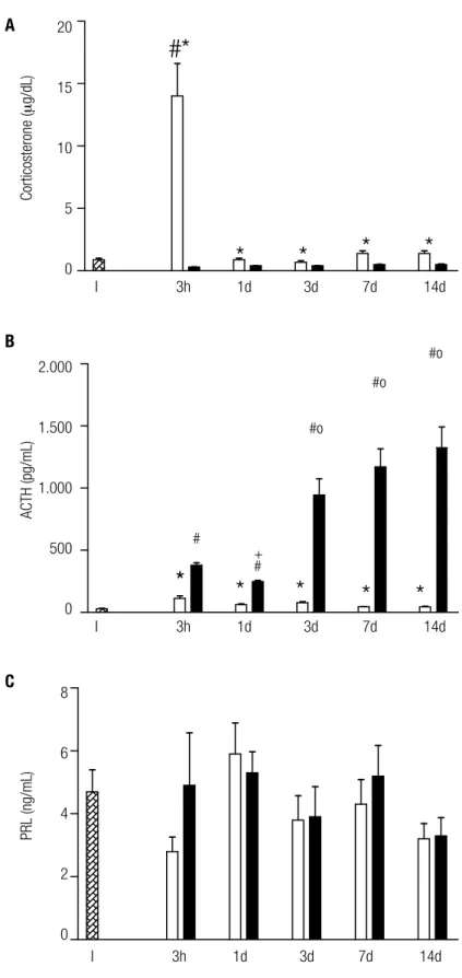
Corticosterone concentration
Cop
yright
© ABE&M t
odos os dir
eit
os r
eser
vados
.
ma corticosterone levels between 1, 3, 7, and 14 days (Figure 1A). Sham groups showed higher corticostero ne levels than ADX groups throughout the study (p < 0.004). ADX groups showed undetectable corticoste rone levels at all periods studied.
ACTH concentration
ACTH plasma levels after 3h, 1 and 3 days of Sham sur gery were similar, and all of them were higher than tho se observed after 7 and 14 days (p < 0.05; Figure 1B). Plasma ACTH showed a triphasic response to bilateral ADX, with a transient increase after 3h (p < 0.0001), followed by a decrease after 1 day (p < 0.0001), and a further increase after 3, 7, and 14 days (p < 0.001). Plas ma ACTH levels were higher in ADX groups compared with the Sham group in all periods studied (p < 0.008).
Prolactin (PRL) concentration
The Sham group did not show difference in PRL le vels after 3 hours (2.8 ± 0.9), 1d (5.9 ± 3.2 ng/mL), 3d (3.8 ± 1.7 ng/mL), 7d (4.3 ± 2.9 ng/mL), and 14d (3.2 ± 1.6 ng/mL). The same was also observed in plasma PRL levels in ADX rats after 3h (4.9 ± 4.1 ng/ mL), 1d (5.3 ± 2.3 ng/mL), 3d (3.9 ± 1.9 ng/mL), 7d (5.2 ± 3.5 ng/mL), and 14 days (3.3 ± 1.9 ng/mL) (Figure 1C). No difference was observed between ADX and Sham groups at the same moment, at all time points of the study.
DISCUSSION
The hypothalamic paraventricular nucleus (PVN) has been shown to be important for PRL secretion, and may play a role in the suppression of tuberoinfundi bular dopamine neurons (27,28). Furthermore, the presence of CRH receptors have been demonstrated in dopamine neurons, suggesting a possible role of CRH in the regulation of PRL secretion (29). Stress media tors – CRH and vasopressin – are essential not only for ACTH release in response to stress, but also in ACTH release after ADX. Therefore, we hypothesized that prolactin secretion could be affected in the short and longterm after ADX. In the present study, we obser ved no changes in plasma prolactin concentrations in the short and longterm after ADX. As expected, these animals showed undetectable corticosterone levels, and the classic ACTH triphasic response to the removal of glucocorticoid feedback.
ADX is an interesting experimental model, since it alters CRH/vasopressin expression in the PVN, indu ces hydroeletrolytic imbalance, and represents a stress model that may modulate PRL secretion. Our data de monstrated a triphasic response pattern of ACTH levels induced by ADX: at irst, there was a marked increase in ACTH release, leading to depletion of pituitary ACTH storage, which explains the decrease in circulating ACTH levels during the second phase. Finally, com pensatory changes occur, the most important being an increase in ACTH synthesis, as conirmed by increased pituitary ACTH content and proopiomelanocortin mRNA expression (19,24,25). These indings indicate a profound alteration in the HPA axis induced by ADX in the short and longterm.
Different stress models might determine the role of hypothalamic factors in ACTH and/or prolactin responses. Previous data have demonstrated increased serum PRL stimulated by ether stress, with maximum effect one minute after the onset of stress (30). In addi tion, reports have shown that ADX potentiates prolac tin release in response to ether stress (31); this incre ment usually occurs after 5 to 10 minutes, and remains signiicantly higher until 30 min after stress compared with controls (32). In the present study, prolactin levels did not differ between intact animals and those in the shortterm after ADX (3 hours). However, as mentio ned, physiological responses to ether stress are usually observed within few minutes (30,31). In this context, the lack of PRL response 3 hours after ADX may not relect PRL immediate response. In addition, we can not rule out that, in our model, PRL levels had already returned to basal levels. Previously, our group descri bed an increase in OT concentration 3 hours after ADX (19,24,25). It is important to point out that subcuta neous administration of oxytocin induces a reduction in basal, as well as in stressinduced prolactin secretion, in male rats (3335). Therefore, increased OT levels observed at this moment could also compensate the ex pected increase of prolactin plasma secretion after ether and surgical stress (18).
Cop
yright
© ABE&M t
odos os dir
eit
os r
eser
vados
.
# p < 0.01 (ADX vs. intact group), * p < 0.008 (Sham vs. ADX); + p < 0.0001 (ADX 1D vs. ADX 3H, 3D, 7D, and 14D); p < 0.0001 (ADX 3H vs. ADX 3D, 7D, and 14D).
Figure 1. Plasma corticosterone (A), ACTH (B), and prolactin (C) levels in intact animals (I; hatched boxes), 3 hours (h); 1, 3, 7, and 14 days (d); after sham surgery (empty boxes) or adrenalectomy (ADX; illed boxes). The numbers of animals in each group varied from 5 to 25.
20
15
10
5
0
2.000
1.500
1.000
500
0
8
6
4
2
0
I 3h 1d 3d 7d 14d
I 3h 1d 3d 7d 14d
I 3h 1d 3d 7d 14d
Corticosterone (
m
g/dL)
ACTH (pg/mL)
PRL (ng/mL)
#*
*
*
*
*
*
*
*
#
#o
#o
#o
+
*
*
*
A
B
C
Cop
yright
© ABE&M t
odos os dir
eit
os r
eser
vados
.
with severe dehydration in rats that underwent ADX after 3, 7, and 14 days (24). Moreover, chronic hypoos molality inhibits PRL secretion induced by the stress of restraint, indicating the involvement of magnocellular AVP neurons in these responses (36).
Although AVP induces prolactin release in vivo (37),
we did not observe changes in plasma AVP throughout the time course after ADX surgery (24), suggesting that this peptide might not have a role in prolactin se cretion in this experimental model. We and others have shown an increase in plasma PRL induced by increased water and salt ingestion or after hypertonic EVE infu sion (21,38). However, the increase in PRL and OT levels after isotonic or hypertonic EVE were blocked by glucocorticoid treatment. These results are similar to those of a previous report that showed that gluco corticoids exert an inhibitory effect on parvocellular OTexpressing neurons (39), PRL and OT secretion induced by volume and concentration changes.
Lesions in the PVN of rats strongly inhibited ether induced ACTH secretion. In contrast, PVN lesion failed to inhibit etherinduced PRL release, suggesting that the peptidergic neurons essential for stressinduced PRL release are modulated by neurons other than PVN neurons (7). In addition, prolactinreleasing peptide (PrRP), expressed in the nucleus tractus solitarii and in
the ventrolateral medulla oblongata, has been shown to induce prolactin secretion and plays an important role in the stress response (40).
In conclusion, there are different HPA axis points of equilibrium after the glucocorticoid negative feedback is removed. Although prolactin is a hormone responsive to different stress factors, it does not seem to be altered in the short or longterm after ADX, suggesting that the peptidergic neurons essential for basal prolactin release are not activated during time course after ADX.
Acknowledgments: the authors are grateful to Maria Valci dos San tos Silva and Marina Hollanda for their technical assistance. MTC LagunaAbreu was supported by a Fundação de Amparo à Pesqui sa do Estado de São Paulo (Fapesp) grant 02/060014.
Disclosure: no potential conlict of interest relevant to this article was reported.
REFERENCES
1. Freeman ME, Kanyicska B, Lerant A, Nagy G. Prolactin: struc-ture, function, and regulation of secretion. Physiol Rev. 2000;80(4):1523-631.
2. Ben-Jonathan N, LaPensee CR, LaPensee EW. What can we learn from
rodents about prolactin in humans? Endocr Rev. 2008;29(1):1-41.
3. Bernichtein S, Touraine P, Gofin V. New concepts in prolactin bio-logy. J Endocrinol. 2010;206(1):1-11.
4. Egli M, Leeners B, Kruger TH. Prolactin secretion patterns: basic mechanisms and clinical implications for reproduction. Repro-duction. 2010;140(5):643-54.
5. Ben-Jonathan N, Hnasko R. Dopamine as a prolactin (PRL)
inhibi-tor. Endocr Rev. 2001;22(6):724-63.
6. Banky Z, Nagy GM, Halasz B. Analysis of pituitary prolactin and adrenocortical response to ether, formalin or restraint in lactating rats: rise in corticosterone, but no increase in plasma prolactin le-vels after exposure to stress. Neuroendocrinology. 1994;59(1):63-71.
7. Makara GB, Kovacs KJ. Lesioning of the hypothalamic
paraven-tricular nucleus inhibits ether-induced ACTH but not prolactin re-lease. Neurobiology (Bp). 1997;5(3):403-11.
8. Gala RR, Haisenleder DJ. Restraint stress decreases afternoon plasma prolactin levels in female rats. Inluence of neural antago-nists and agoantago-nists on restraint-induced changes in plasma prolac-tin and corticosterone. Neuroendocrinology. 1986;43(2):115-23. 9. Jurcovicova J, Kvetnansky R, Dobrakovova M, Jezova D, Kiss A,
Makara GB. Prolactin response to immobilization stress and he-morrhage: the effect of hypothalamic deafferentations and poste-rior pituitary denervation. Endocrinology. 1990;126(5):2527-33. 10. Jaroenporn S, Nagaoka K, Kasahara C, Ohta R, Watanabe G, Taya
K. Physiological roles of prolactin in the adrenocortical response to acute restraint stress. Endocr J. 2007;54(5):703-11.
11. Ren L, Li X, Weng Q, Trisomboon H, Yamamoto T, Pan L, et al. Effects of acute restraint stress on sperm motility and secretion of pituitary, adrenocortical and gonadal hormones in adult male rats. J Vet Med Sci. 2010;72(11):1501-6.
12. Ramalho MJ, Reis LC, Antunes-Rodrigues J, Nonaka KO, De Cas-tro e Silva E. Reduced prolactin release during immobilization stress in thyrotoxic rats: role of the central serotoninergic system. Horm Metab Res. 1995;27(3):121-5.
13. Vaha-Eskeli K, Erkkola R, Irjala K, Viinamaki O. Effect of thermal stress on serum prolactin, cortisol and plasma arginine vasopres-sin concentration in the pregnant and non-pregnant state. Eur J Obstet Gynecol Reprod Biol. 1991;42(1):1-8.
14. Huhman KL, Mougey EH, Moore TO, Meyerhoff JL. Stressors, in-cluding social conlict, decrease plasma prolactin in male golden hamsters. Horm Behav. 1995;29(4):581-92.
15. Malarkey WB, Hall JC, Pearl DK, Kiecolt-Glaser JK, Glaser R. The inluence of academic stress and season on 24-hour concentra-tions of growth hormone and prolactin. J Clin Endocrinol Metab. 1991;73(5):1089-92.
16. Nishizuka M, Shivers BD, Leranth C, Pfaff DW. Ultrastructural characterization of prolactin-like immunoreactivity in rat medial basal hypothalamus. Neuroendocrinology. 1990;51(3):249-54. 17. Griffond B, Deray A, Jacquemard C, Fellmann D, Bugnon C.
Pro-lactin immunoreactive neurons of the rat lateral hypothalamus: immunocytochemical and ultrastructural studies. Brain Res. 1994;635(1-2):179-86.
18. Samson WK, Schell DA. Oxytocin and the anterior pituitary gland. Adv Exp Med Biol. 1995;395:355-64.
19. Laguna-Abreu MT, Margatho L, Germano CM, Antunes-Rodri-gues J, Elias LL, de Castro M. The effect of adrenalectomy on Fos expression in vasopressinergic and oxytocinergic neurons in res-ponse to stress in the rat. Stress. 2007;10(4):332-41.
20. Dohanics J, Smith MS, Blackburn RE, Verbalis JG. Osmotic inhibition of prolactin secretion in rats. J Neuroendocrinol. 1994;6(3):291-8. 21. Durlo FV, Castro M, Elias LL, Antunes-Rodrigues J. Interaction of
Cop
yright
© ABE&M t
odos os dir
eit
os r
eser
vados
.
23. Antoni FA. Hypothalamic control of adrenocorticotropin secre-tion: advances since the discovery of 41-residue corticotropin--releasing factor. Endocr Rev. 1986;7(4):351-78.
24. Laguna-Abreu MT, Koenigkam-Santos M, Colleta AM, Elias PC, Moreira AC, Antunes-Rodrigues J, et al. Time course of vasopres-sin and oxytocin secretion after stress in adrenalectomized rats. Horm Metab Res. 2005;37(2):84-8.
25. Castro M, Figueiredo F, Moreira AC. Time-course of hypothalamic CRH and pituitary ACTH contents, and pituitary responsiveness to CRH stimulation after bilateral adrenalectomy. Horm Metab Res. 1995;27(1):10-5.
26. Haanwinckel MA, Antunes-Rodrigues J, De Castro e Silva E. Role of central beta-adrenoceptors on stress-induced prolactin release in rats. Horm Metab Res. 1991;23(7):318-20.
27. Campbell RE, Grove KL, Smith MS. Distribution of corticotropin releasing hormone receptor immunoreactivity in the rat hypotha-lamus: coexpression in neuropeptide Y and dopamine neurons in the arcuate nucleus. Brain Res. 2003;973(2):223-32.
28. Grattan DR, Kokay IC. Prolactin: a pleiotropic neuroendocrine hormone. J Neuroendocrinol. 2008;20(6):752-63.
29. Wong ML, Licinio J, Pasternak KI, Gold PW. Localization of cor-ticotropin-releasing hormone (CRH) receptor mRNA in adult rat brain by in situ hybridization histochemistry. Endocrinology. 1994;135(5):2275-8.
30. el Feki A, Sakly M. Impairment of stress-induced secretion of prolactin during development: effects of adrenalectomy, TRH and sulpiride. J Physiol (Paris). 1988;83(1):36-42.
31. Harms PG, Langlier P, McCann SM. Modiication of stress-indu-ced prolactin release by dexamethasone or adrenalectomy. En-docrinology. 1975;96(2):475-8.
32. Deis RP, Leguizamon E, Jahn GA. Feedback regulation by proges-terone of stress-induced prolactin release in rats. J Endocrinol. 1989;120(1):37-43.
33. Lumpkin MD, Samson WK, McCann SM. Hypothalamic and pitui-tary sites of action of oxytocin to alter prolactin secretion in the rat. Endocrinology. 1983;112(5):1711-7.
34. Mormede P, Vincent JD, Kerdelhue B. Vasopressin and oxytocin reduce plasma prolactin levels of conscious rats in basal and stress conditions. Study of the characteristics of the receptor in-volved. Life Sci. 1986;39(19):1737-43.
35. Muir JL, Pister HP. Inluence of exogenously administered oxyto-cin on the corticosterone and prolactin response to psychological stress. Pharmacol Biochem Behav. 1988;29(4):699-703.
36. Kjaer A, Knigge U, Jorgensen H, Warberg J. Selective inhibition of magnocellular vasopressin neurons by hypoosmolality: effect on histamine- and stress-induced secretion of adrenocorticotropin and prolactin. Neuroendocrinology. 1998;67(5):330-5.
37. Pan JT, Mai LM. Dopamine antagonism does not potentiate the effects of oxytocin and vasopressin on prolactin secretion. Life Sci. 1990;47(26):2443-50.
38. Bliss DJ, Lote CJ. Prolactin release in response to infusion of iso-tonic or hyperiso-tonic saline. J Endocrinol. 1982;92(2):273-8. 39. Di S, Malcher-Lopes R, Halmos KC, Tasker JG.
Nongeno-mic glucocorticoid inhibition via endocannabinoid release in the hypothalamus: a fast feedback mechanism. J Neurosci. 2003;23(12):4850-7.