ENERGY TRANSDUCTION BY
RESPIRATORY COMPLEX I
Ana Paula Gonçalves Batista
Dissertation presented to obtain a PhD degree in Biochemistry at
the Instituto de Tecnologia Química e Biológica, Universidade
Nova de Lisboa
Supervisors
Prof. Miguel Teixeira & Dr. Manuela M. Pereira
Opponents
Prof. Ulrich Brandt & Prof. Carlos Salgueiro
Second Edition, September 2010
Prof. Miguel Teixeira, Dr. Ricardo O. Louro, Prof. Graça Soveral, Prof. Ulrich Brandt, Ana P. Batista, Dr. Manuela M. Pereira, Prof. Carlos Salgueiro, Prof. Carlos Romão.
Metalloproteins and Bioenergetic Unit Biological Energy Transduction Laboratory Instituto de Tecnologia Química e Biológica Av. da República
Estação Agronómica Nacional 2780-157 Oeiras
Portugal
This dissertation comprises research work performed under the su-pervision of Prof. Miguel Teixeira and Dr. Manuela M. Pereira, in the Metal-loproteins and Bioenergetics Unit from the Instituto de Tecnologia Química e Biológica, Universidade Nova de Lisboa.
The studies presented in this dissertation were to provide a contribu-tion to the understanding of the energy transducing mechanism of respiratory complex I.
The thesis is organized in three parts. The introduction part is divided in three sections describing the energy transduction process, the NADH:quinone oxidoreductases and the sodium/proton antiporters. The second part comprises the experimental results obtained during this work, and is divided in four chapters. Three of the chapters describe the structural and functional studies performed on Rhodothermus marinus complex I, whereas the other describes the studies performed on complexes I from Escherichia coli
I would like to thank all the people that supported and helped me during
these last years:
Prof. Miguel Teixeira and Dr. Manuela M. Pereira, my supervisors, for their
enormous support, enthusiasm, dedication, advices and scientific discussions.
A special thank to Manuela for her friendship, critical sense, and for giving
me strength and confidence during these years.
Dr. Ana Coelho and co-workers, especially Catarina Franco, for their advices,
help and discussions on the mass spectrometry studies.
Prof. Hartmut Michel, Dr. Guohong Peng and co-workers (especially Marco
Marcia), and Dr. Janet Vonck from the Max-Planck Institute for Biophysics,
Frankfurt am Main, for the structural studies of the Rhodothermus marinus
complex I.
Prof. Bernhard Brutschy and Lucie Sokova from the Institute of Physical and
Theoretical Chemistry, Johann Wolfgang Goethe-Universitat, Frankfurt am
Main, for the LILBID MS studies of Rhodothermus marinus complex I.
Dr. Andreia S. Fernandes, for helping and teaching me so much during the
period that we worked together and, especially for her friendship.
Dr. Ricardo O. Louro, for the helpful scientific discussions, innumerous
ad-vices and support.
ysis.
All my colleagues and friends at the Metalloproteins and Bioenergetics Unit,
for providing a stimulating and friendly environment: Célia Romão, João
Vi-cente, Filipa Sousa, Patrícia Refojo, Vera Gonçalves, Ana Filipa Pinto, Sandra
Santos, Lara Paulo, Ana Teresa Bernardo, Liliana Pinto, Carolina Júlio, Miguel
Ribeiro and Bruno Marreiros. And previous members of the group, especially
Smilja Todorovic, João Rodrigues, Andreia Veríssimo and Pedro Almeida.
Everyone in the groups of Dr. Cláudio Gomes, Dr. Lígia Saraiva, Dr. Ricardo
Louro and Dr. Inês Pereira for the helpful and friendly environment in the
“3rd floor” of ITQB.
Bárbara, Lígia, Patrícia, Raquel, Sofia and Vera, my friends with whom I
shared the good and the bad moments during these years. I am deeply
grate-ful to my “girls” for the massive encouragement, friendship and support.
Special thanks also to Jota, Sandra, Hugo and Gabriel for all the great
mo-ments, in and out of the lab.
All my friends outside the “ITQB world”.
My family, especially my parents, brothers and grandparents.
Miguel for his love, unconditional support, and for always being there.
Fundação para a Ciência e Tecnologia is acknowledgment for financial
sup-port, by awarding a PhD Grand SFRH/BD/25288/2005 and for funding
POCI/BIA-PRO/58374/2004, REDE/1517/RMN/2005, REEQ/336/BIO
The work presented in this dissertation is based on the following publications:
1. A.P. Batista*, A.S. Fernandes*, R.O. Louro, J. Steuber, M.M. Pereira, “Energy conservation by Rhodothermus marinus respiratory complex I”, Bio-chim Biophys Acta 1797 (2010) 509-515.
2. A.P. Batista, C. Franco, M. Mendes, A.V. Coelho, M.M. Pereira, “Subunit composition of Rhodothermus marinus respiratory complex I”, Analytical Bio-chemistry 407 (2010) 104-110.
3. A.P. Batista and M.M. Pereira, “Decoupling of the catalytic and transport activities of complex I from Rhodothermus marinus by a sodium/proton anti-porter inhibitor”, (2010), submitted.
4. A.P. Batista and M.M. Pereira, “Sodium influence on energy transduction by complexes I from Escherichia coli and Paracoccus denitrificans”, (2010), submit-ted.
Publications not included in this thesis:
5. A.P. Batista, A. Kletzin and M.M. Pereira, “The dihydrolipoamide dehy-drogenase from the crenarchaeon Acidianus ambivalens”, FEMS Microbiol Lett 281 (2008) 147-154.
The aim of the work presented in this dissertation was to provide a
contribution to the understanding of the energy transducing mechanism of
respiratory complex I. This enzyme is present in most bacteria and in all
mi-tochondrial systems and it is characterized by its large number of subunits, its
prosthetic groups (flavin and iron-sulfur centers), and its NADH:quinone
oxidoreductase activity sensitive to specific inhibitors and coupled with
charge translocation across the membrane.
Complex I from the halothermophilic bacterium Rhodothermus marinus
was the main model system used and its detailed structural and functional
characterization were performed. Electron microscopy studies, suggested that
the enzyme has the typical L-shaped structure. Further studies were
per-formed to clarify the possible presence (and function) of two non-canonical
subunits in R. marinus enzyme, since two additional genes were previously found among the canonical complex I genes, a gene encoding for a pterin-4α -carbinolamine dehydratase (PCD) and a gene encoding for a Nha-type
Na+/H+ antiporter, suggesting that their products could be part of the
com-plex. Using an original approach that combined several protein separation
strategies with different identification methods (ESI-MS/MS, MALDI-TOF
MS and Edman degradation analysis), the identification of the canonical
pro-teins and also of the PCD was possible. On the contrary, the Nha-type
Na+/H+ antiporter was not identified indicating its absence in the
composi-tion of complex I. Studies performed to determine the total mass of the
en-zyme, that included LILBID MS and native gel electrophoresis suggested an
identification of the PCD, a protein proposed to have an auxiliary role in the
metabolism of molybdopterin cofactors, as a novel complex I subunit may
suggest the existence of a possible regulatory mechanism of complex I
involv-ing the PCD and pterin like compounds.
The nature of the coupling charge of R. marinus complex I was inves-tigated using inside-out membrane vesicles, which were active with respect to
NADH oxidation and capable of creating and maintaining an NADH-driven
membrane potential (∆Ψ) positive inside. It was observed that this prokary-otic complex I is able of H+ and Na+ transport, although to opposite
direc-tions. The coupling ion of the system was shown to be the H+ being
trans-ported to the periplasm, contributing in this way to the establishment of the
electrochemical potential difference, while Na+ is translocated to the
cyto-plasm. Proton transport was monitored using pH dependent fluorescence
probes and Na+ transport was measured by 23Na-NMR spectroscopy. This
was the first time that such a technique was used to monitor substrate-driven
Na+ transport by membrane vesicles. Additional studies have shown that
al-though neither the catalytic reaction nor the establishment of the ∆pH re-quired the presence of Na+, the presence of this ion increased the proton
transport. Combining all these results, a model for the coupling mechanism
of complex I was proposed, suggesting the presence of two different energy
coupling sites, one that works only as a proton pump (Na+ independent), and
the other functioning as a Na+/H+ antiporter (Na+ dependent). This model
was further supported by studies performed in the presence of the Na+/H+
antiporter inhibitor, 5-(N-ethyl-N-isopropyl)-amiloride (EIPA). In these con-ditions both H+ and Na+ transports are inhibited but show different
exis-different inhibition behaviors in the presence and absence of Na , which also
reinforce the hypothesis for the presence of two H+ translocating sites. A half
maximal inhibition (IC50) value of ≈ 22 µM was estimated for the Na +
inde-pendent H+ translocating site and a IC50 value of ≈ 10 µM for the Na +
de-pendent H+ translocating site. More interestingly, the NADH:quinone
oxi-doreductase activity is also inhibited by EIPA (IC50= 230 µM); however the H+ and Na+ transports are inhibited at concentrations of EIPA at which the
catalytic activity is not affected, meaning that the catalytic and transports
ac-tivities can be decoupled. Strictly indirect coupling mechanisms, possible
through conformational changes, are thus suggested to be operating on
respi-ratory complex I.
A deeper insight into the coupling mechanism of this enzyme was
provided by studying the influence of sodium ions on energy transduction by
complexes I from Escherichia coli and Paracoccus denitrificans. Using the same approaches as before, it was observed that the Na+/H+ antiport activity is not
exclusive of Rhodothermus complex I, since the E. coli enzyme is also capable of such a transport, but is not a general property given that the P. denitrificans
enzyme does not perform sodium translocation. Due to the fact that R. mari-nus and E. coli enzymes reduce menaquinone while P. denitrificans complex I reduces ubiquinone, it was suggested that the Na+/H+ antiport activity may
be correlated with the type of quinone used as substrate. Although the full
mechanistic details remain to be clarified by atomic structures of the entire
complex I, the results here reported open new perspectives in the studies of
the process of energy transduction by this respiratory enzyme, which may be
O objectivo do trabalho descrito nesta dissertação foi contribuir para
a compreensão do mecanismo de transdução de energia do complexo I
respiratório. Este enzima está presente na maioria das bactérias e em todos os
sistemas mitocondriais e é caracterizado pelo seu grande número de
subunidades, os seus grupos prostéticos (flavina e centros de ferro-enxofre), e
pela actividade NADH: quinona oxidoredutase sensível a inibidores
específicos e acoplada à translocação de iões através da membrana.
O principal sistema modelo utilizado neste trabalho foi o complexo I
da bactéria termohalófila Rhodothermus marinus tendo-se efectuado uma caracterização estrutural e funcional detalhada do mesmo. Estudos de
microscopia eletrónica, sugerem que o enzima tem a estrutura típica em
forma de L. A determinação da composição em subunidades do complexo
teve como base esclarecer a eventual presença (e função) de duas subunidades
não canónicas no enzima de R. marinus, uma vez que dois genes adicionais foram previamente encontrados entre os genes canónicos do complexo I; um
deles codifica para uma pterina-4α-carbinolamina desidratase (PCD) e o outro codifica para um antiporte de Na+/H+ do tipo Nha, sugerindo que os
respectivos produtos possam fazer parte do complexo. A utilização de uma
abordagem original que combinou diferentes estratégias de separação de
proteínas com diferentes métodos de identificação (ESI-MS/MS,
MALDI-TOF MS e degradação de Edman), permitiu a identificação das subunidades
canónicas do complexo I e também do PCD. Contrariamente, o antiporte de
Na+/H+ do tipo Nha não foi identificado, sugerindo a sua ausência na
composição do complexo I. A ausência desta proteína foi reforçada pela
moleculares das quinze subunidades (as canónicas mais o PCD). A
identificacão do PCD, uma proteína sugerida como tendo um papel auxiliar
no metabolismo do cofactor molibdopterina, como uma nova subunidade do
complexo I sugere a existência de um possivel mecanismo de regulação do
complexo I envolvendo o PCD e compostos pterinicos.
A natureza da carga que está acoplada à actividade NADH: quinona
oxidoredutase, catalisada pelo complexo I de R. marinus, foi investigada utilizando vesiculas membranares invertidas, sendo estas activas em relação à
oxidação do NADH e capazes de criar e manter um potencial de membrana
(∆Ψ) positivo dentro. Foi demonstrado que este complexo procariota é capaz de transportar H+ e Na+, embora em direcções opostas. O ião de
acoplamento do sistema é o H+ sendo transportado para o periplasma,
contribuindo desta forma para o estabelecimento da diferença de potencial
electroquímico, enquanto o Na+ é translocado para o citoplasma. O
transporte protónico foi monotorizado utilizando-se sondas de fluorescência
dependentes de pH, enquanto o transporte de Na+ foi medido por
espectroscopia 23Na-RMN, sendo a primeira vez que esta técnica foi utilizada
para monitorizar o transporte de Na+ através de vesículas membranares.
Estudos adicionais mostraram que, embora nem a reacção catalítica, nem o
estabelecimento do ∆pH necessitem de Na+, a presença deste ião aumenta o transporte de protões. Combinando todos estes resultados, um modelo para o
mecanismo de acoplamento do complexo I foi proposto, sugerindo a
presença de dois locais diferentes de transdução de energia, um que funciona
apenas como uma bomba protónica (independente de Na+), e o outro
funcionando como um antiporte de Na+/H+ (dependente de Na+). Este
modelo foi suportado por estudos realizados na presença do inibidor de
antiportes de Na+/H+, 5-(
composto, mas apresentam diferentes perfis de inibição. Na presença de 10
µM de EIPA, diminuções de ≈ 30% e ≈ 50% foram observadas para o transporte de H+ e Na+, respectivamente, o que exclui a existência de apenas
um local de transporte comum. Além disso, o transporte de H+ apresenta
diferentes perfis de inibição na presença e na ausência de Na+, o que também
reforça a hipótese da presença de dois locais diferentes de transporte
protónico. O valor ao qual o transporte se encontra inibido em 50% (IC50),
foi estimado como sendo ≈ 22 µM para o local de transporte protónico independente de Na+, enquanto um valor de IC50 ≈ 10 µM foi estimado para o local de transporte protónico dependente de Na+. Mais interessante ainda é
o facto da actividade NADH:quinona oxidoredutase ser também inibida pelo
EIPA (IC50=230 µM), embora os transportes de H
+ e Na+ sejam inibidos a
concentrações de EIPA em que a actividade catalítica não está afectada. Estes
dados significam que as actividades catalítica e de transporte podem ser
desacopladas. Um mecanismo de acoplamento indirecto, possivelmente
através de alterações conformacionais, foi assim sugerido para o
funcionamento do complexo I respiratório.
Um maior conhecimento sobre o mecanismo de acoplamento deste
enzima foi obtido através do estudo da influência dos iões sódio na
transdução de energia dos complexos I de Escherichia coli e Paracoccus denitrificans. Seguindo a mesma linha de pensamento e aplicando as mesmas técnicas espectroscópicas, observou-se que a actividade de antiporte Na+/H+
não é exclusiva do complexo de Rhodothermus, uma vez que o enzima de E. coli
também é capaz de tal transporte; contudo, não é uma propriedade geral dado
substrato. Embora o mecanismo do complexo I continue por esclarecer, os
resultados aqui reportados abrem novas perspectivas no estudo do processo
de transdução de energia por este enzima respiratório, que poderá ser mais
versátil do que se poderia antecipar.
PART I - INTRODUCTION
CHAPTER 1 ENERGY TRANSDUCTION IN BIOLOGICAL
MEMBRANES
1.1 – ENERGY TRANSDUCTION: AN OVERVIEW
1.1.1 – Life and Energy 7
1.1.2 – Thermodynamics in Bioenergetics 8
1.1.3 – Energy transduction in biological membranes 9
1.1.3.1 – Thermodynamics of electron transfer 11
1.1.3.2 – Thermodynamics of ATP synthesis 13
1.1.3.3 – The Chemiosmotic Theory 14
1.1.3.4 – Ion/electron coupling mechanisms of respiratory complexes 19
1.1.4 – References 22
1.2 – NADH:QUINONE OXIDOREDUCTASES 1.2.1 – Introduction 27
1.2.2 – Complex I 1.2.2.1 – Structure and subunit composition 28
1.2.2.2 – Evolution 33
1.2.2.3 – Prosthetic groups 37
1.2.2.4 – Quinones 39
1.2.2.5 – Inhibitors 42
1.2.2.6 – Coupling ion: proton versus sodium 43
1.2.2.7 – Reaction mechanisms 44
1.2.2.8 – Rhodothermus marinus complex I 52
1.2.3 – Alternative NAD(P)H dehydrogenases 53
1.2.4 – Na+-translocating NADH:quinone oxidoreductase 54
1.3.2 – Multiple resistance and pH related antiporter 67
1.3.3 – References 71
PART II - RESULTS
CHAPTER 2 STRUCTURAL STUDIES OF Rhodothermus mari-nus COMPLEX I 2.1 – MALDI-TOF MS AND ESI MS/MS STUDIES OF R. marinus COMPLEX I 2.1.1 – Summary 792.1.2 – Introduction 79
2.1.3 – Material and Methods 80
2.1.4 – Results and Discussion 85
2.1.5 – Acknowledgments 94
2.1.6 – References 94
2.1.7 – Supplementary Material 97
2.2 – LILBID MS STUDIES OF R. marinus COMPLEX I 2.2.1 – Summary 107
2.2.2 – Introduction 107
2.2.3 – Material and Methods 108
2.2.4 – Results and Discussion 109
2.2.5 – Acknowledgments 114
2.2.6 – References 114
2.3 – CRYSTALLIZATION AND EM STUDIES OF R. marinus COMPLEX I 2.3.1 – Summary 119
2.3.5 – Conclusions 124
2.3.6 – Acknowledgments 124
2.3.7 – References 124
CHAPTER 3 ENERGY CONSERVATION BY Rhodothermus marinus COMPLEX I 3.1 – ENERGY CONSERVATION BY R. marinus COMPLEX I 3.1.1 – Summary 131
3.1.2 – Introduction 131
3.1.3 – Material and Methods 132
3.1.4 – Results 136
3.1.5 – Discussion 144
3.1.6 – Acknowledgments 147
3.1.7 – References 148
3.1.8 – Supplementary Material 150
3.2 – 23Na-NMR SPECTROSCOPY: DATA ANALYSIS 3.2.1 – Summary 155
3.2.2 – Introduction 155
3.2.3 – Material and Methods 156
3.2.4 – Results and Discussion 157
3.2.5 – Acknowledgments 163
3.2.6 – References 163
3.2.7 – Supplementary Material 165
4.4 – Results 174
4.5 – Discussion 182
4.6 – Acknowledgments 187
4.7 – References 187
4.8 – Supplementary Material 189
CHAPTER 5 ENERGY TRANSDUCTION BY COMPLEXES I FROM Escherichia coli AND Paracoccus denitrificans 5.1 – Summary 193
5.2 – Introduction 193
5.3 – Material and Methods 195
5.4 – Results 199
5.5 – Discussion 206
5.6 – Acknowledgments 210
5.7 – References 210
PART III- CONCLUSIONS
CHAPTER 6 FINAL DISCUSSION 6.1 – Energy transduction by respiratory complex I 2176.2 – Final remarks 225
PART I
Energy Transduction in
Biological Membranes
Abbreviations
A.: Aquifex; B.: Bacillus; E.: Escherichia; K.: Klebsiella; P.: Paracoccus; R.: Rhodothermus; T.:
Thermus; T.: Thermosynechococcus; V.: Vibrio; Y.: Yarrowia.
∆G: Gibbs energy change; ∆Gº: standard Gibbs energy change (kJ. mol-1); R: gas
constant (8.3 J mol-1 K-1); ∆E: reduction potential difference (mV); ∆E
m,7: standard
reduction potential difference at pH 7 (mV); E: reduction potential (mV); Em,7:
standard reduction potential at pH 7 (mV); F: Faraday constant (96 485 J V-1 mol-1);
n: number of electrons transferred; ∆µXm+
~ : ion electrochemical potential difference
(J.mol-1); ∆Ψ: membrane potential (mV); m: valence of an ion; pmf: proton-motive
force (mV); smf: sodium-motive force (mV).
bL heme: low potential heme b; bH heme: high potential heme b; cyt. c: cytochrome c;
complex I: NADH:quinone oxidoreductase type I; complex II: succinate:quinone oxidoreductase; complex III: quinol:cytochrome c oxidoreductase; complex IV: cytochrome c:oxygen oxidoreductase; Ech: energy-converting hydrogenase; EPR: electron paramagnetic resonance; HIPIP: high potential iron-sulfur protein; Mrp: multiple resistance and pH related antiporter; NDH-II: alternative NAD(P)H dehydrogenase; Na+-NQR: Na+ translocating NADH:quinone oxidoreductase;
Nqo/Nuo/Ndh/Fpo: complex I subunits; Nqr: Na+-NQR subunits; PCD:
pterin-4α-carbinolamine dehydratase; Q: quinone; QH2: quinol; SQNf: fast relaxing
semiquinone; SQNs: slow relaxing semiquinone; SQNx: very fast relaxing
semiquinone.
Chapter 1: Energy Transduction in Biological Membranes
1.1 – Energy Transduction: an overview 5
1.2 – NADH:quinone oxidoreductases 25
1.1
Chapter 1: Energy Transduction in Biological Membranes Section 1.1 – Energy transduction: an overview
1.1.1 – Life and Energy 7
1.1.2 – Thermodynamics in Bioenergetics 8
1.1.3 – Energy transduction in biological membranes 9
1.1.3.1 – Thermodynamics of electron transfer 11
1.1.3.2 – Thermodynamics of ATP synthesis 13
1.1.3.3 – The Chemiosmotic Theory 14
1.1.3.4 – Ion/electron coupling mechanisms of respiratory
complexes 19
1.1.1–LIFE AND ENERGY
Life depends on continuous flow of energy and on mechanisms that
control this energy flow. In biological systems the main form of chemical
energy is adenosine triphosphate (ATP). The synthesis of this nucleotide is
mainly associated with membrane-bound enzymes, the so-called electron
transfer or respiratory complexes, which are located in the cytoplasmatic
membrane of prokaryotic cells, in the inner membrane of mitochondria and
in the thylakoid membranes of chloroplasts [6]. The endergonic synthesis of
ATP is indirectly driven by the flow of electrons through the electron transfer
chain which is essentially conserved among the three domains of life, despite
the different nature of their primary energy sources (substrate oxidation,
absorption of light). The energy release by the electron transfer must be
transduced to a form that can be used for the ATP synthesis (Figure 1.1.1).
This section will address the biological mechanisms for energy
transduction and ATP synthesis. It starts by a brief description of the
thermodynamic principles of biological energy changes and ends with the
description of the coupling process of electron transfer and ATP synthesis -
the Chemiosmotic Theory – and the different ion/electron coupling
mechanisms of respiratory complexes.
1.1.2–THERMODYNAMICS IN BIOENERGETICS [6-8]
Biological energy changes obey the laws of thermodynamics, meaning
that energy is conserved and spontaneous changes occur in directions that
increase the overall disorder of the universe (system plus surrounding).
Gibbs energy indicates the spontaneity of a process and can be
described by the following expression:
) Reactants (
) Products (
ln º
Π Π +
∆ =
∆G G RT (eq.1)
through which the maximum energy that can be obtained from a process (the energy available to perform work) can be calculated. If ∆G is zero, the system is at equilibrium, which means that it cannot obtain energy from the process.
If ∆G<0, the process can release energy (exergonic process) as the system proceeds spontaneously to the equilibrium. If ∆G>0, the process is headed way from the equilibrium, and it will have to acquire energy (performed work)
in order to occur. The latter process is endergonic and is thermodynamically
unfavorable, however it will take place if associated to another process with a ∆G<<0, which will reverse the overall sign of ∆G. An example of such a process is the phosphorylation of glucose, a reaction from the glycolytic
pathway [9]. This reaction is unfavorable and does not occur spontaneously.
However, when coupled to ATP hydrolysis the combined reaction is
thermodynamically favorable (Figure 1.1.2).
To take advantage of the thermodynamic combination of favorable
and unfavorable processes, the system must have a mechanism of associating
the two reactions. In the particular case described above, the two reactions
share a common intermediate (Pi) and are catalyzed by the same enzyme
(hexokinase). It is described that after glucose binding, conformational
affinity to ATP, which will allow the transfer of phosphate from ATP directly
to glucose.
Besides this example, there are other coupling mechanisms in
biological systems such as: sequential reaction coupling, in which metabolites
works as a product in one reaction and as a reactant in other reaction (both
reactions catalyzed by a different enzyme); mechano-chemical coupling, as in
the case of muscle contraction; reduction-oxidation (redox) coupling, in
which electrons act effectively as common intermediates; ion gradient
coupling, where reactions are coupled through a shared gradient of molecules
across the membrane. The mechanisms describing the coupling of the
electron transfer and ATP synthesis will be further discussed below.
1.1.3–ENERGY TRANSDUCTION IN BIOLOGICAL MEMBRANES
The mitochondrial respiratory chain has NADH (or succinate) as the
initial electron donor and O2 as the final electron acceptor. Electron transfer
does not occur by a direct reaction between NADH (or succinate) and O2 but
Figure 1.1.2 – Example of an unfavorable reaction (R1) that takes place when associated with a more favorable one (R2). Reaction 1: ∆Gº=+13.8 kJ.mol-1;
Reaction 2: ∆Gº=-30.5 kJ.mol-1, Reaction 3: ∆Gº=-17.2 kJ.mol-1 (Adapted
rather by a stepwise flow of electrons through the electron transfer
(respiratory) complexes.
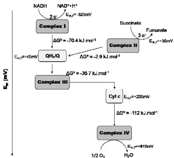
The canonical description of the aerobic respiratory chain considers
the existence of four transmembranar complexes: complex I (NADH:quinone
oxidoreductase); complex II (succinate:quinone oxidoreductase); complex III
(quinol:cytochrome c oxidoreductase) and complex IV (cytochrome c:oxygen
oxidoreductase) (Figure 1.1.3). Complexes I and II are the entry point for
electrons while complex IV is the terminal oxidase, since it is the last complex
involved in the electron transfer, reducing O2 to water. Electrons flow from
complexes I and II to complex III and from complex III to IV via electron
carriers, a hydrophobic quinone (Q) and a soluble cytochrome c (cyt. c),
respectively.
Prokaryotes may have branched respiratory chains with alternative
pathways [10]. The fact that many prokaryotes are facultative aerobes allows
them to reduce besides oxygen a large set of compounds, such as nitrate,
sulfate, fumarate [11]. NADH oxidation can be performed by two other
enzymes besides complex I, the so-called alternative NADH dehydrogenase
(NDH-II) and sodium-translocating NADH:quinone oxidoreductase (Na+
NQR) (see section 1.2 – NADH:quinone oxidoreductases) [12].
Quinol:oxygen oxidoreductases, which are directly reduced by quinol
bypassing complex III, define another variant electron pathway [11]. The
diversity of terminal oxygen reductases is also a clear example of the flexibility
of prokaryotic respiratory chains. Typical cases are Paracoccus (P.) denitrificans
and Rhodothermus (R.) marinus respiratory chains which possesses at least three
different oxygen reductases [13-16]. Several electron carriers can also
intervene in these respiratory chains. Apart from three different types of
quinones which differ in physical and functional proprieties, also the electron
carrier between complex III and IV can vary, being the usual cytochrome c, a
high potential iron-sulfur protein (HiPIP) [17] or small copper proteins (e.g.
halocyanin) [18].
It is possible to estimate the thermodynamic efficiency of electron
transfer but it is necessary first to take into account some considerations.
Biological systems are open systems (not at equilibrium) exchanging both
energy and matter with the surrounding, which require a non-equilibrium
thermodynamic treatment. However, considering individual reactions or
group of reactions as closed systems it is possible to apply equilibrium
thermodynamics. Thus, it is possible to estimate the efficiency of each of the
respiratory chain complexes individually, as well as, the efficiency of the
overall respiratory chain. To simplify, the process of energy transduction will
be described for a canonical aerobic respiratory chain.
1.1.3.1–Thermodynamics of electron transfer [1, 6, 7]
Respiratory chains operate as a sequence of oxidation-reduction
reactions in which electrons are transferred from one component (enzymatic
redox reaction is a function of the difference in reduction potential (∆E) between the oxidant and the reductant couples. In general,
E nF
G=− ∆
∆ (eq.2)
where n is the number of electrons transferred and F is the Faraday constant
(96 485 J.V-1.mol-1). As with Gibbs energy changes (eq.1), the ∆E obtained
under conditions other than the standard ones (∆Em,7
1, where all the
components are present at a concentration of 1M) depends on the
concentrations of the oxidized and reduced species. Considering eq.1 and eq.2, the actual ∆E for a redox reaction is given by,
[
]
[
Oxidized]
Reduced ln 7 , nF RT E
E =∆ m −
∆ (eq.3)
and each of the redox couples can be described as
[
]
[
Oxidized]
Reduced ln 7 , nF RT E
E = m − (eq.4)
Appling these equations, it is possible to estimate the efficiency of the
electron transfer process (Figure 1.1.4). The oxidation of NADH to NAD+,
the reaction with the lowest reduction potential in the chain, occurs at a
reduction potential of -320 mV, while the reduction of O2 to water has a
reduction potential of +815 mV, the highest value. The net reaction has a ∆Em,7 of ≈ 1,14 V, which corresponds to a ∆Gº of ≈ -219 kJ.mol
-1. If NADH
oxidation and O2 reduction occurred in a single step the amount of energy
released would be so large that the cell would no longer be viable. This
problem is overcome by a stepwise electron transfer process in which the
energy is released in a stepwise manner. As a consequence, in the respiratory
chain, the electron transfer is not simply determined by reduction potentials.
Specificity of interaction between the redox components must be present in
order to avoid short-circuiting reactions.
1.1.3.2 – Thermodynamics of ATP synthesis [6, 7]
The phosphorylation of ADP to ATP is catalyzed by the enzyme ATP
synthase and the energy associated with this reaction can be determined by
the following equation (based on eq.1)
[
]
[
][ ]
iATP ATP
P ADP
ATP RT
G
G =∆ º + ln
∆ (eq.5)
The ∆G0
ATP is ≈ +30.5 kJ.mol
-1. This means that the synthesis of ATP
is an unfavorable reaction and that energy has to be supplied for it to occur.
This energy is obtained from the electron transfer process, which is
thermodynamically very efficient (∆Gº≈ -219 kJ.mol-1). The energy released
by complexes I, III and IV reactions is sufficient to the synthesis of ATP
Figure 1.1.4 – Diagram of the respiratory complexes according to their subtract reduction potentials (in mV). For Em,7 values see reference [1]. ∆Gº are
molecules (Figure 1.1.4). On the contrary, the redox reaction performed by
complex II does not release enough energy and it functions only to inject
electrons into the respiratory chain.
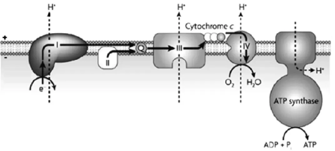
1.1.3.3 – Electron transfer and ATP synthesis coupling mechanism – The Chemiosmotic Theory
Thermodynamics describes energetically how respiratory electron
transfer reactions can allow the synthesis of ATP but does not explain how
the exergonic and endergonic processes are coupled. Peter Mitchell suggested
an indirect mechanism that couples the two processes – the chemiosmotic
theory [19]. This hypothesis postulates that the electron transfer and
phosphorylation are not chemically linked; instead they are coupled by a
transmembrane current of ions. Since the membrane has a low intrinsic
conductance for ions, a difference of ion concentration and of electric
potential is able to develop between the two sides of the membrane. The
energy released by the electron transfer is thus transduced into and stored as
the electrochemical potential of ions. Dissipation of this potential releases the
stored energy, which in turn is used, by ATP synthase, to drive the
phosphorylation of ATP. It was shown that ions move back across the
membrane through specific ion channels in ATP synthase (Figure 1.1.5).
In developing and improving his theory, Mitchell has suggested that
the electrochemical potential is also needed to power other membrane
processes such as flagellum rotation and/or solute transports [3].
The basic principles of the chemiosmotic theory are accepted
nowadays, although the model proposed by Mitchell was to some extent
different from the one considered today. Some of the main differences are
related with: 1. the ion/electron coupling mechanisms by respiratory
mechanism (see below part B. Electrochemical potential as the driving force
for ATP synthesis).
A. Thermodynamics of ion electrochemical potential [6, 7]
The energy available in an ion electrochemical potential can be
determined taking into account the two forces acting on it: the ion
concentration difference and the electrical potential difference between the two sides of the membrane (the membrane potential, ∆Ψ). The ∆G involved
in the transport of a cation (X) of valence m from compartment A to
compartment B, against both a concentration gradient (CB>CA) and a
difference of electrical potential (compartment B is electropositive relative to
compartment A) can be described by,
[ ]
[ ]
+ ∆Ψ= ∆ =
∆ + ++ mF
X X RT G
A m
B m
Xm ln
~
µ (eq.6)
where ∆ m+
X
µ~ is the ion electrochemical potential difference and is
numerically equal to ∆G. It is usual to express eq.6 in units of electric potential, volts or millivolts (instead of J.mol-1):
[ ]
[ ]
+ ∆Ψ= ∆ = ∆ + + + m X X F RT F G
F m A
B m
Xm ln
~
µ
(eq.7)
At equilibrium (∆G=0), equation 6 can be rearranged, relating the equilibrium distribution of an ion to the membrane potential:
[ ]
[ ]
Am B m X X mF RT + + − =
∆Ψ ln (eq.8)
Protons and sodium ions are coupling ions in membrane associated
bioenergetics processes. From a thermodynamic point of view the proton
electrochemical potential difference and the sodium electrochemical potential
difference are equivalent, and composed of both a chemical and an electrical
component. Based on eq.6 the proton electrochemical potential difference is
described by
[ ]
[ ]
+ ∆Ψ= ∆ =
∆ + ++ F
H H RT G A B H ln ~
µ (eq.9)
Considering pH=- log10[H +] ∆Ψ + ∆ − = ∆ =
∆ + G RT pH F
H 2.3
~
µ (eq.10)
In units of electrical potential, the proton electrochemical potential
difference is designated as proton-motive force (pmf) and eq. 10 can be
rearranged as ∆Ψ + ∆ − = ∆ = ∆ = + pH F RT F G F
pmf H 2.3
~
µ
(eq.11)
For an analogous electrochemical difference involving the
[ ]
[ ]
+ ∆Ψ= ∆ =
∆ + ++ F
Na Na RT G A B Na ln ~
µ (eq.12)
and sodium-motive force (smf)
∆Ψ + ∆ − = ∆ = ∆ = + pNa F RT F G F
smf Na 2.3
~
µ
(eq.13)
Most of the known biological systems are proton-motive systems,
however some microorganisms (such as thermophilic anaerobes, marine
bacteria and some bacterial pathogens) use sodium as a coupling ion in
addition to proton or even instead of it [20]. In the latter type of systems
(sodium dependent), sodium-specific versions of proton-translocating
enzymes are present in addition to sodium-translocating pumps which are
absent in proton-motive systems. Examples of these pumps are:
sodium-translocating decarboxylases; sodium-sodium-translocating NADH:quinone
oxidoreductase and sodium-translocating ferredoxin:NAD+ oxidoreductase
[21].
The use of the proton or the sodium as the coupling ion is suggested
to be a consequence of the adaptation of the organism to a changing
environment [20, 22]. Inter-conversion of the different ion motive forces is
achieved by secondary transport mechanisms, such as sodium/proton
antiporters.
B. Electrochemical potential difference as the driving force for ATP synthesis [6]
ATP synthases can be divided into two main types: F-type (F0F1-ATP
synthase) and V-type (vacuolar ATP synthase). F-type comprises F0F1-ATP
synthase of bacteria, chloroplasts and mitochondria and is composed of two
functional units F0 and F1 (Figure 1.1.6). F0 is the transmembranar ion
organism. For example, Escherichia (E.) coli
F0 contains a large subunit a, two copies of
subunits b and probably 12 copies of a
smaller subunit c. F1 is the peripheral part of
the complex, composed of five different subunits (α3, β3, γ, δ, ε) that are easily
dissociated from F0. The subunits α and β
are arranged alternately around the γ subunit. The enzyme has three catalytic sites located at the interface of α and β subunits [23].
The way how ATP synthase uses the
electrochemical potential to synthesize ATP
has been the subject of enormous
discussions over the past years. Paul D.
Boyer was one of the most enthusiastic researchers in this field and his work
helped to open new perspectives concerning biological coupling mechanisms.
After his suggestion that “the energy from oxidations was not used to make
the ATP molecule, but instead was used to bring about the release of the
tightly bound ATP” [24], the possibility of indirect coupling mechanisms
operating in biological systems was put forward.
During ATP synthesis the three catalytic sites act in sequence,
changing conformation and affinity towards substrates and products. First
ADP and Pi bind, then a conformational change takes place and a tightly
bound ATP is produced; finally an additional conformational change occurs
to release this ATP. The conformational changes are accomplished by the
mechanical process of rotation of the F0 portion of the enzyme, which is
driven by the ion (proton or sodium) movement across the membrane (from
Figure 1.1.6 - Schematic representation of the structure of the ATP synthase. It has a membranar part (F0) and a peripheral part
(F1). The ion channel is
located at the F0 and the three
catalytic sites at the F1
the positive side to the negative side) by dissipation of the electrochemical
potential (Figure 1.1.6) [23]. ATP synthase may also operates in reverse,
acting as an ion pump, thereby using the energy derived from the hydrolysis
of ATP to establish an ion concentration difference.
The coupled rotation between the F1 and F0 parts was proven by the
connection of a fluorescent-labeled actin filament to the F0 part (c subunit),
which was able to rotate by using the energy of ATP hydrolysis [25]. This
experiment demonstrated that the mechanical rotation is an essential feature
for the energy coupling between ion transport through the F0 sector and ATP
hydrolysis and synthesis at the F1 sector.
The coupling mechanism of the ion transport and the rotation of the
c-subunits is not completely known. Ion specificity (proton versus sodium) is
determined by the structure of the ion-binding site; sodium binding usually
requires up to six ligands while protons just need, in principle, a single
ionisable group [26]. This may explain the observation that in the absence of
sodium, a sodium ATP synthase can translocate protons, while a proton ATP
synthase is apparently unable of sodium translocation [26].
1.1.3.4 – Ion/electron coupling mechanisms of respiratory complexes
The coupling mechanisms of the three respiratory complexes involved
in energy transduction (complexes I, III and IV) are not completely known.
However, it is recognized that they have different mechanisms of action.
In the original formulation of the chemiosmotic theory, Mitchell
envisaged oxidation chains as vectorial pathways so arranged that at each
respiratory complex an electron/ion carrier is reduced at the membrane
surface. It then diffuses to the other side of the membrane surface, where
membrane by an electron carrier while protons are released. This was known
as Mitchell´s redox loops and shows the direct coupling between a redox
reaction and ion concentration differences across the membrane (Figure
1.1.7-A) [3].
Based on the redox loop mechanism, Mitchell formulated a first
proposal for the coupling mechanism that operates on canonical complex III,
the Q-cycle [27-29]. Taking into account the X-ray structure of complex III,
together with physical and chemical characterization of its cofactors, this
mechanism was later re-formulated giving origin to the mechanism
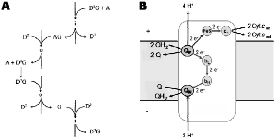
recognized nowadays (Figure 1.1.7-B).
Complex III (that comprises a 2Fe-2S center, two b-type hemes, one
c-type heme and two quinone binding sites) catalyses the oxidation of two
quinol molecules (QH2) with the reduction of two soluble cytochromes c and
one quinone (Q). This redox reaction is directly coupled to proton uptake
from the negative side of the membrane (N-side) and to proton release in the
positive side of the membrane (P-side) (Figure 1.1.7-B). Briefly, a molecule of
Figure 1.1.7- Scheme of the ion/electron coupling mechanism operating in complex III. A) Mitchell´s redox loop, where i and o represent the two sides of the membrane and A, G, AG and D1,2,3 represent a proton, an electron, a
QH2 diffuses to the binding site Qp, close to the P-side, and is oxidized to
quinone. The two protons from the quinol molecule are released to the
P-side. One of the QH2 electrons is transferred to the c-type heme (c1), via
2Fe-2S center (FeS) and then to the soluble electron carrier cytochrome c (Cyt. c).
The other electron is transferred to the bL heme (low potential heme b),
passes to the bH heme (high potential heme b) and reduces a quinone, located close to the N-side, to a semiquinone species. When a second molecule of
QH2 is oxidized at the Qp site, two protons are again released to the P-side
and one of the electrons follows the first pathway reaching the Cyt. c. The
second electron is transferred again through the b-type hemes to the
semiquinone species which is reduced to quinol, with the two protons
required for this being taken up from the N-side [30].
Mitchell had proposed a similar
type of mechanism for complex IV, the
redox zoop, which is an extension of the
redox loop by the addition of a second
electron-conducting arm. In this
respiratory complex the proton carrier
would be oxygen (or oxygen species)
instead of quinone [3]. Although the
proton/electron coupling mechanism of
complex IV remains unsolved, a redox
zoop mechanism can be excluded since it
was determined that for each full
catalytic cycle, four electrons are
transferred and up to eight protons are
taken up from the negative side of the
membrane, four of which are used in
Figure 1.1.8 – Scheme of the overall function of O2
substrate reduction and up to additional four are transported across the
membrane (Figure 1.1.8) [4]. Proton transport excludes the operation of a
redox loop mechanism in complex IV. In alternative it is suggested that this
respiratory complex works as a redox-driven proton pump.
As in the case of complex IV, the ion/electron coupling mechanism
of complex I is not known. Different types of mechanisms, based on the
redox loops and on the redox-driven proton pumps, have been proposed and
will be revised in the next section (see section 1.2-NADH:quinone
oxidoreductases).
1.1.4 – REFERENCES
[1] D. Voet, J.G. Voet, Biochemistry, Second ed., John Wiley and Sons, INC, New York, 1995.
[2] D.L. Nelson, M.M. Cox, Lehninger Principles of Biochemistry, Third ed., Worth Publishers, New York, 2000.
[3] P. Mitchell, Foundations of vectorial metabolism and osmochemistry, Biosci Rep 11 (1991) 297-344; discussion 345-296.
[4] M. Wikstrom, Cytochrome c oxidase: 25 years of the elusive proton pump, Biochim Biophys Acta 1655 (2004) 241-247.
[5] H. Wang, G. Oster, Energy transduction in the F1 motor of ATP synthase,
Nature 396 (1998) 279-282.
[6] D.G. Nicholls, S.J. Ferguson, Bioenergetics 3, Academic Press, London, 2002.
[7] F.M. Harold, The Vital Force: A study of bioenergetics, W.H.Freeman and Company, New York, 1986.
[8] J. Wrigglesworth, Energy and Life, Taylor and Francis, London, 1997. [9] G.L. Zubay, Biochemistry, Fourth ed., Wm. C. Brown Publishers, Dubuque,
1998.
[10] S. Berry, Endosymbiosis and the design of eukaryotic electron transport, Biochim Biophys Acta 1606 (2003) 57-72.
[11] G. Unden, J. Bongaerts, Alternative respiratory pathways of Escherichia coli: energetics and transcriptional regulation in response to electron acceptors, Biochim Biophys Acta 1320 (1997) 217-234.
[12] S. Kerscher, S. Drose, V. Zickermann, U. Brandt, The three families of respiratory NADH dehydrogenases, Results Probl Cell Differ 45 (2008) 185-222.
[14] M.M. Pereira, J.N. Carita, R. Anglin, M. Saraste, M. Teixeira, Heme centers of Rhodothermus marinus respiratory chain. Characterization of its cbb3 oxidase,
J Bioenerg Biomembr 32 (2000) 143-152.
[15] M.M. Pereira, M. Santana, C.M. Soares, J. Mendes, J.N. Carita, A.S. Fernandes, M. Saraste, M.A. Carrondo, M. Teixeira, The caa3 terminal
oxidase of the thermohalophilic bacterium Rhodothermus marinus: a HiPIP:oxygen oxidoreductase lacking the key glutamate of the D-channel, Biochim Biophys Acta 1413 (1999) 1-13.
[16] A.F. Verissimo, M.M. Pereira, A.M. Melo, G.O. Hreggvidsson, J.K. Kristjansson, M. Teixeira, A ba3 oxygen reductase from the thermohalophilic
bacterium Rhodothermus marinus, FEMS Microbiol Lett 269 (2007) 41-47. [17] M.M. Pereira, A.M. Antunes, O.C. Nunes, M.S. da Costa, M. Teixeira, A
membrane-bound HIPIP type center in the thermohalophile Rhodothermus marinus, FEBS Lett 352 (1994) 327-330.
[18] G. Schafer, M. Engelhard, V. Muller, Bioenergetics of the Archaea, Microbiol Mol Biol Rev 63 (1999) 570-620.
[19] P. Mitchell, Coupling of phosphorylation to electron and hydrogen transfer by a chemi-osmotic type of mechanism, Nature 191 (1961) 144-148.
[20] G. Speelmans, Na+ and energy transduction in the thermophile Clostridium
fervidus, vol. Ph. D. thesis, University of Groningen, 1993, pp. 3-38.
[21] P. Dimroth, Primary sodium ion translocating enzymes, Biochim Biophys Acta 1318 (1997) 11-51.
[22] J.S. Lolkema, G. Speelmans, W.N. Konings, Na(+)-coupled versus H(+)-coupled energy transduction in bacteria, Biochim Biophys Acta 1187 (1994) 211-215.
[23] P.D. Boyer, Energy, life, and ATP, Biosci Rep 18 (1998) 97-117.
[24] P.D. Boyer, R.L. Cross, W. Momsen, A new concept for energy coupling in oxidative phosphorylation based on a molecular explanation of the oxygen exchange reactions, Proc Natl Acad Sci U S A 70 (1973) 2837-2839.
[25] Y. Sambongi, Y. Iko, M. Tanabe, H. Omote, A. Iwamoto-Kihara, I. Ueda, T. Yanagida, Y. Wada, M. Futai, Mechanical rotation of the c subunit oligomer in ATP synthase (F0F1): direct observation, Science 286 (1999) 1722-1724.
[26] A.Y. Mulkidjanian, M.Y. Galperin, E.V. Koonin, Co-evolution of primordial membranes and membrane proteins, Trends Biochem Sci 34 (2009) 206-215.
[27] P. Mitchell, The protonmotive Q cycle: a general formulation, FEBS Lett 59 (1975) 137-139.
[28] P. Mitchell, Protonmotive redox mechanism of the cytochrome b-c1 complex in the respiratory chain: protonmotive ubiquinone cycle, FEBS Lett 56 (1975) 1-6.
[29] P. Mitchell, Possible molecular mechanisms of the protonmotive function of cytochrome systems, J Theor Biol 62 (1976) 327-367.
[30] B.L. Trumpower, The protonmotive Q cycle. Energy transduction by coupling of proton translocation to electron transfer by the cytochrome bc1
1.2
Chapter 1: Energy Transduction in Biological Membranes
Section 1.2 – NADH:quinone oxidoreductases
1.2.1 – Introduction 27
1.2.2 – Complex I
1.2.2.1 – Structure and subunit composition 28
1.2.2.2 – Evolution 33
1.2.2.3 – Prosthetic groups 37
1.2.2.4 – Quinones 39
1.2.2.5 – Inhibitors 42
1.2.2.6 – Coupling ion: proton versus sodium 43
1.2.2.7 – Reaction mechanisms 44
1.2.2.8 – Rhodothermus marinus complex I 52
1.2.3 – Alternative NAD(P)H dehydrogenase 53
1.2.4 – Na+-translocating NADH:quinone oxidoreductase 54
1.2.1 - INTRODUCTION
As previously described in section 1.1, the oxidation of NADH and
the reduction of quinone is a highly exergonic reaction (∆Gº≈-70.4 kJ.mol-1,
considering ubiquinone as the electron acceptor). The energy released by this
redox reaction may be used to transport ions across the membrane and thus
contribute to the ∆µXm+
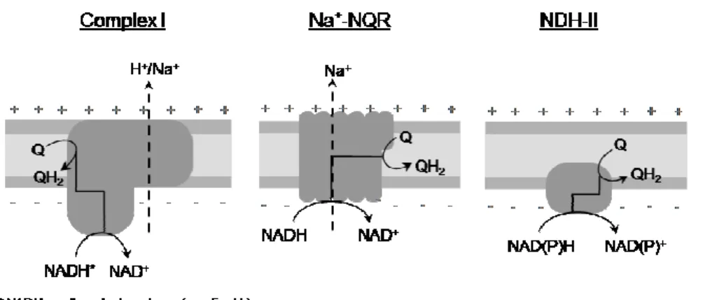
~ . The redox reaction can be performed by three
different types of NADH:quinone oxidoreductases (Figure 1.2.1): complex I
(NDH-I), a multisubunit transmembrane enzyme coupling quinone reduction
to charge translocation across the membrane; a multisubunit
sodium-translocating NADH:quinone oxidoreductase (Na+-NQR) that couples
quinone reduction to sodium transport; and an alternative NAD(P)H
dehydrogenase (NDH-II) which does not perform energy conservation. No
homologies regarding subunits and prosthetic groups composition, inhibitors
and mechanism exist between the three types of enzymes. This section will
introduce the three enzymes with special focus on complex I and its
ion/electron coupling mechanism.
Figure 1.2.1 – Schematic representation of the three types of respiratory NADH:quinone oxidoreductases. The three enzymes are capable of quinone reduction. Complex I and Na+-NQR couple the quinone reduction to charge
1.2.2 - COMPLEX I1
Complex I couples the electron transfer from NADH to quinone with
charge translocation across the membrane, contributing for the establishment
of the ∆µ~X+
) ;
(X=H+ Na+ . Homologues of complex I exist in bacteria, archaea
and eukarya (mitochondria and chloroplasts). This enzyme is characterized by
its large number of subunits, its prosthetic groups (flavin and iron-sulfur
centers) and its sensitivity to specific inhibitors, such as rotenone. The
electron donor of most bacterial and mitochondrial complexes is NADH,
while the archaeal complex I works apparently as a F420H2 dehydrogenase [11].
The electron donor of cyanobacterial and chloroplastidal complexes is not
known, yet [11]. Common to the three domains of life are 11 subunits
suggested to be responsible for the charge translocation and the quinone
binding.
1.2.2.1 – Structure and subunit composition
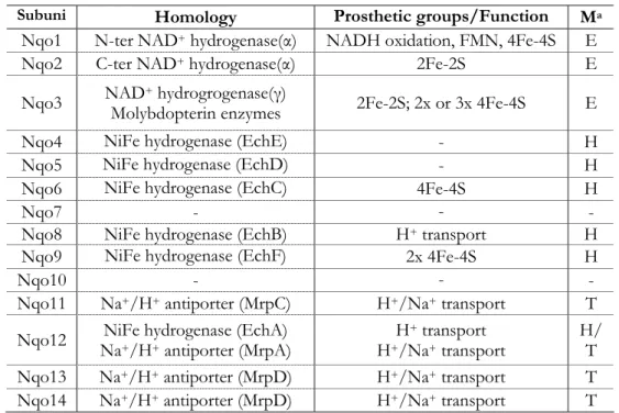
Bacterial complex I is generally composed by 14 subunits designated
as Nqo1-14 (from NADH:quinone oxidoreductase – P. denitrificans
nomenclature [13]) or as NuoA-N (from NADH:ubiquinone oxidoreductase
– E. coli nomenclature [14, 15]). This enzyme has a molecular mass of ≈500
kDa. The genes coding for its subunits may be organized in a single operon
[13, 14, 16-18] or may be disperse throughout the genome (ordered in more
than one operon [7, 19] and/or as isolated genes [7]). In several organisms,
unknown reading frames (URFs) are observed between the genes coding for
complex I subunits [7, 13, 18]. More than one gene coding for a specific
1 Throughout the text, and unless otherwise stated, the subunit nomenclature of Paracoccus
subunit can be found in some organisms [19] and there are also cases in
which two genes are fused [20, 21].
The canonical 14 subunits are organized in two different segments, a
peripheral and a membranar arm, in an L-shaped structure. The peripheral
part is composed by 7 subunits (Nqo1-6 and 9) and contains the binding site
for the electron donor and all known prosthetic groups [10]. The remaining 7
subunits (Nqo7-8 and 10-14) compose the membrane part and are most likely
involved in quinone reduction and charge translocation. The L-shaped
structure of bacterial complex I has been determined by electron microscopy
for E. coli [22] and Aquifex (A.) aeolicus enzymes (Figure 1.2.3) [19]. A
horseshoe shape was also reported for the E. coli enzyme [23], however this
result could not be reproduced and was interpreted as a possible artifact [24].
Only recently, progresses toward a X-ray structure were successful
and the structure of the entire complex I from Thermus (T.) thermophilus was
determined at a 4.5 Å resolution [2]. The structure of the membrane part
from E. coli complex I and the hydrophilic domain from T. thermophilus
enzyme have also been solved to 3.9 Å and 3.3 Å resolution, respectively [2,
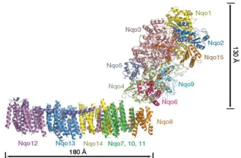
10]. The current model for T. thermophilus complex I consists of an atomic
model for the hydrophilic domain and an α-helical model for the membrane
domain (Figure 1.2.2). The peripheral arm has an Y shape with a length of
130 Å [2, 10]. One of the Y arms is formed by the subunits Nqo1 and Nqo2
and the other by the C-terminal domain of Nqo3. The central part is formed
by the N-terminal domain of Nqo3 and by the subunits Nqo5 and Nqo9. An
additional subunit, named Nqo15, was also observed in T. thermophilus
enzyme, which is not conserved in other complexes I. At the Y end are the
subunits Nqo4 and Nqo6, forming an interface with the membrane domain
[10]. Nine iron-sulfur centers were identified and from the distance between
that bridges a distance of 84 Å (see below section 1.2.2.3 – Prosthetic groups)
[10]. The last center of the chain (center N2) is about 20-25 Å from the
surface of lipid bilayer [2]. An atomic model of the membrane part is still not
available; however, the electron density of E. coli and T. termophilus enzyme
crystals were of sufficient quality to allow the built up of an α-helical model
for the membrane arm. It consists of up to 63 transmembrane (TM) helices
presenting a curve shape with a total length of 180 Å. The model has also
revealed a three times repetitive pattern of 14 TM helices, which were
assigned to the subunits Nqo12, 13 and 14. Interestingly, two of these TM
helices are discontinuous, as in some transporters and channels [25].
Unexpectedly, the subunit Nqo12 also contains a 110 Å long amphipathic α
-helice, spanning almost the entire length of the membrane domain (Figure
1.2.2). The assignment of the individual membrane subunits within the
domain was based on fragmentation studies in which the E. coli complex was
dissociated into sub-complexes [26]. The subunits Nqo12 and Nqo13 are
located in a distal part of the membrane arm and as so separated from the
redox centers of the hydrophilic domain. These subunits can be individually
removed from the entire complex without affecting the other subunits,
demonstrating that neither subunit is involved in contacts between the
membrane and the peripheral arms [26]. Two additional sub-complexes were
detected. One of them was composed by the subunits Nqo7, 11 and 14, and
the other by the subunits Nqo8 and 10. The presence of a transient
sub-complex formed by Nqo12, 13 and 14 places the one composed by Nqo7, 11
and 14 between the subunit Nqo13 and the Nqo8, Nqo10 sub-complex [26].
The assignment of the membrane subunits was also supported by other
experimental studies such as cross-linking experiments that have shown a
direct interaction between subunits Nqo6 and Nqo7 [27]; site-directed
mutagenesis which indicated the involvement of Nqo8 in connecting the
membrane and the peripheral arms, presumably by interacting with Nqo6
and/or Nqo4 [28, 29]; and photoaffinity labeling studies with a specific
complex I inhibitor that suggested that the Nqo6 and Nqo8 subunits work
together in the coupling reaction between electron transfer and ion transfer
[29]. The absence of the Nqo8 subunit in the structure of E. coli membrane
bound subunits also supports the close association between this subunit and
Nqo6 and/or Nqo4 [2]. The distal position of the subunits Nqo12 and
Nqo13 (≈100 Å away from the redox centers) was corroborated by electron
microscopy, whereas a shorter membrane domain was observed when the
two subunits were absent [30, 31].
Mitochondrial complex I is composed of more than 40 subunits and
has a molecular mass of ≈1000 kDa [32]. The nomenclature of the subunits is
molecular mass (e.g. 51 kDa). Some subunits are also designated as ND [32].
Fourteen of the subunits are homologues to the bacterial ones and are
sufficient to perform all the bioenergetics functions; thus they are called the
minimal functional unit [11]. The homologous seven membrane subunits are
mitochondria-encoded (homologues to Nqo7-8 and 10-14) whereas the
peripheral subunits are, predominantly, nuclear-encoded (homologues to
Nqo1-6 and 9) [11]. The function of most of the extra subunits, all
nuclear-encoded, is unknown [32]. As bacterial complex I, the mitochondrial complex
has an L-shaped structure as observed for Yarrowia (Y.) lipolytica [33] and
bovine [34] complexes I (Figure 1.2.3).
Archaea, cyanobacteria and chloroplasts contains homologues of 11
bacterial nqo genes, whereas the three genes encoding the electron input
module (Nqo1-3) are missing [11]. In the case of the archaeal complex I a
F420H2 dehydrogenase module was suggested [11, 35, 36], while in
cyanobacteria and plastids the electron input module is still unknown.
However, it is predicted that they display ferredoxin or NAD(P)H:quinone
oxidoreductase activity [37]. Single particle electron microscopy analysis has
shown that the complete complex I from the cyanobacterium
Thermosynechococcus (T.) elongatus is also L-shaped [31]. The presence of a
structure with a shorter membrane domain, whereas the subunits NhdF and
NdhD (Nqo12 and Nqo13 homologues, respectively) were missing reinforces
the similar basic shape and assembly of prokaryotic and eukaryotic complexes
I. Table 1.2.1 presents examples of homologous subunits in the three
domains of life and respective localization.
Table 1.2.1 – Nomenclature and localization of homologous subunits of bacterial (Paracoccus denitrificans [13], Escherichia coli [15] and Synechocystis), eukaryal (Bos taurus) and archaeal (Methanosarcina mazei [35]) complexes I.
Subunits
Bacteria Eukarya Archaea
P. denitrificans E. coli Synechocystisb B. taurus M. mazeic
Localization
Nqo1 NuoF - 51 - peripheral
Nqo2 NuoE - 24 - peripheral
Nqo3 NuoG - 75 - peripheral
Nqo4 NuoDa NdhH 49 FpoD peripheral
Nqo5 NuoCa NdhJ 30 FpoC peripheral
Nqo6 NuoB NdhK PSST FpoB peripheral
Nqo7 NuoA NdhC ND3 FpoA membranar
Nqo8 NuoH NdhA ND1 FpoH membranar
Nqo9 NuoI NdhI TYKY FpoI peripheral Nqo10 NuoJ NdhG ND6 FpoJ membranar Nqo11 NuoK NdhE ND4L FpoK membranar Nqo12 NuoL NdhF ND5 FpoL membranar Nqo13 NuoM NdhD ND4 FpoM membranar Nqo14 NuoN NdhB ND2 FpoN membranar
- - - - FpoO peripheral
- - - - FpoF peripheral
aThe genes of NuoC and NuoD are fused to one gene. bNdh from NADH dehydrogenase.
cFpo from F
420H2:phenazine oxidoreductase.
1.2.2.2 – Evolution
Amino acid sequence analysis revealed that most of the complex I
which are related with electron transfer and proton translocation processes.
Depending on their homologies the subunits can be grouped into three
functional modules: the electron input module, the hydrogenase module and
the transporter module.
The subunits Nqo4, 5, 6, 8 and 9 are homologous to subunits of the
membrane-bound NiFe-hydrogenases [11, 38] and so are known as the
hydrogenase module. This NiFe-hydrogenase catalyzes the reaction
H2↔2H
++2e, and this reaction is most likely coupled with ion translocation
across the membrane [38, 39]. The hydrogenase complex is composed by six
subunits (Ech A to F from energy-converting hydrogenase – Methanosarcina
barkeri nomenclature) [38], four hydrophilic proteins and two integral
membrane proteins. Nqo6 is closely related to a soluble small subunit that
displays a conserved amino acid sequence characteristic for the binding of a
tetranuclear iron-sulfur center. The Nqo9 is homologous to a two
[4Fe-4S]2+/1+ ferredoxin [40] while Nqo8 is homologous to one of the membrane
proteins.
Subunit Nqo12 was assigned as an hydrogenase homologue [11, 40]
but it was observed that this subunit is also related to one particular class of
Na+/H+ antiporters, the multiple resistance and pH (Mrp) antiporters (see
section 1.3 – Sodium/proton antiporters) [41]. Also subunits Nqo 11, 13 and
14 are related with these antiporters [5, 41]. The Mrp antiporters are encoded
by an operon that contains seven genes (mrpA to G). Nqo12 is closely related
to MrpA and Nqo13 and 14 are related to MrpD [41]. Nqo11 has been
proposed to be homologous to MrpC [5]. Nqo11, 12, 13 and 14 should thus
constitute the transporter module.
The electron input module is not conserved within the complex I
family, which most probably is related to the different nature of the electron
![Table 1.2.1 – Nomenclature and localization of homologous subunits of bacterial (Paracoccus denitrificans [13], Escherichia coli [15] and Synechocystis), eukaryal (Bos taurus) and archaeal (Methanosarcina mazei [35]) complexes I](https://thumb-eu.123doks.com/thumbv2/123dok_br/15769923.641173/53.892.108.682.366.775/nomenclature-localization-homologous-paracoccus-denitrificans-escherichia-synechocystis-methanosarcina.webp)
![Figure 1.2.4 – Evolutionary scheme for complex I based on Mathiesen and Hagerhall proposal [5]](https://thumb-eu.123doks.com/thumbv2/123dok_br/15769923.641173/56.892.236.782.458.836/figure-evolutionary-scheme-complex-based-mathiesen-hagerhall-proposal.webp)
![Figure 1.2.5 – Arrangement of prosthetic groups and respective subunit localization, according to Ohnishi [6]](https://thumb-eu.123doks.com/thumbv2/123dok_br/15769923.641173/58.892.251.484.116.509/figure-arrangement-prosthetic-respective-subunit-localization-according-ohnishi.webp)
![Figure 1.2.8 – Direct coupling mechanisms proposed by Esposti and Ghelli (A) [3], Dutton et al (B) [9] and Brandt (C) [12]](https://thumb-eu.123doks.com/thumbv2/123dok_br/15769923.641173/67.892.131.652.493.883/figure-direct-coupling-mechanisms-proposed-esposti-ghelli-dutton.webp)
