Materials Research, Vol. 9, No. 2, 243-246, 2006 © 2006
*e-mail: [email protected]
Article presented at the XV CBECIMAT, Natal - RN, November/2002
Characterization by X Ray Diffraction of Mechanically
Alloyed Tripotassium Sodium Sulfate
Marcus Aurélio Ribeiro Miranda, José Marcos Sasaki, Antônio Sérgio Bezerra Sombra,
Cléber Costa Silva, Cláudio Márcio Rocha Remédios*
Departamento de Física, Universidade Federal do Ceará – UFC,
C. P. 6030, 60455-760 Fortaleza - CE, Brazil
Received: January 24, 2003; Revised: March 30, 2006
Nanocrystalline powder of tripotassium sodium sulfate (K3Na(SO4)2 - KNS) was successfully obtained by mechanical alloying a mixture of potassium sulfate (K2SO4) and sodium sulfate (Na2SO4) with a proportion of 3:1 in a planetary mill. X ray powder diffraction (XRPD) was used to characterize this material. Powders produced with high milling times: 15, 30, 45 and 60 hours, have shown a single phase. For 60 hours of milling time, the powder was formed with very small grains (60 nm in average). We also prepared several samples with low milling times: 30, 60, 120, 150 and 180 minutes. The results for this series show that 120 minutes of milling is enough to produce a single crystalline phase of KNS. Therefore, we showed that a nanocrystalline powder of tripotassium sodium sulfate is easily obtained by mechanical alloying and that the grain size can be controlled by the amount of milling time.
Keywords:sulfate, mechanical alloying, X ray, powder diffraction
1. Introduction
The interest in the physical properties of nanoparticles has increased in the last decades because of the different properties encountered in these nanocrystalline materials when compared to their corresponding bulk. They present, in general, properties with innumerable technological applications, for example: there is a drastic variation of electric properties of materials in the nanosize regime attributed to the quantum confinement of charge carriers and associated modification of the band structure1,2. Furthermore, there
are some nanoparticles with reduced value of conductivity in contrast to those of the single crystal or coarse-grained materials3,4. In some
semiconductors, a decrease in the value of conductivity is expected due to the narrowing of valence and conduction bands resulting in an increase of forbidden energy gap. Reduced particle size is also needed for improved sintering abilities, that means decreased sinter-ing temperatures, increased density of sintered powders6 and shorter
reaction times, as compared with the classical ceramic synthesis. The crystal of tripotassium sodium sulfate (abbreviated as KNS) is the most investigated member of the glaserite family. This family also includes K3Na(CrO4)2, K3Na(SeO4)2 and K3Na(MoO4)2. The members of this family are characterized by showing a sequence of phase transitions1 and a predicted and not observed ferroelastic phase
transition1 around 75 K for KNS. The space group of the KNS is
P 3 m1 and the lattice parameters3 are a = 5.6801 Å and c = 7.309 Å.
Single crystals of KNS can be obtained by various methods includ-ing slow evaporation of an aqueous solution4. In this work we are
interested in structurally characterize the nanocrystalline powder of KNS produced by mechanical alloying using X ray powder dif-fraction. Special attention is given to the grain size as a function of milling time.
2. Experimental
In this work, mechanical alloying has been successfully used to produce nanocrystalline powder of tripotassium sodium sulfate (KNS). This procedure is analogous to the one used for other mate-rials, hydroxylapatite5 for example. Commercial powders of K
2SO4
(Vetec 99%) and Na2SO4 (Vetec 99%) were used in the preparation of KNS. The reaction used was the following:
3K2SO4 + Na2SO4→ (IMPACTS) → 2K3Na(SO4)2 (1)
The reagents were ground on a Fritsch Pulverisette planetary mill with the stechiometric ratio 3:1 given in Equation 1. Mechanical alloying was carried out using sealed stainless steel vials and balls under air. Two sets of samples were produced; 30, 60, 120, 150 and 180 minutes, samples with low milling time; 15, 30, 45 and 60 hours, samples with high milling time.
The X ray diffraction (XRD) patterns were obtained at room temperature (300 K) in an X´Pert PRO Phillips powder diffractometer using the Bragg-Bretano geometry with Cu-Kα radiation. We used three seconds for each step of counting time, an angular step of 0.02° and with the tube at 40 KV and 40 mA.
The analysis of the grain size (Lhkl) of the sulfate has been done to all samples using the Scherrer´s equation6,
cos
L
hkl=
k
b
m
i
(2)where k is the shape coefficient (values between 0.9 and 1.0), λ is the wavelength of the radiation, β is the full width at half maximum (FWHM) of the peaks of each phase and θ is the diffraction angle. For this purpose the β parameter has been corrected in order to rep-resent only the effect of the grain size in the FWHM. Assuming a Gaussian function for the diffraction peaks and for the instrumental broadening, one can subtract the instrumental broadening using the following equation:
c inst
2 2
exp
b
=
s
-
s
(3)where ϖexp corresponds to the experimental FWHM obtained for
each sample. We have used the LaB6 (SRM 660-National Institute of Standard Technology) powder standard pattern to determine the instrumental line width (ϖinst = 0.08°) of the equipment close to the
244 Miranda et al. Materials Research
In order to perform this calculation we have chosen two peaks, one at 24.1° and another at 31.3° and according to the space group P 3 m1 of KNS these peaks correspond respectively to hkl = 002 and hkl = 110, i.e. along and perpendicular to the c crystallographic axis of the diffracting grains. The shape coefficient k was assumed to be equal to 1, which means an approximately spherical shape of the grain.
3. Results and Discussion
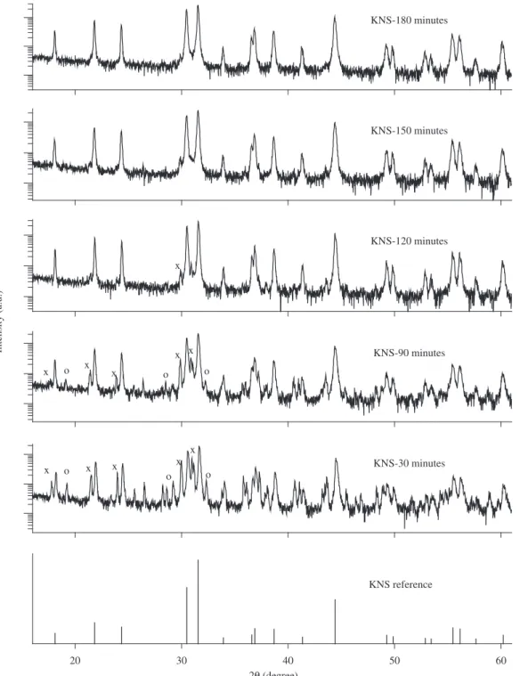
In the Figure 1 it is shown the diffraction patterns of the K3Na(SO4)2 (KNS) powder in log scale to enhance the low intensity peaks. In this figure, one has the reference pattern obtained from the
literature (ICDD7) denoted by bars and the X ray powder diffraction
of the (KNS) prepared for low milling time. It can seen that for times below 120 minutes there are traces of the original reagents used in the preparation (K2SO4 and Na2SO4). Peaks corresponding to the reagents are indicated by ‘o’ and ‘x’ in the figure.
Moreover, the X ray data in Figure 1 also shows that the formation of the KNS is very fast. Even for a very low milling time the peaks associated to KNS have already appeared. There is no formation of any other phases; the diffraction pattern is completely covered by the peaks associated with the KNS, K2SO4, marked as ‘x’, and the Na2SO4 phases, marked as ‘o’. All patterns are obtained in ICDD7.
The crystalline grain size of these samples are shown in Table 1.
20 30 40 50 60
KNS reference KNS-30 minutes
2Q (degree)
Intensity (a.u.)
o o
x
KNS-90 minutes
o o
x x
x x
x x
x x x
o
o
KNS-120 minutes
x
KNS-150 minutes KNS-180 minutes
Vol. 9, No 2, 2006 Characterization by X Ray Diffraction of Mechanically Alloyed Tripotassium Sodium Sulfate 245
Table 1. Average grain size of the samples with low milling time. Average error is 1 nm.
Milling time 30 minutes 90 minutes 120 minutes 150 minutes 180 minutes
Size fwhm Size fwhm Size fwhm Size fwhm Size fwhm
110 – direction 117 nm 0.0784 95 nm 0.0961 153 nm 0.0600 178 nm 0.1173 121 nm 0.0755 002 – direction 133 nm 0.0680 111 nm 0.0812 170 nm 0.0531 80 nm 0.1125 164 nm 0.0617
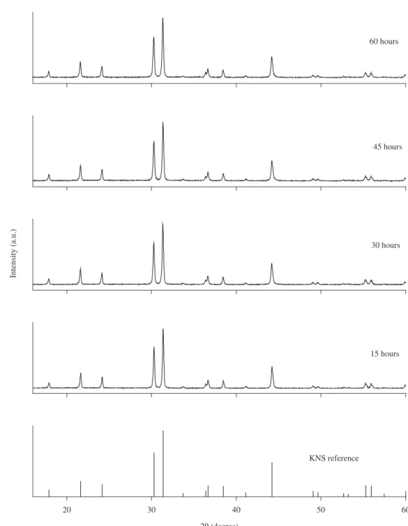
Figure 2. X ray powder diffraction pattern of KNS powder prepared with high milling time (15-60 hours) and the reference pattern of KNS.
Figure 2 shows the diffraction patterns of the KNS for high mill-ing time plus the reference pattern. No trace of another phase than the KNS has been found.
20 30 40 50 60
2Q (degree)
KNS reference
15 hours 30 hours 45 hours 60 hours
Intensity (a.u.)
246 Miranda et al. Materials Research
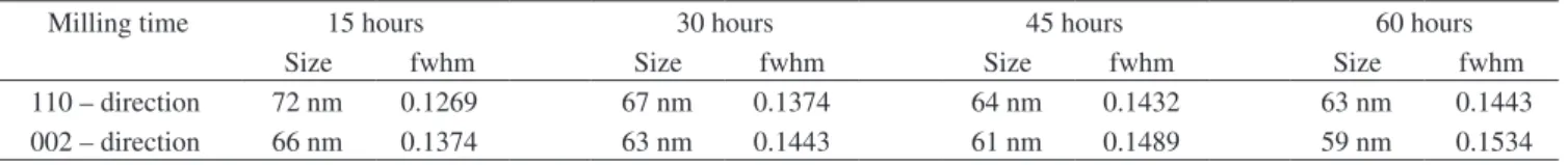
the milling time is increased and it is also noted that the grains are smaller than the ones of the samples prepared with low milling time. So, this is an efficient method to obtain nanocrystalline powder of tripotassium sodium sulfate, and it can be also achieved with a low milling time.
References
1. Bernardin FE, Hammack WS. Pressure-induced disordering of sodium potassium sulfates and chromates. Physical Review B. 1996;
54(10):7026-7033.
2. Kaczmarski M, Mroz B. Raman study of the ferroelastic phase transition in K3Na(SeO4)2. Physical Review B. 1998; 57(21):589-598.
3. Okada K, Ossaka J. Structures of Potassium Sodium Sulphate and Tripotassium Sodium Dulphate. Acta Crystallography. 1980;
B36:919-921.
4. Natarajan M, Secco EA. Infrared Spectra of KnaSO4 and K3Na(SO4)2.
Journal of Solid State Chemistry. 1983; 47(2):231-235.
5. Silva CC, Pinheiro AG, Figueiró SD, Góes JC, Sasaki JM, Miranda MAR, Sombra ASB. Piezoelectric properties of collagen-nanocrystalline hydroxyapatite composites. Journal of Materials Science. 2002; 37(10):1-10.
6. Azároff LV. Elements of X-ray Crystallography, McGraw-Hill Book
Company, New York; 1968.
7. ICDD, International Center for Diffraction Data, 12 Campus Blvd., Newton Square, Pennsylvania USA, 1995; 19073-3723.
Table 2. Average grain size of the samples with high milling time. Average error is 1 nm.
Milling time 15 hours 30 hours 45 hours 60 hours
Size fwhm Size fwhm Size fwhm Size fwhm