5S rDNA characterization in twelve Sciaenidae fish species (Teleostei,
Perciformes): Depicting gene diversity and molecular markers
Fernanda A. Alves-Costa
1, Cesar Martins
2, Fernanda Del Campos de Matos
2, Fausto Foresti
2,
Claudio Oliveira
2and Adriane P.Wasko
11
Departamento de Genética, Instituto de Biociências, Universidade Estadual Paulista, Botucatu,
SP, Brazil.
2
Departamento de Morfologia, Instituto de Biociências, Universidade Estadual Paulista, Botucatu,
SP, Brazil.
Abstract
In order to extend the genetic data on the Sciaenidae fish family, the present study had the purpose to characterize PCR-generated 5S rDNA repeats of twelve species of this group through PAGE (Polyacrylamide Gel Electrophore-sis) analysis. The results showed the occurrence of at least two different 5S rDNA size classes in all the species. Moreover, 5S rDNA repeats of one of the studied species -Isopisthus parvipinnis - were cloned and subjected to nu-cleotide sequencing and Southern blot membrane hybridization analyses, which permitted to confirm the existence of two major 5S rDNA classes. Phylogenetic analysis based on the nucleotide sequences of different 5S rDNA re-peats ofI. parvipinnis lead to their separation into two major clusters. These results may reflect the high dynamism that rules the evolution rate of 5S rDNA repeats. The obtained data suggest that 5S rDNA can be useful in genetic analyses to identify species-specific markers and determine relationships among species of the Sciaenidae group.
Key words:Sciaenidae, fish,Isopisthus parvipinnis, 5S rDNA, molecular markers.
Received: August 22, 2006; Accepted: May 4, 2007.
Introduction
The 5S ribosomal multigene family (5S rDNA) of higher eukaryotes is comprised of tandemly repeated units of hundreds to thousands of copies that consist of a highly conserved coding sequence of 120 base pairs (bp) and a variable non-transcribed spacer sequence (NTS) (reviewed in Long and David, 1980). Studies on 5S rDNA organiza-tion could provide useful data for the understanding of ge-nome organization and dynamics of repetitive sequences, and also provide genetic markers for the identification of species, subspecies, population, strain, or hybrids (e.g.
Martins and Wasko, 2004). Although 5S rDNA repeats have been characterized in several vertebrate species, pres-ent data are mostly restricted to mammals and amphibians. To date, few analyses have been conducted on fishes, spe-cially taking into account the great number of species of this group (e.g.Martins and Wasko, 2004).
The Sciaenidae (Perciformes) family contains ap-proximately 70 genera and 270 fish species (Schwarzhans,
1993; Nelson, 1994) that are distributed in Indian, Pacific and Atlantic Oceans (Longhurst and Pauly, 1987; Sasaki, 1996). Although Sciaenidae is considered a monophyletic group, a great diversity of body shape and mouth position can be observed in several species, associated with differ-ent feeding patterns and life histories (Chao and Musick, 1977). Moreover, most species of the group represent im-portant fishery resources with a high commercial value. However, despite the economic importance and the great species diversity, genetic studies are still scarce in this fish group. The available genetic data refer to molecular popu-lation genetic (Turneret al., 1998; Lankford et al., 1999; Goldet al., 2001; Cordes and Graves, 2003; O’Malley et al., 2003; Santoset al., 2003) and phylogenetic analyses (Chao, 1978; Sasaki, 1989; Vinsonet al., 2004).
To improve the genetic data on Scianidae fish, the present study characterized the PCR-generated 5S rDNA repeat patterns of twelve species of the group. Moreover, 5S rDNA repeats of one of the species - Isopisthus parvipinnis- were cloned and subjected to nucleotide se-quencing, Southern blot-membrane hybridization and phylogenetic analyses.
Materials and Methods
Fish samples, DNA extraction and PCR
Samples of twelve different species of the Sciaenidae fish family (Nebris microps, Paralonchurus brasiliensis, Stellifer stellifer, S. rastrifer, S. microps, Isopisthus parvipinnis, Cynoscion jamaicensis, C. virescens, Menticirrhus americanus, Micropogonias furnieri, Ctenosciaena gracilicirrhus,andLarimus breviceps) from Ubatuba (São Paulo State, Brazil) were analyzed.
Genomic DNA was extracted from gill tissue, accord-ing to the method described by Waskoet al.(2003). A set of primers (Primer A 5’-TACGCCCGATCTCGTCCGAT C-3’ and primer B 5’-CAGGCTGGTATGGCCGTAAG C-3’), corresponding to nucleotides 24-44 and 1-21, re-spectively, of the 5S coding region, were designed from the 5S rRNA sequence of rainbow trout (Komiya and Take-mura, 1979) to amplify the 5S rRNA genes and their non-transcribed spacer regions (Martins and Galetti, 1999). PCR amplifications were carried out in a total volume of 50 µL, using 20-100 ng of genomic template DNA, 150 pmol of each primer, 1.25 mM of each dNTP, 1x PCR buffer containing 1.5 mM MgCl2, and 1U ofTaq DNA polymerase (GE Healthcare Life Sciences), using a PTC-200 Programmable Thermal Controller (MJ Research, INC). The optimum cycling times were as follows: 94 °C (5 min) denaturation, 35 cycles of 1 min at 95 °C, 30 s at 63 °C, and 1 min at 72 °C. A final 5 min extension was per-formed at 72 °C. The 5S rDNA-PCR products were visual-ized in 6% polyacrylamide gels by silver nitrate staining.
Cloning, nucleotide sequencing and sequence analyses
The PCR products of two DNA samples ofIsopisthus parvipinniswere selected to be cloned and sequenced, and to be used in Southern blot-membrane hybridization and phylogenetic analyses, since this species presented the most common banding pattern among the analyzed Sciae-nidae fish species, which was also observed in Stellifer microps, Stellifer rastrifer, Cynoscion jamaicensis, and
Cynoscion virescens. The PCR products ofI. parvipinnis
were inserted into pGEM-T (Promega), which was used to transform competent cells ofE. coliDH5αstrain
(Invitro-gen), according to the manufacturer’s instructions. Positive recombinant clones were recovered and stored in 75% glycerol at -80 °C for subsequent analysis. Several clones were sequenced on an ABI Prism 377 automatic DNA se-quencer (Applied Biosystems) with DYEnamic ET Termi-nator Cycle Sequencing (GE Healthcare Life Sciences), following the manufacturer’s instructions. Nucleic acid se-quences were subjected to BLASTN (Altschulet al., 1990) searches at the National Center for Biotechnology Informa-tion (NCBI), and the sequence alignment was performed using the computer program Clustal W (Thompsonet al., 1994) and by eye. Neighbor-Joining (NJ) phylogenetic
analyses employing the Kimura-two-parameter genetic dis-tance model (Kimura, 1980) were conducted using MEGA version 3.1 software (Kumar et al., 2004). Bootstrap resampling using 1000 replicates (Felsenstein, 1985) was applied to assess support for individual nodes.
Southern blot hybridization
Genomic DNA samples of Isopisthus parvipinnis
(8µg) were completely and partially digested withHindIII.
This restriction enzyme was chosen since it cuts once inside the 5S rRNA gene of most teleost fish species (Martins and Wasko, 2004). The digestion products were subjected to 1% agarose gel electrophoresis and Southern-transferred to a Hybond-N nylon membrane (Southern, 1975). The hy-bridization of the filter-immobilized DNA was performed using as probes clones containing repeat units of I. parvipinnis 5S rDNA.Probe labeling, hybridization and detection steps were performed with the kit ECL-Direct nu-cleic Acid Labeling and Detection System (GE Healthcare Life Sciences) following the manufacturer’s instructions.
Results and Discussion
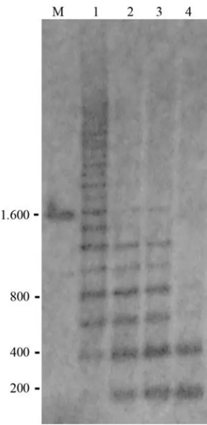
PCR amplification of 5S rDNA repeats of twelve Sciaenidae species generated distinct polyacrylamide gel electrophoresis banding patterns (Figure 1). The different sizes of the amplified fragments may reflect the intense dy-namism that rules the evolution of the 5S rDNA tandem ar-rays in the fish genome. As the 5S rDNA repeats consist of a 120 bp conserved coding sequence, the observed differ-ences among several Sciaenidae species is related to the NTS regions that can be extremely variable due to
inser-Figure 1- 5S rDNA PCR products of Sciaenidae species visualized on 6% polyacrylamide gel: (1) Larimus breviceps, (2) Nebris microps, (3)
Paralonchurus brasiliensis, (4) Stellifer stellifer, (5) Menticirrhus americanus, (6)Micropogonias furnieri, (7)S. microps, (8)Ctenosciaena gracilicirrhus, (9) S. rastrifer, (10) Isopisthus parvipinnis, (11)
tions/deletions, minirepeats, and pseudogenes (Nelson and Honda, 1985; Leahet al., 1990; Sajdaket al., 1998).
On the other hand, it was possible to identify similar 5S rDNA amplification patterns for some species of the same genus, such asStellifer micropsandS. rastrifer,and
Cynoscion jamaicensisandC. virescens(Figure 1). More-over,Isopisthus parvipinnisalso presented a PCR amplifi-cation pattern that resembles the pattern evidenced for the four former species (Figure 1). It was also possible to note that the amplified fragments obtained forStellifer stellifer
were very similar to the ones obtained forNebris microps
(Figure 1). Similar sized 5S-PCR products may reflect a higher genetic similarity among some Sciaenidae species, as already evidenced for other fish (e.g. Wasko et al.,
2001). However, we could not discard the possibility that similar DNA fragment sizes may also present large nucleo-tide differences. Although mitochondrial DNA sequence analysis has shown a distant relationship between
CynoscionandStellifer(Vinson et al., 2004), previously published morphometric data (Aguirre and Shervette,
2005) are in accordance with the present results that sug-gest a closer relationship between these two genera.
amplified products clearly discriminate other fish species, such as the Atlantic salmon (Salmo salar), the brown trout (Salmo truta), and their hybrids (Pendáset al., 1995). Sev-eral species of the genusBrycon(Waskoet al., 2001), and
Solea soleaandReinhardtius hippoglossoides(Céspedeset al., 1999) also show 5S rDNA PCR species-specific pat-terns. PCR amplification of 5S rDNA repeats thus repre-sents a potential and simple methodology that can be applied to identify several fish species.
PAGE analysis of the 5S rDNA PCR products also showed at least two amplified fragments for all the Sciaenidae species. Each fragment could correspond to a distinct 5S rDNA class, as already detected for other fish species through PCR electrophoresis and nucleotide se-quencing analyses (Martins and Galetti, 2001; Martins and Wasko, 2004). Although the occurrence of two distinct 5S rDNA classes represents the most common feature in fish (Martins and Wasko, 2004), some species may also present more than two 5S rDNA types, as shown for some of the an-alyzed Sciaenidae species with three or more clearly dis-tinct PCR generated fragments (Figure 1).
In order to characterize the 5S rDNA nucleotide se-quence and genomic organization for one of the analyzed species and also to verify the occurrence of two distinct classes of this repetitive ribosomal DNA in Sciaenidae fish, the two amplified fragments ofIsopisthus parvipinnis- with around 200 and 400 base pairs - were cloned, sequenced, and subjected to Southern blot-membrane hybridization and phylogenetic analyses. The nucleotide sequencing analysis of these two fragments supports the existence of two differ-ent 5S rDNA size classes, named 5S rDNA type I (201-205 bp) and 5S rDNA type II (413-426 bp) (Figure 2). Both classes showed a conserved 120 bp 5S rRNA gene se-quence and a variable NTS ranging between 80 bp (5S rDNA type I) to 280 bp (5S rDNA type II). These variations were characterized by insertions/deletions, and base substi-tutions. These 5S rDNA classes were separated in the phylo-genetic analysis in 100% of the generated trees (Figure 3). Yet, one single sequence of 5S rDNA type I was quite differ-ent and could be considered a subclass since it appeared sep-arated in 98% of the generated trees (Figure 3). Although the occurrence of two 5S rDNA repeat classes represents the most common feature in fishes (Martins and Galetti 2001; Martins and Wasko 2004), the presence of variant subclasses also seems to be frequent in this vertebrate group. The pres-ence of two major repeated 5S rDNA classes inI. parvipinnis
was confirmed by Southern blot hybridization which re-vealed the presence of tandem repeats of 200 and 400 bp, in agreement with the PCR products and the nucleotide se-quencing data (Figure 4).
We take these results as reflecting the high dynamism that governs the evolution rate of the 5S rDNA repeats in the genome of Isopisthus parvipinnis and also in other Sciaenidae species. Moreover, the present data indicate that 5S rDNA can be useful for general and applied genetic
analyses of different sciaenids, in order to identify spe-cies-specific markers and determine species relationships.
Acknowledgments
The authors thank Prof. Dr. Adilson Fransozo for pro-viding the Sciaenidae samples, Ms. Ursulla Pereira Souza for samples identification, and Dr. Anderson L. Alves for helpful assistance in DNA sequencing. This research was supported by grants from FAPESP (Fundação de Amparo à Pesquisa do Estado de São Paulo) and CNPq (Conselho Nacional de Desenvolvimento Científico e Tecnológico). Figure 3- Neighbor-joining three based on the 5S rDNA nucleotide se-quences ofIsopisthus parvipinnis. The numbers at each node indicate the percentage recovery (> 50%) of the particular node (1000 bootstrap repli-cates).
References
Aguirre WE and Shervette VR (2005) Morphological diversity of theCynosciongroup (Perciformes, Sciaenidae) in the Gulf of Guayaquil region, Ecuador: A comparative approach. En-viron Biol Fish 73:403-413.
Altschul SF, Gish W, Miller W, Myers EW and Lipman DJ (1990) Basic local alignment search tool. J Mol Biol 215:403-410. Céspedes A, Garcia T, Carrera E, Gonzalez I, Fernandez A,
Hernández PE and Martin R (1999) Identification of sole
(Solea solea) and greenland halibut (Reinhardtius
hippoglossoides) by PCR amplification of the 5S rDNA gene. J Agric Food Chem 47:1046-1050.
Chao NL and Musick JA (1977) Early life history, functional mor-phology and feeding habits Sciaenidae of York River, Vir-ginia. Fish Bull 75:657-702.
Chao NL (1978) A basis for classifying Western Atlantic Sciae-nidae (Teleostei, Perciformes). NMFS Technical Reports 415:1-64.
Cordes JF and Graves JE (2003) Investigation of congeneric hy-bridization in and stock structure of weakfish (Cynoscion regalis) inferred from analyses of nuclear and mitochondrial DNA loci. Fish Bull 101:443-450.
Felsenstein J (1985) Confidence limits on phylogenies: An ap-proach using the bootstrap. Evolution 39:783-791. Gold JR, Burridge CP and Turner TF (2001) A modified
step-ping-stone model of population structure in red drum,
Sciaenops ocellatus(Sciaenidae), from the northern Gulf of Mexico. Genetica 111:305-317.
Kimura M (1980) A simple method for estimating evolutionary rate of base substitution through comparative studies of nu-cleotide sequences. J Mol Evol 16:111-120.
Komiya H and Takemura S (1979) Nucleotide sequence of 5S ri-bosomal RNA from rainbow trout (Salmo gairdnerii) liver. J Biochem 86:1067-1080.
Kumar S, Tamura K and Nei M (2004) MEGA 3: Integrated soft-ware for molecular evolutionary genetics analysis and se-quence alignment. Brief Bioinform 5:150-163.
Lankford TE, Targett TE and Gaffney PM (1999) Mitochondrial DNA analysis of population structure in the Atlantic croaker, Micropogonias undulates (Perciformes, Scia-enidae). Fish Bull 97:884-890.
Leah R, Frederiksen S, Engberg J and Sorensen PD (1990) Nucle-otide sequence of a mouse 5S rRNA variant gene. Nucleic Acids Res 18:7441.
Long ED and David ID (1980) Repeated genes in eukaryotes. Annu Rev Biochem 49:727-764.
Longhurst AR and Pauly D (1987) Ecology of Tropical Ocean. Academic Press Inc., San Diego, 407 pp.
Martins C and Galetti Jr PM (1999) Chromosomal localization of 5S rDNA genes in Leporinus fish (Anostomidae, Characiformes). Chrom Res 7:363-367.
Martins C and Galetti Jr PM (2001) Two 5S rDNA arrays in Neo-tropical fish species: Is it a general rule for fishes? Genetica 111:439-446.
Martins C and Wasko AP (2004) Organization and evolution of 5S ribosomal DNA in the fish genome. In: Williams CR (ed) Focus on Genome Research. Nova Science Publishers, Hauppauge, pp 289-318.
Nelson DW and Honda BM (1985) Genes coding for 5S ribo-somal RNA of the nematodeCaenorhabditis elegans. Gene 38:245-251.
Nelson JS (1994) Fishes of the world. 3rd ed. John Wiley & Sons Inc., New York, 600 pp.
O’Malley KG, Abbey CA, Ross K and Gold JR (2003) Micro-satellite DNA markers for kinship analysis and genetic map-ping in red drum, Sciaenops ocellatus (Sciaenidae, Teleostei). Mol Ecol Notes 3:155-158.
Pendás AM, Morán P, Martínez JL and Garcia-Vásquez E (1995) Applications of 5S rDNA in Atlantic salmon, brown trout, and in Atlantic salmon x brown trout hybrid identification. Mol Ecol 4:275-276.
Sadjak SL, Reed KM and Phillips RB (1998) Intraindividual and interspecies variation in the 5S rDNA of coregonid fish. J Mol Evol 46:680-688.
Santos S, Schneider H and Sampaio I (2003) Genetic differentia-tion ofMacrodon ancylodon(Sciaenidae, Perciformes) pop-ulations in Atlantic coastal waters of South America as re-vealed by mtDNA analysis. Genet Mol Biol 26:151-161. Sasaki K (1989) Phylogeny of the family Sciaenidae, with notes
on its zoogeography (Teleostei, Perciformes). Mem Fac Fish Hokkaido Univ 36:1-137.
Sasaki K (1996) Sciaenid fishes of the Indian Ocean (Teleostei, Perciformes). Mem Fac Sci Kochi Univ Ser D (Biol) 16/17:83-95.
Schwarzhans W (1993) A Comparative Morphological Treatise of Recent and Fossil Otoliths of the Family Sciaenidae (Perciformes), Piscium Catalogus: Part Otolothi Piscium, v. 1. Verlag Dr. Friedrich Pfeil, München, 245 pp.
Southern EM (1975) Detection of specific sequences among DNA fragments separated by gel electrophoresis. J Mol Biol 98:503-517.
Thompson JD, Higgins DG and Gibson TJ (1994) Clustal W: Im-proving the sensitivity of progressive multiple sequence alignment through sequence weighting, position-specific gap penalties and weight matrix choice. Nucleic Acids Res 22:4673-4680.
Turner TF, Richardson LR and Gold JR (1998) Polymorphic microsatellite DNA markers in red drum (Sciaenops ocellatus). Mol Ecol 7:1771-1788.
Vinson C, Gomes G, Schneider H and Sampaio I (2004) Sciae-nidae fish of the Caeté River estuary, northern Brazil: Mito-chondrial DNA suggests explosive radiation for the Western Atlantic assemblage. Genet Mol Biol 27:174-180.
Wasko AP, Martins C, Wright JM and Galetti Jr PM (2001) Mo-lecular organization of 5S rDNA in fishes of the genus
Brycon. Genome 44:893-902.
Wasko AP, Martins C, Oliveira C and Foresti F (2003) Non-destructive genetic sampling in fish, an improvement method for DNA extraction from fish fins and scales. Here-ditas 138:161-165.
Internet Resources
National Center for Biotechnology Information (NCBI) (http:www.ncbi.nlm.nih.gov/blast).
Associate Editor: Pedro Manoel Galetti Junior