Evaluation of different therapeutic Carnoy's formulations on hard
human tissues: A Raman microspectroscopy, microhardness, and
scanning electron microscopy study
Francisco Samuel Rodrigues Carvalho
a,b,*, Victor Pinheiro Feitosa
c,
Paulo Goberl
^
anio de Barros Silva
d, Eduardo Costa Studart Soares
e,
Thyciana Rodrigues Ribeiro
f, Cristiane S
a Roriz Fonteles
f, F
abio Wildson Gurgel Costa
g aDivision of Oral and Maxillofacial Surgery, Federal University of Ceara, Fortaleza, BrazilbDivision of Oral and Maxillofacial Morphology and Oral and Maxillofacial Surgery, UNIFOR, Fortaleza, Brazil cPaulo Picanço School of Dentistry, Fortaleza, Ceara, Brazil
dUnichristus University Center, Fortaleza, Ceara, Brazil
eDivision of Oral and Maxillofacial Surgery, Walter Cantídio University Hospital, Federal University of Ceara, Fortaleza, Brazil fSchool of Dentistry, Federal University of Ceara, Ceara, Fortaleza, Brazil
gDivision of Oral Radiology, Walter Cantídio University Hospital, Federal University of Ceara, Fortaleza, Brazil
a r t i c l e
i n f o
Article history:
Paper received 29 November 2017 Accepted 14 February 2018 Available online 21 February 2018
Keywords:
Carnoy's solution Hard tissues Treatment
Raman microspectroscopy In vitro study
a b s t r a c t
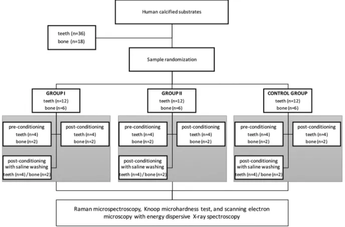
Purpose: To evaluate different therapeutic Carnoy's solution formulations on hard human tissues. Materials and methods: An in vitro study was performed with human teeth (n¼36) and bone fragments (n¼18), randomly divided into two experimental groups (Group I¼Carnoy solution with chloroform; Group II¼Carnoy solution without chloroform) and a control group (saline solution). The groups were subdivided into pre-conditioning, post-conditioning, and post-conditioning with saline washing. Raman microspectroscopy, Knoop microhardness test, and scanning electron microscopy with energy dispersive X-ray spectroscopy were used.
Results: There was demineralization of dental structures regarding mineral/matrix and carbonate/phosphate ratios (GI versus GII, p<0.05). The presence of chloroform resulted in a statistically significant reduction of the teeth surface microhardness (p¼0.036), but not exceeding 0.01mm. Both GI and GII showed significant structural changes by using scanning electron microscopy with energy dispersive X-ray spectroscopy. Conclusion: Carnoy's solution altered the organic and inorganic matrix of the human calcified specimens analyzed in vitro, and its effect was more pronounced when chloroform was present.
©2018 Published by Elsevier Ltd on behalf of European Association for Cranio-Maxillo-Facial Surgery.
1. Introduction
Benign lesions (odontogenic and non-odontogenic), but locally aggressive and with a high percentage of recurrence, may occur within the maxillofacial region. (Abou Neel et al., 2016) Conservative treat-ment of these jaw lesions can lead to high recurrence rates; however, a radical approach usually is followed by severe functional and aesthetic impairments (Costa et al., 2010; Pogrel, 2015). Adjunct therapies allied to surgical treatments, such as the use of Carnoy's solution or liquid nitrogen as a cryosurgical agent, have been widely used since the 1980s, aiming to improve clinical outcomes (Costa et al., 2011).
Voorsmit, Stoelinga and van Haelst in 1981 (Voorsmit et al., 1981) were thefirst investigators that used Carnoy's solution in intraosseous-related jaw lesions, recommending the use of this cauterizing agent in the treatment of odontogenic keratocysts, as this lesion shows important infiltration patterns and high rates of recurrence (Voorsmit et al., 1981). Since itsfirst use in thefield of surgical interventions, Carnoy's solution has also been reported in the adjunct treatment of other pathologies such as glandular odontogenic cyst (Cano et al., 2012), ossifyingfibroma of the jaws (Gurol et al., 2001; Rajeshkumar et al., 2013), and ameloblastomas (Lee et al., 2004).
Based on previously published studies (Costa et al., 2010; Voorsmit et al., 1981; Hellstein et al., 2007; Cutler and Zollinger, 1933), Voorsmit established a clinical protocol for applying *Corresponding author. Rua Monsenhor Furtado, s/n. Rodolfo Teofilo, Fortaleza,
CEP: 60.430-350, Ceara, Brazil.
E-mail address:[email protected](F.S.R. Carvalho).
Contents lists available atScienceDirect
Journal of Cranio-Maxillo-Facial Surgery
j o u r n a l h o m e p a g e : w w w . j c m f s . c o m
https://doi.org/10.1016/j.jcms.2018.02.006
Carnoy's solution over a period of 5 min on the bony defect, which promoted necrosis of approximately 1.5 mm in depth (Voorsmit, 1985). Initially, the Carnoy's solution consisted of 9 ml of 95% ethanol, 3 ml of glacial acetic acid, and 1 g of ferric chloride (Hellstein et al., 2007). Subsequently, there were two important changes in its formulation: (1) presence of 6 ml of absolute alcohol instead of ethanol; and (2) addition of chloroform (Cutler and Zollinger, 1933). However, the addition of chloroform has been discussed in the literature (Hellstein et al., 2007; Dashow et al., 2013). Since 2000, the US Food and Drug Administration has prohibited its use in cosmetic products because of its carcinoge-nicity as observed in animal studies and a probable risk to human health (US Food and Drug Administration, 1992).
The evidence of bony penetrability and therapeutic effect have been demonstrated in experimental (Voorsmit et al., 1981) and clinical studies (Dashow et al., 2013), respectively. In some clinical situations, there is a possibility of this chemical agent covering teeth root surfaces after its application in surgical cavities closely adjacent to vital teeth. However, there is a lack of knowledge regarding the in vitro effects of Carnoy's solution, with or without chloroform, on the tooth root surface. In comparison with cryo-therapy, which has yielded important data in the study byPollan et al. (1974), which described the effects of liquid nitrogen on root teeth obtained from dogs, there is no in vitro or in vivo similar investigations involving Carnoy's solution to date.
In addition to uncertain effects of Carnoy's solution when in contact with dentin from root surfaces, possible mechanisms explaining the clinical outcomes after its use in human bone tissue remain unclear. In thefield of Carnoy's solution applied in oral and maxillofacial surgery, it could be beneficial if chemical analysis was performed. For this purpose, there are specific methodologies such as Raman microspectroscopy.
In 1928, the Indian physicist Sir Chandrasekhara Venkata Raman observed and analyzed the phenomenon of light inelastic scat-tering through matter using a microspectroscopy technique called the Raman effect (Kann et al., 2015). Raman microspectroscopy is recognized as a valuable analytical technique for measuring the chemical composition of complex biological samples, such as biofluids, cells, and tissues. In addition, it has been considered as a modern molecular fingerprint of different substrates, providing quantitative information regarding its chemical composition (Kong et al., 2015). This methodology is considered to be a noninva-sive, chemically selective modality that has been reported in oral surgery related scientific publications (Zizzari et al., 2016; Carvalho et al., 2017;Owosho et al., 2014).
We have recently conducted physicochemical and rheological characterizations of different Carnoy's solutions (Carvalho et al., 2017); however, in vitro effects on mineralized oral tissues were not reported. Thus, the objective of the present study was to perform a novel investigation of possible physico-chemical alter-ations of different calcified human substrates (teeth and bone tissue) that have undergone different Carnoy's solution protocols through Raman microspectroscopy, the Knoop microhardness test, and scanning electron microscopy approaches. To date, there are no published studies that have conducted a similar investigation with distinct Carnoy's solution formulations.
2. Materials and methods
2.1. Study design and samples
An in vitro study evaluating two Carnoy's solution formulations was performed after its approval by the Ethics Committee on Human Research at the Federal University of Ceara, Brazil (protocol #1.610.791). The samples consisted of 36 lower third molars and 18
fresh jaw bone tissue fragments obtained from the Oral and Maxillofacial Surgery Unit at the Walter Cantídio University Hos-pital (Fortaleza, Ceara, Brazil). The samples were obtained from volunteers who signed an appropriate written informed consent.
All teeth were obtained from healthy patients (American Society of Anesthesiologists classification I) who underwent surgical removal of lower third molars, and the teeth were immediately stored in 0.9% physiological solution at 4C for a maximum period of
7 days until the experimental protocol. Bone specimens were stored in 0.9% physiological solution and subjected to the experiments on the same day that they were obtained. We used 0.9% physiological solution aiming to preserve the characteristics of cementum (Hawkins et al., 1997) and bone structure (Suvorova et al., 2007). In addition, no root surface treatment was performed, such as scaling or conditioning with acidic solutions, aiming to maintain the structural integrity of the cementum and a possible maintenance of remaining periodontal fibers. All teeth were equally allocated between each experimental group and its subgroup (Fig. 1).
The bone fragments were obtained from a single healthy patient (American Society of Anesthesiologists classification I) who un-derwent orthognathic surgery, and they were appropriately stored immediately after the surgical procedure. These fragments were previously sectioned with a metallographic cutter (BuehlereModel ISOMETTM LS; Lake Bluff, IL, USA) using a diamond disk under irrigation with physiological saline solution, and sectioned to obtain specimens with the following dimensions: 0.4 cm in width, 1 cm in height, and 0.5 in cm thickness. Subsequently, these blocks were equally allocated between each experimental group and its subgroup (Fig. 1). The protocol experiments in bone specimens were conducted on the same day as they were obtained.
2.2. Inclusion criteria
To be included in the present study, all teeth should be healthy, as well as their removal would have to be done with the use of forceps or elevators in order to maintain the integrity of the root tooth surface. In addition, it was decided that the teeth would need to present at least two-thirds of root formation aiming to guarantee a representative area for Raman microspectroscopy analysis.
The bone fragments would need to be originated from mandibular orthognathic surgery, the planning for which included the need for its removal.
2.3. Exclusion criteria
Exclusion criteria consisted of dental decay, teeth presenting with dental calculus root formation, teeth that previously received periodontal treatment, root fractures, and root teeth with superfi -cial macroscopic alterations, as well as macroscopically altered bone fragments revealing attached pathological tissue or superfi -cial morphological alterations probably during their removal.
2.4. Preparation of Carnoy's solutions
with a plastic cap and maintained in a cool, light-free room at room temperature until the beginning of the experiments. In addition, all solutions were handled and used on the same day of preparation, and were appropriately discarded soon after finishing the experiments.
2.5. Experimental protocol
The total of specimens used in the present study was required aiming to perform triplicate data acquisition. A collaborator who was unaware of the research protocol and who did not participate in any laboratory phases of the present investigation numbered and randomly allocated all specimens (Randbetween function, Microsoft Excel®
; Microsoft Corp., Redmond, WA, USA) to the studied groups. There were three main groups, and each group was further divided into two subgroups according to the experimental protocol. The main groups were classified as: Carnoy's solution with chloroform (group I), Carnoy's solution without chloroform (group II), and saline solution (control; group III). In each group, specimens were separated into those that received or did not receive washing with 0.9% sodium chloride (0.9% saline solution; Fresenius Kabi Brazil Ltda, Barueri, SP, Brazil) after using Carnoy's solution.
The human specimens were first immersed during a 5-min period in a glass recipient containing 100 ml of the tested solu-tion according to its group. After that, they were removed using plastic tweezers. If the subgroup did not include washing with a physiological solution, the specimens were packed in individual plastic containers at room temperature. However, if the subgroup involved irrigation with a physiological solution prior to storage, the substrates had their surfaces irrigated with saline solution 0.9% during a period of 5 min (Fig. 1). The introduction of a subgroup
with saline washing was to simulate, in an in vitro environment, the procedure that is usually performed in surgical protocols using Carnoy's solutions.
All specimens were sectioned in order to obtain 3 sagittal sections (Fig. 1). These were analyzed by Raman microspectroscopy, Knoop microhardness test, and scanning electron microscopy/ energy-dispersive X-ray spectroscopy. A previously trained inves-tigator who was unaware of the studied groups/subgroups per-formed all analyzes, aiming to provide a blinded study design. In addition, the statistical analysis was initially carried out with groups coded with the letter“A”representing the Carnoy's solution group with chloroform, “B” representing the Carnoy's solution group without chloroform, and“C”representing the control group. The envelope decoding this information was accessed only after the statistical analysis had been concluded.
2.6. Raman microspectroscopy
Initially, all specimens were evaluated by Raman micro-spectrometry (Xplora, Horiba, Paris, France). The Raman spectra werefirst acquired in LabSpec 6 software (Horiba Jobin Yvon Inc, Edison, NJ, USA) and then manipulated in OriginPro 9.0 software (OriginLab Co., Northampton, MA, USA). They were processed with baseline correction, smoothing by the polynomial method (Savitzk-Golay) and peaks position/intensity identification by Gaussian and Lorentzian methods to ensure characterization and deconvolution of graphs (Nouri et al., 2015). The spatial distribution of the organic and inorganic components were determined by the relative intensities of peaks obtained in a Raman microspectrophotometer. An argon laser with 638-nm wavelength and 3.2-mW power were used along with 10x and 100x magnification lens (Olympus American Inc., London, UK) to provide the focus.
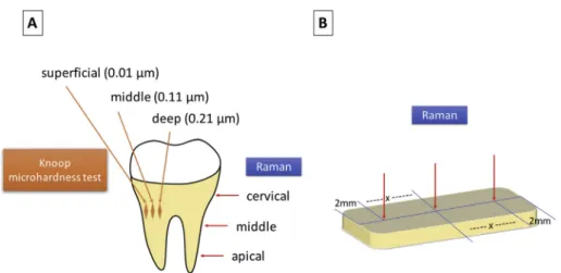
As a standardized procedure adopted in the present study, teeth were submitted to evaluation in three root regions: apical, cervical, and a midpoint between the apical and cervical regions (Fig. 2A). Bone specimens were evaluated in three focal regions (Fig. 2B): two points located 2.0 mm from the lateral aspects of the specimens, and a midpoint located between these points. Raman spectra were obtained at baseline (“pre-conditioning”), after conditioning with Carnoy's solutions (“post-conditioning”), and after post-conditioning with or without saline washing.
Raman spectra were attained in the range between 400 and 4000 cm 1 with 3 accumulations and 10 s' acquisition time per analyzed region. The analyzed Raman spectra variables were as follows: (1) mineral to matrix ratio calculated by intensity of phosphate
n1 band (~958 cm
1) divided by the combined intensities of proline and hydroxyproline bands (854þ 871 cm 1); and (2) carbonate to phosphate ratio measured by the intensity ratio of the carbonate band (~1070 cm 1) to the phosphaten1 band (~958 cm
1) (Gong et al., 2013).The present study evaluated the existing relations between band intensities due to the variability of data acquisition in Raman microspectroscopy. It includes the likelihood of a photon causing a Raman scattering effect, as well as the specimen orientation, vol-ume of laser-excited material, light absorption, cosmic radiation, and fluorescence. Thus, this measure would act to reduce in-terferences from external agents, as recommended in the published literature (Gong et al., 2013; Butler et al., 2016).
2.7. Knoop microhardness test
All teeth were mesiodistally sectioned into equal cross-sectional parts using a water-cooled low-speed Isomet diamond saw (Buehler Ltd., Lake Bluff, IL, USA). The resulting teeth sections were positioned on self-cure acrylic-filled cylindrical molds, with the sectioned sur-face facing outward and then metallographically polished using a series of 400, 500, and 600 silicon carbide abrasive grit papers. The specimens werefinely polished with a water-based diamond paste of 1 to 0.25
mm (Buehler, Lake Bluff, IL, USA) at low speed over a 180-s
interval to provide a flat surface, which was confirmed at x40 magnification. This protocol was adopted because microhardness requires polished dental surfaces aiming to allow an adequated visualization of the indentations (Albuquerque et al., 2016).Knoop microhardness was evaluated using a microhardness tester (Shimadzu HMV-2000, Shimadzu Corporation, Kyoto, Japan). Settings for load and penetration were 25 g and 15 s, respectively.
Knoop penetrations were made on the acrylic surface of each sample (apical, cervical, and middle root regions adopted in the present study) at depths of 0.01, 0.11, and 0.21
mm (
Fig. 2A). Three measurements were performed for each distance, and a mean value was calculated.2.8. Scanning electron microscopy/Energy dispersive spectroscopy
After finishing the previously described tests, the specimens were mounted onto stubs with double-faced conductive adhesive tape, metalized with gold powder (Suvorova et al., 2007) (Quorum Metallizer QT150ES, Quorum Technologies, Laughton, UK), vacuu-mized, and then analyzed using a scanning electron microscopy (SEM inspect-50, FEI, Hillsboro, OR, USA). This was used in conjunction with an energy-dispersive X-ray spectroscopy detector (Oxford Instruments, Abingdon, Oxfordshire, UK) installed in the vacuum chamber of the scanning electron microscope. The surface images were captured through a Philips XL30 microscope, and all analyses were performed using the manufacturer's software (AztecEnergy, Oxford Instruments, Abingdon, Oxfordshire, UK). This protocol aimed to evaluate possible surface changes and composi-tion of the studied specimens.
2.9. Statistical analysis
Raman microspectroscopy data are presented as mean±standard error of the mean. The difference between groups was evaluated using one-way analysis of variance followed by the Bonferroni posttest. Data from the Knoop hardness test were analyzed using the Student t test. All analysis were performed using the GraphPad Prism 5.0 software, and the significance level for all variables wasfixed at 5%. In addition, scanning electron microscopy data were analyzed by observation of qualitative structural alterations on the evaluated surfaces and quantifying the proportion of sample components.
3. Results
3.1. Raman microspectroscopy
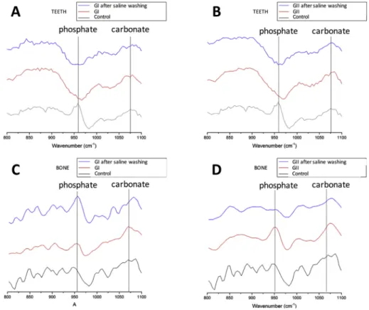
The analysis of isolated bands and peaks provided significant changes in phosphate v1 (~960 cm-1) and carbonate (~1070 cm-1) bands (Fig. 3). Thisfinding was noticeable because of the loss of the phosphate v1 band after conditioning with both Carnoy's solutions on the dental substrate (Fig. 3A and B), as well as the loss of the
Fig. 2.Schematic representation of the sites in teeth and bone specimens on which the tests were performed. (A) Raman spectroscopy (apical, middle, and cervical thirds) and Knoop microhardness test (superficial region, 0.01mm; middle region, 0.11; and deep region, 0.21mm) on teeth specimens. (B) Raman spectroscopy performed on bone substrates,
peak after washing with saline solution in the substrate bone treated with Carnoy's solution without chloroform (Fig. 3D). After analyzing the isolated bands, the values obtained by the Raman parameters of the human substrates conditioned with Carnoy's solution with chloroform and that without chloroform (Tables 1 and 2) were evaluated. The mineral matrix/organic matrix and carbonate/phosphate (Fig. 4) were evaluated in the cementum and bone surface (Fig. 5) of the specimens.
In the root teeth surface, a reduced mineral matrix/organic matrix ratio was observed when both studied Carnoy's solutions were used (Fig. 4A). Considering this parameter in relation to group II, a statis-tically significant difference (p¼0.025) was observed between pre-conditioning, post-pre-conditioning, and post-conditioning followed by saline washing. In this group, a statistically significant difference was observed between pre-conditioning versus post-conditioning with saline washing (p¼0.003). In the group in which Carnoy's solution was formulated with chloroform (group I), a statistically significant difference was observed between pre-conditioning versus post-conditioning (p¼0.004), as well as pre-conditioning versus post-conditioning with saline washing (p¼0.001).
There was an increase of cementum carbonate/phosphate ratio in Carnoy's solution either with or without chloroform (Fig. 4B). In group I, there was a statistically significant difference for this parameter regarding pre-conditioning versus post-conditioning (p¼ 0.036), and pre-conditioning versus post-conditioning with saline washing (p¼0.001). In group II, a statistically significant difference (p ¼ 0.004) was observed between pre-conditioning, post-conditioning, and post-conditioning with saline washing. In this experimental group, a statistically significant difference was observed between pre-conditioning versus post-conditioning with saline washing (p¼0.001).
3.2. Knoop microhardness
There was a decrease in Knoop microhardness between pre- and post-conditioning values when the test was performed at a distance of 0.01
mm.
Table 3 shows differences among group I (pre-condi-tioning, 38.48±4.411; post-conditioning, 24.06±1.413), group II (pre-conditioning, 38.48±4.411; post-conditioning, 32.96±2.733), and the control group (pre-conditioning, 34.08 ± 5.994; post-conditioning, 40.25 ± 9.369). There was a statistically significant difference (p¼0.036) in group I (Fig. 6).3.3. Scanning electron microscopy/Energy-dispersive spectroscopy
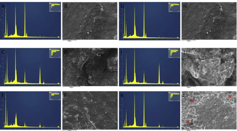
It has been known that calcium (Ca) content in normal cementum is usually higher than phosphorus (P) amount (Fig. 7A) (Almehdi et al., 2013). However, group I (Fig. 7C) showed CaeP ratio reduced and inverted, reduction of the organic component (carbon - C), and an increase in the concentration of iron ions (Fe). In this group, post-conditioning with saline washing rendered a normal CaeP ratio but inverted, reestablishment of the organic component (C), and reduction of the concentration of Fe ions (Fig. 7E). Similarly, group II showed na inversion of the CaeP ratio and organic component (C) preservation; however, there was an increase in the concentration of Fe ions (Fig. 8I). Post-conditioning with saline washing rendered a normal proportion between Ca and P contents, decrease in the organic component (C), and reduced concentration of Fe ions (Fig. 7I). In addition, smear-layer was observed after conditioning with both Carnoy solutions (Fig. 7D and H), but group II showed a reduction in its electrodensity (Fig. 7H). Post-conditioning with saline washing did not show the presence of smear-layer either in group I or II (Fig. 7F and L); however, a more
Table 1
Raman spectra data according to mineral/matrix and carbonate/phosphate ratios on teeth specimens.
Control PRE Control POST Group I PRE Group I POST Group I POSTþwashing
Group II PRE Group II POST Group II POSTþwashing
Mineral/Matrix ratio
Mean 0.1911 0.06497 0.2153 0.1289 0.04592 0.2102 0.04253 0.0814
Standard error of the mean 0.04581 0.03064 0.05293 0.06336 0.02739 0.04383 0.003497 0.03088
Carbonate/Phosphate ratio
Mean 7.991 10.2 3.661 9.735 13.85 3.49 8.088 10.92
Standard error of the mean 8.692 13.37 3.881 10.79 13.02 4.335 8.976 7.917
Control, control group; Group I, Carnoy's solution with chloroform; Group II, Carnoy's solution without chloroform; PRE, pre-conditioning; POST, post-conditioning; POSTþwashing, post-conditioning followed by saline washing.
Fig. 4.Raman spectroscopy parameters in teeth specimens. (A) Mineral/matrix ratio. (B) Carbonate/phosphate ratio.ap<0.05 versus pre-conditioning; analysis of variance/Bon-ferroni test. GI, group I. GII, group II. *p¼0.025; #p¼0.004.
Fig. 5.Raman spectroscopy parameters in bone specimens. (A) Mineral/matrix ratio. (B) Carbonate/phosphate ratio.ap<0.05 versus pre-conditioning; analysis of variance/Bon-ferroni test. GI, group I. GII, group II.
Table 3
Mean Knoop microhardness according to teeth analyzed regions.
Control PRE Control POST Group I PRE Group I POST Group II PRE Group II POST
0.01mm (Superficial)
Mean 34.08 40.25 38.48 24.06 38.48 32.96
Standard error of the mean 5.994 9.369 4.411 1.413 4.411 2.733
0.11mm (Medium)
Mean 29.75 35.72 33 37.69 40.12 39.23
Standard error of the mean 5.289 8.076 2.115 7.558 3.396 2.396
0.21mm (Deep)
Mean 41.73 33.02 34.41 42.4 43.12 39.64
Standard error of the mean 11.52 6.281 0.6339 6.994 3.25 3.799
Control, control group; Group I, Carnoy's solution with chloroform; Group II, Carnoy's solution without chloroform; PRE, pre-conditioning; POST, post-conditioning. Table 2
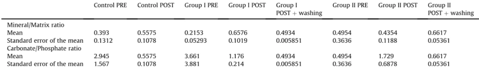
Raman spectra data according to mineral/matrix, carbonate/phosphate ratios on bone specimens.
Control PRE Control POST Group I PRE Group I POST Group I POSTþwashing
Group II PRE Group II POST Group II POSTþwashing
Mineral/Matrix ratio
Mean 0.393 0.5575 0.2153 0.6576 0.4934 0.4954 0.4354 0.6617
Standard error of the mean 0.1312 0.1078 0.05293 0.1019 0.005851 0.3636 0.1188 0.05361
Carbonate/Phosphate ratio
Mean 2.945 0.5575 3.661 1.176 0.4934 0.4954 1.729 0.6617
Standard error of the mean 1.567 0.1078 3.881 0.214 0.005851 0.3636 0.6878 0.05361
eroded appearance was found on the surface treated with Carnoy's solution without chloroform (Fig. 7J).
In bone, similarly to cementum, the physiological amount of Ca is usually greater than the level of P (Almehdi et al., 2013), as observed inFig. 8A. Group I showed a reduced and inverted CaeP ratio, and the increase of organic component (C) and Fe concen-tration (Fig. 8C). Post-conditioning with saline washing rendered the abnormal CaeP ratio but with inverted proportions between both chemical contents, increased organic component (C), and reduction of Fe concentration (Fig. 8E). Group II showed a slight inversion of the CaeP ratio and an increase in the organic compo-nent (C) content; however, there was a highlighted increase in Fe concentration (Fig. 8I). Post-conditioning with saline washing rendered a normal CaeP ratio, a highlighted increase of the organic component (C), and reduction of Fe concentration (Fig. 8I). The smear-layer was observed in both groups (Fig. 8D and H); however, it was intense in group II (Fig. 8H). Post-conditioning with saline washing did not show its presence (Fig. 8F and J), but an evident eroded surface aspect was observed in group II after saline washing (Fig. 8J).
4. Discussion
The ideal management of aggressive jaw lesions, especially benign odontogenic tumors, has been widely discussed in the literature. The treatment would be able to provide complete lesional exeresis without significant post-operative morbidity (Costa et al., 2010; Pogrel, 2015). In this context, Carnoy's solu-tion has been considered an effective adjunct therapy (Albuquerque et al., 2016), and data regarding this treatment have been reported usually based on in vivo studies. For example, a systematic review with a meta-analysis showed that Carnoy's solution combined with enucleation resulted in the lowest recurrence rate (Al-Moraissi et al., 2017). Thus, such clinical evidence reinforces the importance of the present investigation, since in vitro Carnoy's solution data have not yet been published. To date, this is the first in vitro study using both Carnoy's formulations for which there are reported data from Raman microspectroscopy, scanning electron microscopy with energy dispersive spectroscopy, and microhardness of human tooth and bone specimens.
Fig. 6.Knoop microhardness mean±standard deviation values between control group, GI (group I), and GII (group II). (A) Superficial region (0.01mm). (B) Medium region (0.11mm).
(C) Deep region (0.21mm). *p<0.05 versus pre-conditioning; Studentttest. GI, group I. GII, group II.
The analysis of chloroform influence on Carnoy's solution properties when applied to oral hard tissues was an important aspect addressed in the current literature. Data from this study showed some chemical, physical, and ultrastructural differences in specimens subjected to Carnoy's solution with chloroform. Hellstein et al. (2007) evaluated the tissue damage after using Carnoy's solution in specimens containing bone, connective tissue, and mucosa, and they did not observe a chloroform-related addi-tional tissue effect. However, a clinical study (Dashow et al., 2013) showed a beneficial effect of formulations using chloroform on the rate of recurrence of odontogenic keratocyst, which significantly reduced the rate and the average time of recurrence in comparison with Carnoy's solution without chloroform. In the present in vitro study, the presence of chloroform resulted in more destruction and structural alterations of human mineral components.
Bone tissue (Abou Neel et al., 2016) and dentin and cementum (Schulze et al., 2004) have both organic and inorganic contents. The inorganic component of these hard tissues consists of biological apatite (Ca10(PO4)6(OH)2), which possesses a hexagonal unit cell (Abou Neel et al., 2016). Repetitions of biological apatite unit cells produce crystals of various sizes with calciumephosphate ratio differences (Almehdi et al., 2013), which provides the necessary hardness (Abou Neel et al., 2016). In this study, Raman spectra analysis of bands and peaks showed alterations in phosphate v1 (~960 cm-1), which was characterized by the loss of the phosphate v1 band in both groups on the dental/bone specimens. Scanning electron microscopy with energy dispersive spectroscopy showed that Carnoy's solution altered the proportion between the elemental atomic percentage values of Ca and P, as well as the Knoop microhardness (Gong et al., 2013).
Both Ca and P have shown great importance in the composition of bones and teeth, influencing the stiffness andflexibility of these
tissues (Abou Neel et al., 2016). Structural modifications can occur, for example, by replacing calcium ions with magnesium and so-dium ions, which replaces the hydroxyl sites withfluorides and chlorides, as well as by substituting both phosphate and hydroxyl contents by carbonates. With the substitution of ions, a consider-able variation in the properties of the hydroxyapatite can occur; for example, like the substitution of magnesium inhibits the growth of the crystals, substitution by carbonate increases the solubility, whereas the substitution by fluoride decreases the solubility (Nanci, 2008). Considering that these changes reflect on the prop-erties of the related tissues (Abou Neel et al., 2016; Nanci, 2008), the present study highlighted some alterations after microhardness analysis. Carnoy's solution with chloroform showed the highest reduction in the microhardness value, presenting a statistically significant difference, which reinforced the tissue erosion pattern observed in tissues subjected to Carnoy's solution with chloroform. The authors of the present study believe that thisfinding can be partially explained by the chloroform chemical properties, which enhance the penetration of alcohol in the tissues, promoting tissue dehydration through the dissolution of membrane lipids, and leading to a significant specimen demineralization and tissue damage (Luz et al., 2008; Dias et al., 2016; Tsai, 2006).
Regarding the present study, Raman microspectroscopy was a valuable tool to adequately reflect the chemical patterns of the studied tissues. The known Raman spectrum aspects that define specific cellfindings (Kann et al., 2015) were evaluated according to the regions of interest (fingerprint region,<1800 cm-1; silent region between 1800 and 2800 cm-1; and high-frequency region,
no analyses testing different Carnoy's solution formulations similar to that performed in the present in vitro investigation. In order to evaluate these tissues, isolated peaks of phosphate and carbonate were analyzed; however, the possibility of influence from specific factors (mirroring, size of the evaluated granules, refractive index, and roughness of the specimen) justified the establishment of a relation between peak intensities used in the present methodology (Gong et al., 2013; Morris and Mandair, 2011).
The Carnoy's solutions caused an acidic environment (Nouri et al., 2015; Baker, 1958) that promoted a demineralization process, resulting in the loss of mineral content in both the teeth and bone specimens studied. It is possible that Carnoy's solution related hydroxyapatite degradation may have occurred as the same way as during physiological processes or pathological conditions, such as bone remodeling and carious lesions. It has been assumed that pH reduction favors the degradation of the hydroxyapatite in ortho-calcium phosphate and di-ortho-calcium phosphate dihydrate, whereas a pH increase enhances the displacement of this chemical reaction to the opposite side, favoring the formation of hydroxyapatite (Abou Neel et al., 2016;Seredin et al., 2015). In addition, the acid pattern related to the Carnoy's solutions also gives a greater propensity to calcium loss, resulting in a more carbonated structure (Abou Neel et al., 2016; Seredin et al., 2015). Thisfinding was evident in the teeth specimens in this study, which showed a mineral matrix/ organic matrix ratio decrease and a carbonate/phosphate ratio increase. This fact was also observed by scanning electron micro-scopy associated with a dispersive energy spectrometer.
Three-dimensional aspects of the morphological and organiza-tional characteristics of teeth and bone specimens treated with different Carnoy's solutions with chloroform highlighted a signifi -cant structural destruction along with an altered CaeP ratio, both occurring in post-conditioning and post-conditioning followed by saline washing. Regarding the solution without chloroform, there was an important structural disorganization of the organic compo-nent and a slightly altered mineral content, which was confirmed by energy-dispersive X-ray spectroscopy. Similarfindings related to an inverted CaeP ratio and superficial erosion may be observed in a phosphoric acid challenge-based in vitro study performed by Abou Neel et al. (Abou Neel et al., 2016). In addition, these alterations correlated with the Raman microspectroscopyfindings.
Carnoy's solution with or without chloroform caused an in-crease of Fe concentration in all analyzed specimens. Thisfinding probably occurred because of the possibility of a Ca substitution in the stoichiometric formula of the hydroxyapatite by a metal cation (Abou Neel et al., 2016), such as the Fe ion. It has been known that ferric chloride has an acidic pattern and acts by promoting protein coagulation (Nouri et al., 2015). Since Carnoy's solution showed an effect of increased Fe concentration, it is possible that a progressive substitution of Ca by Fe occurred in teeth and bone hydroxyapatite crystals, increasing the Fe tissue concentration. As Carnoy's solution containing chloroform showed the highest Fe concentration, then a more pronounced coagulative necrosis may be expected. This hypothesis could justify the satisfactory clinical outcome after us-ing Carnoy's solution with chloroform in comparison to Carnoy's solution without chloroform in its formula (Dashow et al., 2013, 2015).
5. Conclusion
Data from this in vitro study showed that Carnoy's solution with or without chloroform demineralized bone and dental tissues, changing their mineral and organic matrix compositions. The presence of chloroform rendered a highlighted structural superfi -cial damage in comparison with the Carnoy's solution without this chemical agent. In addition, mean hardness values were altered
after using either Carnoy's solution with or without chloroform. Although clinical practitioners have widely used both Carnoy's solutions as an adjunct therapy in benign aggressive jaw lesions, further in vitro and animal studies are still necessary in order to expand our knowledge about Carnoy's formulations containing or not containing chloroform.
Conflict of interest and/orfinancial support
None.
References
Abou Neel EA, Aljabo A, Strange A, Ibrahim S, Coathup M, Young AM, et al: Demineralization-remineralization dynamics in teeth and bone. Int J Nanomedicine 11: 4743e4763, 2016
Al-Moraissi EA, Dahan AA, Alwadeai MS, Oginni FO, Al-Jamali JM, Alkhutari AS, et al: What surgical treatment has the lowest recurrence rate following the management of keratocystic odontogenic tumor? A large systematic review and meta-analysis. J Craniomaxillofac Surg 45(1): 131e144, 2017
Albuquerque AFM, Silva PGB, Bezerra TMM, Alves APNN, Pereira KMA, Ribeiro TR, et al: Surgical treatment with or without the use of Carnoy solution in aggressive tumors of odontogenic origin: a systematized critical literature review. Int J Clin Dent 9(2): 87e98, 2016
Almehdi A, Aoki A, Ichinose S, Taniguchi Y, Sasaki KM, Ejiri K, et al: Histological and SEM analysis of root cementum following irradiation with Er:YAG and CO2 lasers. Lasers Med Sci 28(1): 203e213, 2013
Baker JR: Principles of biological microtechnique; a study offixation and dyeing. London: Methuen, 1958
Butler HJ, Ashton L, Bird B, Cinque G, Curtis K, Dorney J, et al: Using Raman spec-troscopy to characterize biological materials. Nat Protoc 11(4): 664e687, 2016 Cano J, Benito DM, Montans J, Rodriguez-Vazquez JF, Campo J, Colmenero C: Glandular odontogenic cyst: two high-risk cases treated with conservative approaches. J Craniomaxillofac Surg 40(5): e131ee136, 2012
Carvalho FSR, Feitosa VP, Fonseca SGC, Araújo TDV, Soares ECS, Fonteles CSR, et al: Physicochemical and rheological characterization of different Carnoy's solu-tions applied in oral and maxillofacial surgery. J Raman Spectrosc 48: 1375e1384, 2017
Costa FWG, Brito GAC, Pessoa RMA, Soares ECS: Histomorphometric assessment of bone necrosis produced by two cryosurgery protocols using liquid nitrogen: an experimental study on rat femurs. J Appl Oral Sci 19(6): 604e609, 2011 Costa FWG, Soares ECS, Batista SHB: Cryosurgery in treatment of benign jaw
lesions: literature review and analyze of 103 cases previously reported. RSBO 7(2): 208e215, 2010
Cutler EC, Zollinger R: The use of sclerosing solutions in the treatment of cysts and fistulae. Am J Surg 19(3): 411e418, 1933
Dashow J, Helman JI, Edwards SP, McHugh J, Ward BB: Keratocystic odontogenic tumor recurrence rates with enucleation and curettage using Carnoy's versus modified Carnoy's solution. J Oral Maxillofac Surg 71(9): e4e5, 2013 Dashow JE, McHugh JB, Braun TM, Edwards SP, Helman JI, Ward BB: Significantly
decreased recurrence rates in keratocystic odontogenic tumor with simple enucleation and curettage using Carnoy's versus modified Carnoy's solution. J Oral Maxillofac Surg 73(11): 2132e2135, 2015
Dias AR, Pereira MA, Mello ES, Zilberstein B, Cecconello I, Ribeiro Junior U: Carnoy's solution increases the number of examined lymph nodes following gastrectomy for adenocarcinoma: a randomized trial. Gastric Cancer 19(1): 136e142, 2016 Ecker J, Horst RT, Koslovsky D: Current role of Carnoy's solution in treating
kera-tocystic odontogenic tumors. J Oral Maxillofac Surg 74(2): 278e282, 2016 Gong B, Oest ME, Mann KA, Damron TA, Morris MD: Raman spectroscopy
demonstrates prolonged alteration of bone chemical composition following extremity localized irradiation. Bone 57(1): 252e258, 2013
Gurol M, Uckan S, Guler N, Yatmaz PI: Surgical and reconstructive treatment of a large ossifyingfibroma of the mandible in a retrognathic patient. J Oral Max-illofac Surg 59(9): 1097e1100, 2001
Hawkins C, Sterrett JD, Russell C: Citric acid demineralization of cementum and dentin: the effect of the storage medium. J Clin Periodontol 24(4): 264e271, 1997
Hellstein J, Hopkins T, Morgan T: The history and mystery of Carnoy solution: an assessment of the need for chloroform. Oral Surg Oral Med Oral Pathol Oral Radiol Oral Endod 103(4): 24, 2007
Ho SP, Senkyrikova P, Marshall GW, Yun W, Wang Y, Karan K, et al: Structure, chemical composition and mechanical properties of coronal cementum in human deciduous molars. Dent Mater 25(10): 1195e1204, 2009
Kann B, Offerhaus HL, Windbergs M, Otto C: Raman microscopy for cellular investigationsefrom single cell imaging to drug carrier uptake visualization. Adv Drug Deliv Rev 89: 71e90, 2015
Kong K, Kendall C, Stone N, Notingher I: Raman spectroscopy for medical diag-nosticsefrom in-vitro biofluid assays to in-vivo cancer detection. Adv Drug Deliv Rev 89: 121e134, 2015
Luz DA, Ribeiro Jr U, Chassot C, Collet ESFS, Cecconello I, Corbett CE: Carnoy's solution enhances lymph node detection: an anatomical dissection study in cadavers. Histopathology 53(6): 740e742, 2008
Morris MD, Mandair GS: Raman assessment of bone quality. Clin Orthop Relat Res 469(8): 2160e2169, 2011
Nanci A: Ten Cate's oral histology: development, structure, and function. Maryland heights. MO: Mosby, 2008
Nouri S, Sharif MR, Sahba S: The effect of ferric chloride on superficial bleeding. Trauma Mon 20(1): e18042, 2015
Owosho AA, Bilodeau EA, Vu J, Summersgill KF: Orofacial dermalfillers: foreign body reactions, histopathologic features, and spectrometric studies. Oral Surg Oral Med Oral Pathol Oral Radiol 117(5): 617e625, 2014
Pogrel MA: The keratocystic odontogenic tumour (KCOT)ean odyssey. Int J Oral Maxillofac Surg 44(12): 1565e1568, 2015
Pollan LD, Kruger GO, Reynolds DC, Mopsik ER: Osseous cryosurgery and its effect on adjacent pulpal tissues. Oral Surg Oral Med Oral Pathol 38(5): 668e674, 1974
Rajeshkumar BP, Rai KK, Geetha NT, Shivakumar HR, Upasi AP: Carnoy's in aggressive lesions: our experience. J Oral Maxillofac Surg 12(1): 42e47, 2013
Schulze KA, Balooch M, Balooch G, Marshall GW, Marshall SJ: Micro-Raman spec-troscopic investigation of dental calcified tissues. J Biomed Mater Res A 69(2): 286e293, 2004
Seredin P, Goloshchapov D, Prutskij T, Ippolitov Y: Phase transformations in a hu-man tooth tissue at the initial stage of caries. PLoS One 10(4), 2015 e0124008 Suvorova EI, Petrenko PP, Buffat PA: Scanning and transmission electron microscopy
for evaluation of order/disorder in bone structure. Scanning 29(4): 162e170, 2007
Tsai C-J: Comparing DNA damage caused by formaldehyde, glutaraldehye, Carnoy's and methacarn in cancer tissuefixations. Bowling Green State University, 2006 US Food and Drug Administration: Section 460.200. In: FDA compliance policy
guides. Washington, DC: Food and Drug Administration, 219, 1992
Voorsmit RA, Stoelinga PJ, van Haelst UJ: The management of keratocysts. J Maxillofac Surg 9(4): 228e236, 1981
Voorsmit RA: The incredible keratocyst: a new approach to treatment. Deutsch Zahnarztl Z 40(6): 641e644, 1985