UNIVERSIDADE FEDERAL DO CEARÁ - UFC Campus SOBRAL FACULDADE DE MEDICINA – FAMED
PROGRAMA DE PÓS-GRADUAÇÃO EM CIÊNCIAS DA SAÚDE
LUZIA HERMINIA TEIXEIRA DE SOUSA
Influência da osteoporose induzida por glicocorticoide na perda
óssea alveolar em ratos com periodontite experimental e o efeito
do tratamento com Atorvastatina
LUZIA HERMINIA TEIXEIRA DE SOUSA
Influência da osteoporose induzida por glicocorticoide na perda
óssea alveolar em ratos com periodontite experimental e o efeito
do tratamento com Atorvastatina
Dissertação apresentada ao
Programa de Pós-graduação em
Ciências
da
Saúde
da
Universidade Federal do Ceará,
como requisito para a obtenção
do Título de Mestre em Ciências
da Saúde.
Orientadora: Profa. Dra. Paula
Goes.
Aos meus pais Francisco e
Socorro pelo apoio incondicional
"Se cheguei até aqui foi porque
me apoiei no ombro dos gigantes"
AGRADECIMENTOS ESPECIAIS
Agradeço, em primeiro lugar, a Deus, pela oportunidade
de viver, pela força, por sua infinita bondade e por não me
deixar fraquejar nunca.
Aos meus pais, Francisco e Socorro, os quais sempre
estiveram ao meu lado e me motivaram à busca dos meus
ideais, e são responsáveis pelo que eu sou.
À minha irmã, Vitória Régia, pelo amor e amizade
reservados a mim durante todas as etapas de minha vida, os
quais certamente foram essenciais para a concretização de
mais um objetivo.
Aos meus familiares, ao meu namorado Estevão Gomes,
que sempre esteve ao meu lado, à minha avó Herminia e meu
tio Mário, que sempre me incentivaram e contribuíram em
mais uma etapa alcançada na minha vida.
A todos os meus amigos, em especial ao Ealber Luna, que
desde a vida acadêmica, permanecem fazendo parte da minha
vida e que, além do apoio imprescindível, também
compartilharam muitos momentos importantes da minha
AGRADECIMENTOS
À minha orientadora, Profa. Dra. Paula Goes, pelo incentivo,
competência, paciência, amizade, além dos constantes e valiosos
ensinamentos em meu processo de aprendizagem.
Aos professores do Programa de Pós-Graduação em Ciências
da Saúde (PPGCS), em especial ao Prof. Dr. Paulo Santos, pela
compressão e ajuda inestimável que muito contribuíram em minha
formação acadêmica.
Às professoras Mirna Marques Bezerra e Hellíada Vasconcelos
Chaves do Laboratório de Farmacologia de Sobral (LAFS) pela
amizade, compreensão e contribuição na realização de diversas
fases desse estudo.
Ao doutorando Mário Lisboa e à Profa. Dra. Flávia Furlaneto
pela colaboração na pesquisa desse trabalho.
Aos doutorandos Danielle do Val, Raul Freitas e Isabela Ribeiro
pelo companheirismo, disposição e dedicação em auxiliar durante
toda a pesquisa.
Às acadêmicas Trycia Magalhães e Larissa França pela
amizade e valiosíssima ajuda em todas as etapas desse trabalho.
Aos colegas do Laboratório (LAFS) Hermany Capistrano,
Karianne Mendonça, Pedro Henrique, Giselle Linhares, Paula Frota,
Profa. Alrieta Teixeira e Eveline Moura pela colaboração nas etapas
da pesquisa.
À funcionária da secretaria do PPGCS, Mary, pela atenção
prestada.
À doutoranda Conceição Martins e a técnica de laboratório
Socorro França (Departamento de Morfologia-UFC) por suas
colaborações nas etapas desse estudo.
À Fundação Cearense de Apoio ao desenvolvimento Científico
e Tecnológico (FUNCAP) e ao Conselho Nacional de
Desenvolvimento Científico e Tecnológico (CNPq) pelo suporte
financeiro a este estudo e pela concessão de bolsa de mestrado.
RESUMO
A Periodontite é uma doença infecto-inflamatória crônica que afeta as estruturas de suporte dos dentes. A osteoporose induzida por glicocorticoides (OIGC) é o efeito adverso principal do uso prolongado do fármaco. A atorvastatina (ATV), um fármaco hipolipemiante, tem se destacado por apresentar efeitos pleiotrópicos: anti-inflamatório, antioxidante e anabólico ósseo. Diante disso, essa dissertação tem como objetivo avaliar a influência da osteoporose induzida por glicocorticoide na perda óssea alveolar em ratos com periodontite experimental (PE) e o efeito anti-inflamatório, antirreabsortivo e antioxidante do tratamento com Atorvastatina. No estudo foram utilizados 48 ratos Wistar
machos que foram divididos em grupos: Naïve, PE, OIGC, OIGC+PE e ATV.
Para indução da osteoporose induzida por glicocorticoide, os grupos OIGC, OIGC+PE e ATV receberam injeções intramusculares de 7 mg/kg de dexametasona (DEXA) 1x/semana por 5 semanas, e os grupos Naïve e PE
receberam injeções intramusculares de 0,5 ml de solução salina (SAL). Em seguida, os grupos PE, OIGC+PE e ATV foram submetidos a PE induzida por ligadura em torno do 2o molar superior esquerdo. O grupo ATV recebeu 27 mg/kg de ATV 30 minutos antes da ligadura e diariamente por 11 dias, os demais (Naïve, PE, OICG e OICG+PE), receberam 2 ml de SAL por via oral por
11 dias, quando então foram eutanasiados. Os animais do grupo Naïve não
receberam ligadura e foram utilizados como controle. A perda óssea alveolar (POA) foi determinada por exames macroscópicos, radiográficos, micro-tomográficos e microscópicos das hemimaxilas dos animais. Os fêmures direitos dos ratos foram removidos para análise radiográfica e biomecânica. A atividade de mieloperoxidase (MPO) na gengiva, e os níveis gengivais de fator de necrose tumoral (TNF)-α, interleucina (IL)-1β, -6, -8, e -10, glutationa reduzida (GSH), superóxido dismutase (SOD) e catalase (CAT) foram analisados para investigar o estresse oxidativo e inflamação. Para avaliar a influência da via WNT/β -catenina, a expressão imunohistoquimica de RANKL, OPG, WNT10b, DKK-1 e β-catenina foram investigadas. O leucograma e os níveis séricos de transaminases, ureia, creatinina e fosfatase alcalina óssea (FAO) também foram realizadas. Os animais do grupo OICG+PE mostraram maior POA (p<0,05), maior redução de FAO (p<0,05), da densidade radiográfica da maxila e módulo de Young no fêmur (p<0,05) quando comparado ao grupo PE. O tratamento com ATV reduziu a POA considerando todos os parâmetros analisados. A ATV reduziu a atividade de MPO, e os níveis de TNF-α, IL-1β, -6, e -8, enquanto aumentou os níveis gengivais de IL-10, GSH, SOD e CAT. ATV reduziu a expressão de RANKL e DKK-1, e aumentou OPG, WNT10b e β-catenina. A administração de ATV aumentou a atividade de FAO no 11o dia comparado a OICG+PE. Nenhuma diferença foi vista nos níveis de transaminases, ureia e creatinina em qualquer dos grupos estudados. Assim podemos concluir que OICG piora a POA, inibe a atividade osteoblástica e altera a perda óssea sistêmica em ratos com PE, e que a ATV previne a POA em ratos submetidos à OICG+PE, por meio de efeito antirreabsortivo, anti-inflamatório e antioxidante, com participação da via de sinalização WNT.
ABSTRACT
Periodontitis is a chronic infectious and inflammatory disease affecting the tooth supporting structures. The glucocorticoid-induced osteoporosis (GIOP) is the major adverse effect of long-term use of the drug. Atorvastatin (ATV) a lipid-lowering drug, has been highlighted by presenting pleiotropic effects: anti-inflammatory, antioxidant and bone anabolic. Therefore, this dissertation aims to evaluate the influence of glucocorticoid-induced osteoporosis in alveolar bone loss in rats with experimental periodontitis (EP) and anti-inflammatory effect, antirreabsortivo and antioxidant treatment with Atorvastatin. For the study, 48 male Wistar rats were divided into groups: Naïve, EP, GIOP, GIOPE + EP and
ATV. For induction of glucocorticoid-induced osteoporosis, the GIOP groups GIOP + EP and ATV received intramuscular injections of 7 mg / kg of dexamethasone (DEXA) 1x / week for 5 weeks, and Naïve and EP groups were
given intramuscular injections of 0.5 ml of saline (SAL). Then the EP groups GIOP + EP and EP were subjected to ATV induced by ligature around the left upper second molar. The ATV group received 27 mg / kg ATV 30 minutes before the ligation and daily for 11 days, the other (Naïve, EP, GIOP and GIOP + EP)
received 2 ml of SAL orally for 11 days, when they were euthanized. Animals
naïve group received no ligation and were used as controls. Alveolar bone loss
(ABL) was determined by macroscopic, radiographic, micro-tomographic and microscopic of the maxillary sides of animals. The right femurs of rats were removed for radiographic and biomechanical analysis. Myeloperoxidase activity (MPO) in the gingiva and gingival levels of tumor necrosis factor (TNF) -α, interleukin (IL) -1β, -6, -8, and -10, reduced glutathione (GSH), superoxide dismutase (SOD) and catalase (CAT) were analyzed to investigate oxidative stress and inflammation. To evaluate the influence of Wnt / β-catenin, the immunohistochemical expression of RANKL, OPG, Wnt10b, DKK-1 and β -catenin were investigated. The leucocyte count and serum levels of transaminases, urea, creatinine, and bone alkaline phosphatase (BALP) were also performed. The animals GIOP + EP group showed higher of ABL (p <0.05) greater reduction of BALP (p <0.05), the radiographic density of the maxilla and Young's modulus of the femur (p <0.05) when compared the EP group. Treatment with ATV reduced the ABL considering all parameters. The ATV reduced MPO activity and TNF-α levels, IL-1β, -6, and -8, while gingival increased levels of IL-10, GSH, SOD and CAT. ATV reduced the expression of DKK-1 and RANKL and increased OPG Wnt10b and β-catenin. The ATV administration increased the BALP activity on the 11th day compared to GIOP + EP. No difference was seen in the levels of transaminases, urea and creatinine in any of the groups studied. Thus we conclude that GIOP worsens ABL inhibit osteoblastic activity and alters systemic bone loss in rats with EP, and ATV prevents ABL in rats subjected to GIOP + EP through antirreabsortive effect, anti-inflammatory and antioxidant with participation of the WNT signaling pathway.
LISTA DE ABREVIATURAS
ATV Atorvastatina
BMP Proteína morfogenética óssea CBFA core-binding factor subunit alpha
CAT Catalase
DKK Dickkopf
EO Estresse Oxidativo
EROs Espécies reativas de oxigênio FAO Fosfatase alcalina óssea GCs Glicocorticoides
GPx Glutationa peroxidase
HMG-CoA 3-hidroxi3-metilglutaratl co-enzima A ICAM Moléculas de adesão intercelular IFN Interferon
IL Interleucina
LEF1 Fator amplificador de linfócitos LPS Lipopolissacarídeos
LRP5/6 Low-density lipoprotein receptor-related protein 5/6
MPO Mieloperoxidase
OIGC Osteoporose Induzida por glicocorticoide OPG Osteoprotegerina
PG Prostaglandina PMNs Polimorfonucleares POA Perda óssea alveolar
RANK Receptor Ativador do Fator Nuclear-κB
RANKL Ligante do Receptor Ativador do Fator Nuclear-κB ROOH Peróxido
RAR Raspagem e alisamento radicular RUNX Runt-related transcription fator
SAL Salina
SFRP Proteína relacionada ao frizzled secretada
SOST Esclerostina
TCF Fator de célula T
TNF Fator de Necrose Tumoral VCAM Moléculas de adesão vascular
VEGF Fator de crescimento endotelial vascular WIF Fator inibidor de WNT
Sumário
1. INTRODUÇÃO ………. 14
1.1 Periodontite……… 14
1.2 Osteoporose Induzida por glicocorticoide (OIGC)... 18
1.3 OICG e Periodontite... 19
1.4 Estatinas... 20
2. OBJETIVOS GERAIS E ESPECÍFICOS……… 22
3. CAPÍTULOS... 23
3.1. Effect of glucocorticoid-induced osteoporosis on alveolar bone loss of rats with experimental periodontitis……… 24
3.2. Effects of Atorvastatin on periodontitis of rats subjected to glucocorticoid-induced osteoporosis ……… 44
4. CONCLUSÃO GERAL………... 70
5. REFERÊNCIAS……….. 71
1. INTRODUÇÃO
1.1. PERIODONTITE
A periodontite é uma doença infecto-inflamatória dos tecidos de suporte do dente, de alta prevalência na população mundial, e é uma das principais causas de perda dentária (Petersen e Ogawa, 2005; Borrell e Papapanou, 2005). A etiologia da periodontite é multifatorial. Estudos têm demonstrado que a presença de periodontopatógenos é fundamental para o desencadeamento dessa doença nos sítios periodontais (Teles et al., 2006; Boehm &
Scannappieco, 2007; Bascones et al., 2009). Fatores de risco genéticos e ambientais podem alterar a resposta do hospedeiro, o que contribui sobremaneira para o início e progressão da periodontite (Jansson, 2006), por desequilibrar a homeostasia dos tecidos periodontais (Pihlstrom et al., 2005). A susceptibilidade do hospedeiro é determinante para o início e progressão da doença, pois a hiperresponsividade de vias imunológicas e exacerbação da inflamação com a liberação de citocinas inflamatórias, podem resultar em destruição tecidual aumentada (Pihlstrom et al., 2005).
Considerando a resposta do hospedeiro, diante da presença de periodontopatógenos, há uma maior liberação de lipopolissacarídeos (LPS) e recrutamento de neutrófilos para o sítio de infecção, os quais liberam enzimas proteolíticas que causam dano tecidual (Giannobile, 2008). A primeira linha de defesa contra os periodontopatógenos presentes no periodonto é formada pelos leucócitos polimorfonucleares (PMN). No sulco gengival, os PMNs tentam eliminar as bactérias através da fagocitose, auxiliado por substâncias inflamatórias e antimicrobianas, tais como lisozima, lactoferrina, fosfatase alcalina, hidrolases ácidas, proteínas catiônicas, espécies reativas de oxigênio (EROs) e mieloperoxidase (MPO) (Bascones et al., 2009).
Os LPS também agem ativando macrófagos, que promovem a liberação de vários mediadores pró-inflamatórios, tais como interleucina (IL)-1β, Fator de Necrose Tumoral (TNF)-α e prostaglandina (PG) E2, importantes na destruição de tecido periodontal (Górska et al., 2003; Giannobile, 2008).
inflamatória torna-se cada vez mais amplificada, levando ao aparecimento de outras células da imunidade adaptativa tais como linfócitos T e B, que em conjunto acabam por repercutir no tecido ósseo aumentando a reabsorção óssea alveolar (Soedarsono et al., 2006).
A MPO é uma enzima que auxilia a geração de espécies reativas de oxigênio (EROs) e está presente nos grânulos azurofílicos dos neutrófilos, e por isso pode ser considerada um indicador do acúmulo de neutrófilos nos tecidos. Fisiologicamente, participa da destruição de microrganismos, mas, em condições patológicas e causar danos teciduais, por aumentar o estresse oxidativo (EO) (Yamalik et al., 2000; Malle et al., 2006).
Consequências do EO incluem danos ou morte de células através de uma variedade de mecanismos (Halliwell e Whiteman, 2004). Mecanismos enzimáticos são responsáveis para a neutralização direta de EROs, e esses mecanismos são constituídos por enzimas que tem por objetivo manter os níveis de EROs normais (Kaklamanos e Tsalikis, 2002). Exemplos destas enzimas são superóxido dismutase (SOD), catalase (CAT) e glutationa peroxidase (GPx) (Kaklamanos e Tsalikis, 2002).
A SOD é uma das mais abundantes enzimas antioxidantes no corpo humano (Akalin et al., 2007). Um de seus mecanismos de ação é a conversão de ânions superóxido em peróxido de hidrogênio (H2O2), operando como um antioxidante, porque evita a formação do radical hidroxila (OH-) (Scandalios et al., 2000). Os níveis de SOD tem se mostrado reduzidos em pacientes com periodontite crônica, quando comparados aos controles (Kim et al., 2010). A CAT é encontrada nos peroxissomas e é capaz de remover H2O2 e radicais superóxido intracelulares com grande eficácia (Battino et al., 1999). Níveis salivares de CAT foram reduzidos em pacientes com periodontite crônica quando comparados com adultos saudáveis (Trivedi et al., 2014). A GPx é responsável pela a proteção de células de mamíferos contra danos oxidativos por reduzir uma variedade de hidroperóxidos, tais como o ROOH e H2O2 (Chapple et al., 2002).
Nuclear-κB (RANK), pelo seu Ligante (RANKL) e pela Osteoprotegerina (OPG) (RANK/RANKL/OPG) (Xing et al., 2005; Brennan et al., 2007; Reid & Holen, 2009). O RANKL, pertencente à superfamília do TNF, é expresso por osteoblastos como uma proteína transmembrana e se liga ao seu receptor RANK na superfície de osteoclastos e de precursores de osteoclastos. Isso resulta na ativação de vias de sinalização que conduzem à formação, diferenciação e ativação de osteoclastos e, consequente, reabsorção óssea (Reid & Holen, 2009). Por outro lado, para regular o metabolismo ósseo, osteoblastos produzem e liberam a OPG que inibe a interação RANKL-RANK, por meio de ligação competitiva com RANKL (Soedarsono et al., 2006; Reid & Holen, 2009). Entretanto, durante um processo inflamatório crônico, como se observa na periodontite, o aumento dos vários mediadores pró-inflamatórios, tais como TNF-α e IL-1β, estimulam a expressão abundante de RANKL em osteoblastos, bem como em células T, B e fibroblastos, favorecendo então a osteoclastogênese (Soedarsono et al., 2006).
Além do eixo RANK-RANKL-OPG, mais recentemente, foi relatado na literatura a participação do via WNT/β-catenina como um modulador do metabolismo ósseo (Wang et al., 2014). Esta via tem sido bastante explorada em estudos de proliferação celular e carcinogênese (Kikuchi, 2003; Nusse, 2005; Clevers, 2006; Sherwood, 2015), entretanto, foi demostrado que ela também é crítica para a diferenciação de células mesenquimais em osteoblastos maduros (Hartmann, 2006).
Os membros da família WNT são proteínas secretadas que após interação com seu receptor impede a destruição da β-catenina, a qual se estabiliza e é translocada para o núcleo e, acompanhada por LEF1 (fator amplificador de linfócitos)/TCF (fator de célula T), funciona como co-ativador para transcrição de genes que levam a diferenciação de osteoblastos (Moon et al., 2004). Esta via por sua vez, é controlada por proteínas, tais como proteína relacionada ao frizzled secretada (SFRP) ou fator inibidor de WNT (WIF) que
capturam e inativam a WNT no compartimento intercelular, ou ainda Dickkopf (DKK) e Esclerostina (SOST) que bloqueiam o receptor LRP5/LRP6 (
Low-density lipoprotein receptor-related protein) de WNT (Wend et al., 2012). Em
levando a um bloqueio dessa via a qual é importante para o metabolismo ósseo levando a alterações na osteoblastogênese (Westendorf et al., 2004). Foi demostrado um aumento dos níveis de SOST e DKK no tecido gengival de pacientes com periodontite crônica evidenciando o envolvimento da via WNT/β -catenina na patogênese da perda óssea na periodontite (Napimoga et al., 2014). Sabendo que o processo inflamatório e a perda óssea são os principais achados clínicos da periodontite, o diagnóstico desta doença na prática clínica tem sido realizado por meio de exames capazes de detectar parâmetros como: aspecto clínico do periodonto, presença de sangramento, profundidade de bolsas, nível clínico de inserção, dentre outros (Pihlstrom et al., 2005). Adicionalmente, como forma auxiliar de avaliação do grau de perda óssea periodontal, exames de imagens são frequentemente requeridos. Imagens radiográficas digitais se destacam frente às imagens convencionais, visto que apresentam maior capacidade de detecção de sítios com perdas ósseas ainda sutis, quando comparadas às imagens radiográficas convencionais (Khocht et al., 2003). O uso de imagens tomográficas vem assumindo posição de destaque na Odontologia e Periodontia, pois propiciam maior riqueza de detalhes quanto à qualidade e quantidade de suporte ósseo (Khocht et al., 2003), o que favorece, consequentemente, diagnóstico e terapia precoces.
1.2. OSTEOPOROSE INDUZIDA POR GLICOCORTICOIDE
Os glicocorticoides (GCs) são uma opção terapêutica eficaz utilizada no tratamento de muitas doenças inflamatórias e auto-imunes. No entanto, mesmo em doses moderadas, os GCs apresentam sérios efeitos adversos, dentre eles a osteoporose (Grossman et al., 2010; Rizzoli e Biver, 2015).
A osteoporose induzida por glicocorticóides (OIGC) é a forma mais comum da osteoporose iatrogênica (Compston, 2010). É caracterizada por fragilidade à fraturas que ocorrem em 30% a 50% de pacientes que fazem uso de GCs sistêmicos a longo prazo (Van Staa et al., 2003).
A patogênese de OIGC é o resultado dos efeitos indiretos mediados pela alteração no metabolismo do cálcio nos rins e no intestino, redução da produção de hormônios gonodais e efeitos prejudiciais sobre o sistema neuromuscular associado aos efeitos diretos dos GCs sobre as células ósseas (Canalis et al., 2007; Hofbauer et al., 2009, 2010).
Os GCs têm ações complexas sobre a expressão gênica das células ósseas dependendo do estágio de diferenciação e crescimento dos osteoblastos. Aumentam a reabsorção óssea por efeitos diretos e indiretos sobre os osteoclastos. Estes agentes inibem aspectos específicos da diferenciação osteoblástica, como a transcrição de osteocalcina, a proteína não colágena mais abundante no osso, e a expressão do colágeno tipo I. Além de diminuírem a síntese, os GCs aumentam a degradação do colágeno por aumentarem a expressão das colagenases e inibirem a expressão do inibidor tecidual das metaloproteinases. Como o colágeno tipo I é a maior proteína estrutural da matriz óssea, uma diminuição na sua expressão e aumento da sua degradação são críticos para a ação inibitória dos GCs na matriz óssea. (Schleimer, 1993; Delany et al., 1995; Canalis, 1996; Lukert, 1996).
Os efeitos sobre os osteoclastos são bifásicos, concentrações fisiológicas são necessárias para o estágio final de diferenciação e função, porém altas doses e exposição prolongada podem inibir a replicação celular. A reabsorção óssea observada in vivo ocorre pelo aumento da atividade osteoclástica com
A terapia com GCs é marcada por um aumento precoce e transiente de osteoclastos (Mazziotti et al., 2006; Weinstein, 2011) seguido da inibição importante de osteoblastos (Zhou et al., 2014). Estudos in vitro e in vivo
pré-clínico e pré-clínico, mostraram que a dexametasona impede a diferenciação osteoblástica, especialmente através da inibição da via de sinalização WNT. Este efeito foi parcialmente atribuído ao aumento da expressão de DKK-1 pela dexametasona (Ohnaka et al., 2004, 2005). Os GCs inibem a expressão Runx2/Cbfa, um fator de transcrição essencial para a diferenciação de osteoblastos e sua expressão é dependente da via WNT (Chang et al.,1998).
Uma forma de avaliação da atividade de osteoblastos aplicada a clínica em pacientes com osteoporose é a dosagem de fosfatase alcalina óssea (FAO). A FAO é uma glicoproteína tetramérica específica do osso, secretada por osteoblastos com papel na mineralização óssea (Vieira, 1999). Ela está presente nos condrócitos hipertróficos e na placa de crescimento epifisária (Price, 1993; Crofton et al., 1999). Apesar da grande semelhança estrutural entre as isoformas, imunoensaios específicos foram desenvolvidos para a isoforma óssea, o que diminui a reação cruzada com a isoforma hepática em 15% a 20% (Watts, 1999).
1.3. OIGC E PERIODONTITE
A doença periodontal esta associada a várias condições sistêmicas. Foi demonstrada a relação entre periodontite e diabetes, parto prematuro, pneumonia nosocomial, doenças cardiovasculares, osteoporose, dentre outras. (Seymour et al.,2003)
O interesse na relação entre osteoporose e periodontite tem crescido consideravelmente (von Wowern et al, 1994; Wactawski-Wende et al., 1996; Tezal et al., 2000; Boyce e Xing, 2007). Elas compartilham alguns fatores de risco comuns, tais como idade avançada, história prévia de perda óssea, algumas doenças sistêmicas, medicamentos, tabagismo e genética familiar (Reddy, 2001).
2005). A baixa densidade óssea em mulheres em fase pós-menopausa foi relacionada à perda alveolar interproximal, que persistiu após o tratamento periodontal convencional (Tezal et al., 2000). No entanto, a relação entre OIGC e periodontite ainda permanece pouco estudada. Alguns estudos suportam a ideia de que a OIGC pode piorar a destruição de tecidos de suporte do dente aumentando a agressividade e acelerando a taxa de progressão da doença periodontal (Wactawski-Wende et al., 1996; Nicopoulou-Karayianni et al., 2009). Alternativas terapêuticas, como a modulação da resposta do hospedeiro, vêm sendo estudadas como uma nova abordagem no tratamento de doenças inflamatórias ósseas como a periodontite (Preshaw et al., 2004; Buduneli, 2007).
1.4. ESTATINAS
As estatinas são uma classe farmacológica que atua sobre a via do mevalonato, inibindo a enzima 3-hidroxi-3-metilglutaratil co-enzima A (HMG-CoA) redutase. As estatinas são redutoras efetivas dos níveis séricos de colesterol, portanto, amplamente prescritas para o tratamento da hipercolesterolemia e aterosclerose (Kim et al., 2011). Além dos efeitos sobre o colesterol, estudos têm mostrado que as estatinas apresentam efeitos pleiotrópicos, incluindo ação anti-inflamatória (Dimitrow et al., 2011) e efeito anabólico ósseo (Mundy et al., 1999; Horiuchi & Maeda, 2006; Goes et al., 2010; Dalcico et al., 2013). Também foi demonstrado o efeito antioxidante das estatinas atuando reduzindo as EROs (Bolayirli et al., 2007). Tais propriedades oferecem grande potencial para estatinas modificarem o curso de doenças inflamatórias crônicas (Barsante et al., 2005) dentre as quais podem ser incluídas a periodontite (Dalcico et al., 2013).
Dentre as estatinas destaca-se a Atorvastatina (ATV), pela sua característica lipofílica e seus poucos efeitos adversos quando comparada a outras estatinas, sendo, portanto amplamente utilizada na prática clínica (Plosker e Lyseng-Williamson, 2007).
2. OBJETIVOS GERAIS
a) Avaliar a influência osteoporose induzida por glicocorticoide na perda óssea alveolar de ratos com periodontite experimental.
b) Avaliar o efeito antirreabsortivo, anti-inflamatório e antioxidante da Atorvastatina (ATV) na perda óssea alveolar de ratos submetidos à osteoporose induzida por glicocorticoide com periodontite experimental.
3. OBJETIVOS ESPECÍFICOS
a) Avaliação da POA por meio de análises macroscópicas, radiográficas, micro-tomográficas e microscópicas;
b) Avaliação da perda óssea no fêmur por meio de análises radiográficas e ensaio biomecânico
c) Avaliação da atividade anti-inflamatória através da atividade de mieloperoxidase (MPO) e quantificação dos níveis de TNF-α, IL-1β, -6, -8, -10 no tecido gengival por ELISA;
d) Avaliação da atividade antioxidante por meio da quantificação dos níveis de enzimas do estresse oxidativo (SOD, CAT e GSH) no tecido gengival por ELISA;
e) Avaliação das vias de sinalização do metabolismo ósseo por meio de análise imunohistoquímica para RANKL, OPG, WNT 10b, DKK e β -catenina;
f) Avaliação de parâmetros sistêmicos por meio do leucograma; dosagem sérica de transaminases, ureia e creatinina;
4. CAPÍTULOS
Esta dissertação esta baseada no Regimento Interno do Programa de Pós-graduação em Ciências da Saúde da Universidade Federal do Ceará – Campus
Sobral que regulamenta o formato alternativo para dissertações de Mestrado e permite a inserção de artigos científicos de autoria ou co-autoria do candidato.
Por se tratar de pesquisa envolvendo animais, os protocolos utilizados neste trabalho foram submetidos à apreciação e foram devidamente aprovados pelo comitê de ética animal com protocolo 78/2014. (Anexo I).
4.1 ARTIGO 1
“Effect of glucocorticoid-induced osteoporosis on alveolar bone loss of
rats with experimental periodontitis”
Autores: Luzia Herminia Teixeira de Sousa, Eveline Valeriano Moura, Ana Larissa Queiroz, Danielle Val, Hellíada Chaves, Mario Lisboa, Flávia Furlaneto, Gerly Anne Brito, Paula Goes
Esse artigo seguiu normas de publicação do periódico: Archives of Oral Biology
(ISSN 0003-9969). Qualis B1: Medicina II
3.2 ARTIGO 2
“Effects of Atorvastatin on periodontitis of rats subjected to glucocorticoid-induced osteoporosis”
Autores: Luzia Herminia Teixeira de Sousa, Eveline Valeriano Moura, Joanna Trycia Alexandre, Mario Lisboa, Flavia Furlaneto, Raul Freitas, Isabela Ribeiro, Danielle Val, Mirna Marques, Hellíada Chaves, Conceição Martins, Gerly Anne Brito, Paula Goes.
Esse artigo seguiu normas de publicação do periódico: Journal of
Periodontology. (ISSN 0022-3492).
Artigo 1
Effect of glucocorticoid-induced osteoporosis on alveolar bone loss of rats with experimental periodontitis
Luzia Herminia Teixeira Sousaa, Eveline Valeriano Mouraa, Ana Larissa Queirozb, Danielle Valc, Hellíada Chavesa,b, Mario Lisboad, Flávia Furlanetoe, Gerly Anne Britod,f, Paula Goesa,g,*
a Post-graduation program of Health Science, Medical School, Federal University of Ceará - Sobral, Avenida Comandante Maurocelio Rocha Pontes, 100 - Derby, Sobral - CE, Brazil, 62.042-280.
b School of Dentistry, Federal University of Ceará – Sobral, R. Cel. Estanislau Frota - Centro, Sobral - CE, Brazil, 62.010-560.
c Post Graduation Program RENORBIO, Federal University of Pernambuco, Av. Professor Morais Rego, 1235 - Cidade Universitária, Recife – PE, Brazil, 50670-901.
d Post-graduation Program of Morphological Science, Department of Morphology, Medical School, Federal University of Ceará – Fortaleza, Rua Delmiro de Farias, s/n - Rodolfo Teófilo Fortaleza – CE, Brazil, 60.430-170.
e Department of Oral & Maxillofacial Surgery and Periodontology, Ribeirão Preto School of Dentistry, University of São Paulo, Ribeirão Preto, São Paulo, Brazil, Av. do Café, 3000 - Vila Amelia, Ribeirão Preto - SP, Brazil, 14050-904.
f Department of Morphology, Medical School, Federal University of Ceará – Fortaleza, Rua Delmiro de Farias, s/n - Rodolfo Teófilo, Fortaleza – CE, Brazil, CEP 60.430-170
g Department of Pathology and Legal Medicine, Medical School, Federal University of Ceará – Fortaleza, Rua Monsenhor Furtado, S/N - Rodolfo Teófilo, Fortaleza – CE, Brazil, 60.441-750.
Fone: +55 85 3366.8301 Fax: +55 85 3366.8300. Email: paulagpinheiro@yahoo.com.br
Running title: Effect of GIOP on alveolar bone loss
ABSTRACT
Objective: To evaluate the effect of osteoporosis induced by glucocorticoid
(GIOP) in rats with experimental periodontitis (EP).
Design: 48 male Wistar rats were divided into: Naïve, EP, GIOP and GIOP+EP
groups. Rats of GIOP and GIOP+EP groups received intramuscularly 7 mg/kg of dexamethasone once a week for 5 weeks. Following, the rats from EP and GIOP+EP groups were subjected to ligature-induced EP for 11 days. Later, the animals were euthanized, and maxillae collected for macroscopic, radiographic, micro-tomographic, and microscopic analysis of alveolar bone loss (ABL). Blood samples were collected for determination of bone-specific alkaline phosphatase (BALP) serum levels and the right femurs were removed for radiographic and biomechanical analysis.
Results: EP caused ABL and reduced BALP serum levels, but did not change the
architecture or biomechanics of femur when compared to Naïve. GIOP did not cause ABL compared to Naïve, but it decreased bone mineral density, bone percentage, trabecular thickness and an increase on bone porosity, compared to Naïve (p<0.05). GIOP significantly reduced BALP serum levels, and radiographic density and Young’s module of femur, compared to Naïve. In GIOP+EP group it was seen a greater increase on ABL, when compared to EP (p<0.05). It also showed a greater reduction on BALP serum levels (p<0.05), on radiographic density and Young’s module of femur, compared to EP (p<0.05)
Conclusions: This data suggest that GIOP aggravates ABL, inhibits the
osteoblast activity and alters systemic bone loss in rats with EP.
Keywords: Periodontitis; Osteoporosis; Glucocorticoids; Alveolar bone loss
Highlights:
GIOP caused a greater decrease on osteoblast activity in animals subjected to EP
GIOP caused bone loss on femur of animals subjected to EP
1. Introduction
Periodontitis is an infectious-inflammatory and high prevalent disease, characterized by destruction of connective tissue and alveolar bone loss (ABL), and it is considered the second major cause of tooth loss (Krustrup & Petersen, 2006). This disease is mainly initiated by oral biofilm however the development of an exacerbated host response plays an important role on tissue breakdown (Pihlstrom, Michalowicz & Johnson, 2005).
Osteoporosis is a common disease characterized by systemic bone loss and impaired bone microarchitecture. It can be a consequence of hormonal imbalance in postmenopausal woman (Jilka, Weinstein, Bellido, Parfitt & Manolagas, 1992), but also it can has a secondary cause, mainly as a result of drug use such as Glucocorticoids (GCs). Glucocorticoid-induced osteoporosis (GIOP) is the most common cause of secondary osteoporosis, the first cause before 50 years and the first iatrogenic cause of the disease (Kok & Sambrook, 2009). In addition, considering the continuous raise on the prevalence of GCs use in the community population (Overman, Yeh & Deal, 2013), it seems interesting understand the biological mechanism underlying GIOP.
In recent decades, numerous studies have focused on the association between osteoporosis and periodontitis at the bone level. The majority of the studies has focused on the effect of postmenopausal osteoporosis in the loss of periodontal attachment (Hernández-Vigueras, Martínez-Garriga, Sánchez, Sanz, Estrugo-Devesa & Vinuesa, 2015; Juluri, Viswanathan, Gopalakrishnan,
Kathariya, Devanoorkar & Prashanth, 2015). However, a little is known about the
effect of GIOP on periodontal tissue and even more during periodontal inflammation. In this way we aimed to evaluate the effect of GIOP on alveolar bone loss of animals with experimental periodontitis.
2. Material and Methods Animals and study design
norvegicus) from central Animal Facility of Federal University of Ceará, weighing
180–220 g, kept in appropriate cages with six animals each. The animals were housed in standard conditions (12h light-dark cycles and temperature-controlled rooms) with food and water ad libitum. The protocols for experimental procedures
and animal treatment were approved by Animal Ethics Committee (number 78/2014) of Federal University of Ceará, Brazil.
A power calculation was performed to determine the sample size. The animal was considered the study unit. The sample size was determined to provide 80% power to recognize a significant difference of 20% among groups and the standard deviation of 15% with a 95% confidence interval (p = 0.05), considering the change in alveolar bone loss (ABL) as the primary outcome variable. Therefore, a sample size of 6 animals per group was required.
After two weeks of adapting in the laboratory environment, the rats were divided into four groups (n=6): Naïve, Experimental Periodontitis (EP), Glucocorticoid-induced osteoporosis (GIOP) and GIOP+EP. Initially, rats from GIOP and GIOP+EP groups received intramuscularly injections of 7 mg/kg dexamethasone (Decadron, Aché - Guarulhos - SP, Brazil), once a week, for 5 weeks (Lucinda, Aaresturup, Peters, Reis, de Oliveira, & Guerra, 2012), and the ones from Naïve and EP groups received 0.5 ml of 0.9% of Saline Solution. Following, the rats were anesthetized with 100 mg/ml ketamin (Cetamin - Syntec®) and 20 mg/ml xylanzine (Xilazin - Syntec®) on the dose of 1 ml/kg – intramuscularly, for EP, which was induced by the placement of a sterile nylon thread ligature (3-0; polysuture NP45330, São Paulo, Brazil) around the cervix of the maxillary left second molar of rats (EP and GIOP+EP groups) (Bezerra et al., 2000). After 11 days all animals were euthanized with 20 mg/kg thiopental (0.5 g Thiopentax; Cristália, São Paulo, SP, Brazil). Rats of Naïve and GIOP group did not receive the ligature.
Micro-computed tomography (CT) scanning and Assessment of alveolar bone loss
(ABL) was measured by a trained and blinded observer, using an imaging software (ImageJ® National Institutes of Health, Washington, DC, USA), as previously described (Goes, Lima, Melo, Rego, & Lima, 2010).
The same specimens used for macroscopic study were radiographed using a digital system (Digora Soredex Digital System®, Portslade-East Sussex, UK). The specimens were posed over the sensor and the radiographic images were acquired using 63 kVp, 8 mA, exposure time of 0.06 s and focal distance of 30 mm. These images were evaluated using Image J® software (Goes, Lima, Melo, Rego, & Lima, 2010).
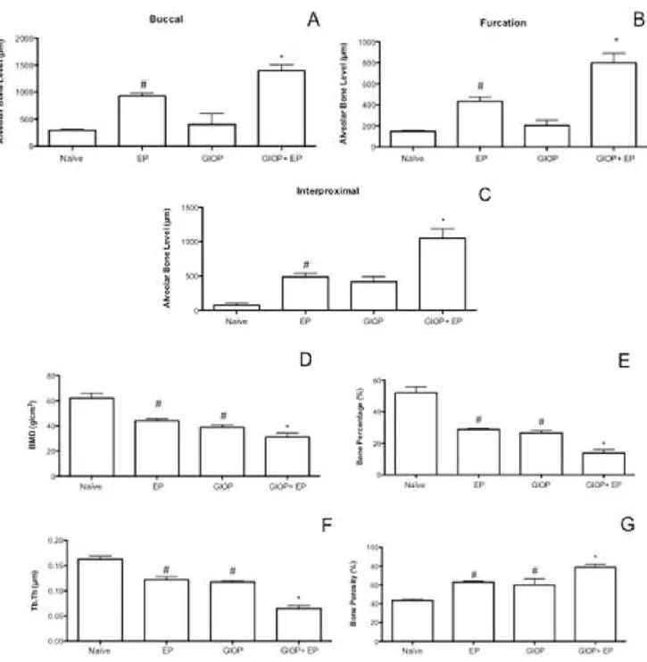
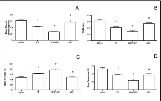
The same non-demineralized specimens were then scanned by a cone beam micro-computed tomography (CT) system (Skyscan 1172, Bruker, Kontich, Belgium). The x-ray generator was operated at an accelerated potential of 50 kV with a beam current of 200 μA and an exposure time of 560 ms per projection. Images were produced with a voxel size of 6x6x6 μm. Using an appropriated software (Data Viewer®, version 1.5.0, Bruker, Kontich, Belgium), the generated 3 dimensional models were rotated into a standard position as described previously (Lisboa, Gondim, Ervolino, Vale, Frota & Nunes, 2015). Linear measurements on ABL were performed at 3 different sites: buccal, furcation and interproximal. For the interproximal site, coronal dataset was analyzed using appropriated software (CT-Analyser®, version 1.13.5.1+, Bruker, Kontich, Belgium). Bone mineral density (BMD), bone percentage, bone porosity, and trabecular thickness (Tb.Th) were also analyzed (Lisboa, Gondim, Ervolino, Vale, Frota & Nunes, 2015). All micro-CT analyses were performed by one blinded and calibrated examiner.
Bone histology observation
Ribeiro, Chaves, Rocha, Lima, & Brito, 2005) using scores from 0–3. The histopathological analysis was performed by a certified histologist.
Serum levels of bone-specific alkaline phosphatase (BALP)
Blood samples were collected from the orbital plexus at the 11th day after EP. Bone-specific alkaline phosphatase (BALP), a thermosensible isoform of total alkaline phosphatase, was evaluated using a thermoactivation method (Whitby & Moss, 1975). The samples were heated to 56 °C for 10 min. Serum levels of BALP were calculated by the difference between heated alkaline phosphatase from total alkaline phosphatase in serum (Labest®, Lagoa Santa, MG, Brazil) (Goes, Melo, Dutra, Lima, & Lima, 2012; Goes, Melo, Silva, Benevides, Alencar & Ribeiro, 2014).
Biomechanical testing and Radiographic Density (RD) of Femur
The rats’ right femur were collected 11 days after ligature placement and fixed, for 24 h, in 10% formaldehyde (Reagen Produtos para Laboratórios Ltda®, Rio de Janeiro-RJ, Brazil). The specimens were radiographed using Digora Soredex System® (Dental Imaging Company Ltd, Portslade-East Sussex, UK) with the same configuration as for the maxilla. These images were evaluated by Image J® software. A region of interest (ROI) was created measuring 0.5 (w) 0.5 (y), in the form of a square, with 473 x 229 pixels, and was posed in the proximal femur diaphysis, and the same evaluator drew all ROI panels. Grey tone differences from both areas were considered as a value of radiographic density. The RD analysis was done similarly to the one of maxilla (Mahl, Tonietto, Giorgi, Girotto, & Fontanella, 2009).
Statistical analysis
The data are presented as mean±standard error of the mean or as median (range), where appropriate. Normality and homoscedasticity of the data were verified. ANOVA followed by the Bonferroni test were used to compare the means, and Kruskal–Wallis and Dunn tests were used to compare the medians. The significance level was set at 5% in all tests. All calculations were performed using the Prism 5 (GraphPad Software Inc., San Diego, CA, USA). All protocols and analyses were performed in a blinded manner.
3. Results
Assessment of alveolar bone loss and Micro-computed tomography (CT) scanning
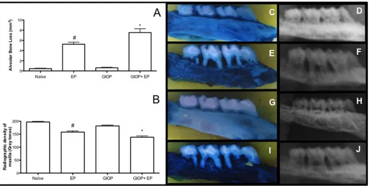
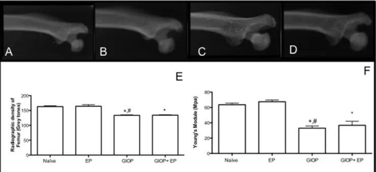
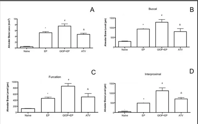
On the macroscopic analysis on hemimaxillae (Figure 1A), Naïve group (Figure 1C) presented an ABL close to zero, there was a non significant difference between the two sides of the maxilla. Eleven days of EP caused a significant (p<0.05) ABL compared to Naïve showing furcation lesions and root exposure (Figure 1E). The response to GIOP (Figure 1G) was similar to that of Naïve group. However, animals subjected to GIOP+EP (Figure 1I) showed greater (p<0.05), ABL when compared to EP.
The results of RD analysis of hemimaxillae can be evaluated on Figure 1B. Radiographs of EP animals showed ABL by 19% (Figure 1F) (p<0.05) when compared Naïve group (Figure 1D). In GIOP group there was no significant change in the RD of hemimaxillae (Figure 1H) compared to Naïve. However hemimaxillae of the animals subjected to GIOP+EP had lower RD, by 12% (Figure 1J) when compared to EP group (p <0.05)
Naïve group. No differences were seen in GIOP group compared to EP group (p>0.05). However, in GIOP+EP group, it was observed greater reduction on BMD, bone percentage and trabecular thickness as well as, greater increase on bone porosity when when compared to EP (p<0.05).
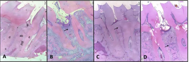
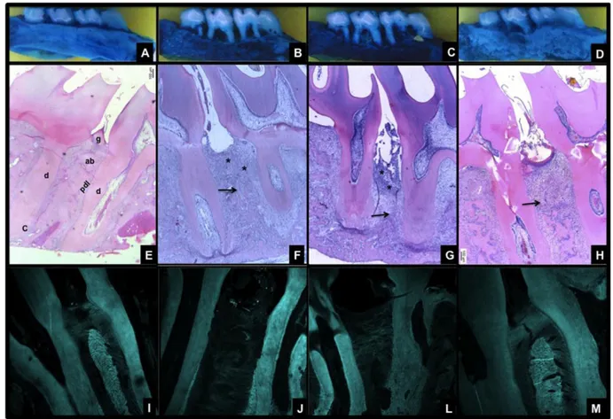
Bone histology
Periodontal histopathological analysis of the region between first and second molars of naïve animals shows the normal structure of gingiva, periodontal ligament (PDL), alveolar bone and cementum (Figure 3A). The periodontium of animals subjected to experimental periodontitis (EP group) demonstrated accentuated inflammatory cell infiltration, breakdown of alveolar bone, collagen fiber derangement within the periodontal ligament, and resorption of cementum, receiving a score of 3 (Figure 3B; Table 1). The periodontal tissue of animals subjected to GIOP did not any change on the architecture (Figure 3C; Table 1). However, the animals subjected to GIOP+EP showed enhanced bone loss and inflammatory infiltrate (Figure 3D; Table 1).
Table 1. Effect of GIOP on Histopathological analysis of hemimaxillae
Naïve EP GIOP GIOP+EP
Histopathological Analysis (Scores)
0 3 # 0
3*
Data is presented as median (extreme value).
EP=experimental periodontitis; GIOP=glucocorticoid-induced osteoporosis;
#Significant compared to Naïve group *Significant compared to EP group;
Figure 3. Effect of GIOP on the microarchitecture of alveolar bone tissue of rats with experimental periodontitis. A) Naïve, B) EP, C) GIOP, D) GIOP+EP. Region between the first and second molars of rats of normal periodontium (A), periodontium of animals submitted to ligature-induced alveolar bone resorption (B), periodontium of animals submitted to GIOP (C), periodontium of animals submitted to GIOP ad EP (D). Dentin (d); Cementum (c); Alveolar Bone (ab); Gingiva (g); Periodontal Ligament (pdl). Inflammatory infiltrate (*) and bone resorption ( ). Bars - 500 μm. Hematoxylin & eosin (H&E). (40x magnification),
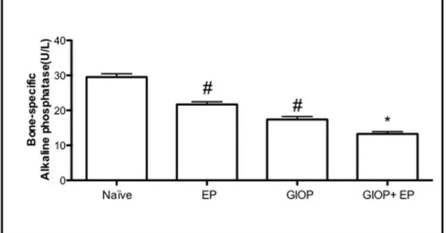
Serum biochemical analysis of BALP
Serum dosage of BALP was analyzed (Figure 4). EP caused a significant decrease, by 27% on BALP serum levels (21.64±2.48 U/L) when compared to Naïve (29.52±5.14 U/L). GIOP also caused a reduction by 41% (17.4±2.65 U/L) on BALP serum levels compared to Naive (p>0.05). However, in GIOP+EP group it was seen a greater reduction by 55% (13.25±2.11 U/L) on BALP serum levels when compared to EP (p<0.05).
Biomechanical testing and RD of Femur
Assessing RD of femur (Figure 5E), those animals undergoing only EP (Figure 5B) showed no significant difference in RD when compared to the femurs of Naïve group (Figure 5A). The GIOP caused a significant reduction of RD in femur (Figure 5C) when compared to Naïve and EP groups. In the group animals submitted to GIOP+EP there was a reduction of RD femur (Figure 5D) when compared to Naïve or EP groups (p <0.05).
We further checked femur biomechanical properties GIOP and experimental periodontitis (Figure 5F). After 11 days of EP did not show difference on Young’s modulus when compared to the femurs of Naïve group (p>0.05). There was a significant decrease in Young’s modulus in the group subjected to GIOP compared to Naïve group, as well as in the group GIOP+EP compared to EP group.
per group. #Significant difference compared to Naïve group. *Significant difference compared to EP group. (p<0.05; ANOVA followed by Bonferroni Test).
4. Discussion
In this study we examined the effect on GIOP on EP. It was seen that EP caused an increase on ABL considering macroscopic, radiographic, micro-tomographic and histological analysis, and also reduced BALP serum levels, however it did not alter the architecture or biomechanics of femur when compared to Naïve group. The animals subjected to GIOP, in turn, did not show ABL when compared to Naïve group, but in micro-CT analysis of volumetric parameters of alveolar bone it was seen decrease on BMD, bone percentage, trabecular thickness and an increase on bone porosity when compared to Naïve group (p<0.05). GIOP promoted significant reduction on BALP serum levels and caused reduction on radiographic density and Young’s module of femur when compared to Naïve group. However, in GIOP+EP group it was seen a greater increase on ABL in macroscopic, radiographic, micro-tomographic analysis, a greater reduction on BALP serum levels, and higher decrease on radiographic density and Young’s module of femur when compared to EP.
Lima, Melo, Rego, & Lima, 2010; Goes, Melo, Dutra, Lima, & Lima, 2012; Dalcico, de Menezes, Deocleciano, Oriá, Vale & Ribeiro, 2013; Goes, Melo, Silva, Benevides, Alencar & Ribeiro, 2014; Lisboa, Gondim, Ervolino, Vale, Frota & Nunes, 2015). In the other hand, consistent with our findings, there is no evidence that periodontitis, by itself, can provoke systemic bone loss (Xu, Chen, Zhang, Zhai, Liu & Qin, 2014).
Systemic risk factors may determine acceleration on the initiation of periodontal disease (Borrell & Papapanou, 2005; Kornman, 2008), as well as, the increase on the rate of progression and severity of periodontitis (Genco & Borgnakke, 2013). Among these risk factors, osteoporosis is one of the six main factors (Genco & Borgnakke, 2013). Therefore, using reproducible experimental models, the present study investigates the influence of glucocorticoid-induced osteoporosis on ABL of rats subjected to experimental periodontitis assessing alveolar bone level, femur bone loss and osteoblastic activity.
Osteoporosis is mainly caused by postmenopausal estrogen deficiency, and its association with periodontitis it well described in literature (Hernández-Vigueras, Martínez-Garriga, Sánchez, Sanz, Estrugo-Devesa & Vinuesa, 2015; Juluri, Viswanathan, Gopalakrishnan, Kathariya, Devanoorkar & Prashanth,
2015). However, osteoporosis has also a secondary cause, related to the long-term use of glucocorticoids (GCs). Considering the periodontal tissue, from the best of our knowledge, this the first time that the effect of GIOP is evaluated on experimental periodontitis in rats.
Griffin, Jawad, Hall, & Doyle, 1993). This initial rapid and greater bone loss can reflect persistence of the prior effects of inflammatory cytokines, such as TNF-α and IL-1, as well as, the osteoclastic cytokine, RANKL (Teitelbaum, 2015). Therefore, these findings can explain the greater ABL in GIOP+EP.
GIOP is also characterized by increased apoptosis of osteoblasts. There is evidence that GCs decrease osteoblastogenesis, impair osteoblastic differentiation and maturation and decrease the number and function of osteoblasts (Canalis, Mazziotti, Giustina, & Bilezikian, 2007). GCs, also favor the differentiation of bone marrow stromal cells toward cells of the adipocyte lineage instead of the osteoblastic linage by blocking Wnt/β-catenin signaling pathway (Canalis, Mazziotti, Giustina, & Bilezikian, 2007; Compston, 2010). In addition, when associated to inflammatory bone disorders, such as periodontitis, the osteoblastic activity may also be suppressed in a greater way. TNF-α, IL-1, -6 and -17 can perturb the WNT and bone morphogenetic protein (BMP) signaling pathways, leading to lower osteoblast differentiation and activity (Walsh, & Gravallese, 2010). TNF-α can also induce osteoblasts apoptosis (Jilka, Weinstein, Bellido, Parfitt, & Manolagas, 1998; Wei, Kitaura, Zhou, Ross, & Teitelbaum, 2005). Thus these findings can justify the decrease on BALP serum levels seen on GIOP and GIOP+EP groups, since BALP is a homodimeric glycoprotein that is anchored to the membrane of osteoblasts and consequently it is considered a general indicator of the bone formation rate in skeletal tissue (Sardiwal, Magnusson, Goldsmith, & Lamb, 2013).
5. Conclusion
In summary, considering our results, we can conclude that GIOP can potentiate the destructive effect of periodontitis on bone tissue, by promoting bone resorption and reducing osteoblast activity. Nevertheless more studies are necessary in order to better understand the biochemical mechanisms of GCs on bone cells during a inflammatory status.
Conflict interests
Funding
This study was supported by grants from CNPq and FUNCAP Brazilian Agencies and authors themselves.
Ethical approval
The experimental procedures described here were approved by the Institutional Animal Care and Use Committee (#78/2014) and performed in accordance with the Animal Care Standards of Federal University of Ceará.
Acknowledgments
The authors also gratefully thank Socorro França Monte of the Department of Morphology, Faculty of Medicine, Federal University of Ceará, Ceará, Brazil, for technical assistance. This study was supported by grants from CNPq and FUNCAP Brazilian Agencies and authors themselves.
References
Bezerra, M. M., Brito, G. A., Ribeiro, R. A., & Rocha. F. A. (2002). Low-dose doxycycline prevents inflammatory bone resorption in rats. Brazilian Journal of
Medical and Biological Research, 35(5), 613-616.
Bezerra, M. M., de Lima, V., Alencar, V. B., Vieira, I. B., Brito, G. A., Ribeiro, R. A., et al. (2000). Selective cyclooxygenase-2 inhibition prevents alveolar bone loss in experimental periodontitis in rats. Journal of Periodontology, 71(6),
1009-1014.
Canalis, E., Mazziotti, G., Giustina, A., & Bilezikian, J. P. (2007) Glucocorticoid-induced osteoporosis: pathophysiology and therapy. Osteoporosis International, 18(10), 1319–1328.
Compston, J. (2010) Management of glucocorticoid-induced osteoporosis. Nature
Reviews. Rheumatology,6(2), 82-88.
Dalcico, R., de Menezes, A. M., Deocleciano, O. B., Oriá, R. B., Vale, M. L., Ribeiro, R. A., et al. (2013). Protective mechanisms of simvastatin in experimental periodontal disease. Journal ofPeriodontology, 84(8), 1145-1157.
de Lima, V., Bezerra, M. M., de Menezes Alencar, V. B., Vidal, F. D., da Rocha, F. A., de Castro Brito, G. A., et al. (2000). Effects of chlorpromazine on alveolar bone loss in experimental periodontal disease in rats. European Journal of Oral
Science, 108(2), 123-129.
de Menezes, A. M., de Souza, G. F., Gomes, A. S., de Carvalho Leitão, R. F., Ribeiro, R. de A., de Oliveira, M. G., et al. (2012). S-nitrosoglutathione decreases inflammation and bone resorption in experimental periodontitis in rats. Journal of
Periodontology, 83(4), 514-521.
Genco, R. J., & Borgnakke, W. S. (2013). Risk factors for periodontal disease.
Periodontology2000, 62(1), 59-94.
Goes, P., Lima, A. P. S., Melo, I. M., Rego, R. O., & Lima, V. (2010). Effect of atorvastatin on ligature-induced periodontitis in Wistar rats: radiographic and macroscopic analysis. Brazilian Dental Journal, 21(3), 193–198.
Goes, P., Melo, I. M., Dutra, C. S., Lima, A. P. S., & Lima, V. (2012). Effect of alendronate on bone- specific alkaline phosphatase on periodontal bone loss in rats. Archives of Oral Biology, 57(11), 537-544.
atorvastatin reduces ligature-induced alveolar bone loss in rats. Journal of
Periodontal Research, 49(1), 45-54.
Hall, G. M., Spector, T. D., Griffin, A. J., Jawad, A. S., Hall, M. L., & Doyle D. V. (1993). The effect of rheumatoid arthritis and steroid therapy on bone density in postmenopausal women. Arthritis and Rheumatism, 36(11), 1510–1516.
Hernández-Vigueras, S., Martínez-Garriga, B., Sánchez, M. C., Sanz, M., Estrugo-Devesa, A., Vinuesa, T. T., et al. (2015). Oral Microbiota, Periodontal Status and Osteoporosis in Postmenopausal Women. Journal of Periodontology. 15, 1-15.
Jilka, R. L., Hangoc, G., Girasole, G., Passeri, G., Williams, D. C., Abrams, J. S., et al. (1992). Increased osteoclast development after estrogen loss: mediation by interleukin-6. Science, 257(5066), 88–91.
Jilka, R. L., Weinstein, R. S., Bellido, T., Parfitt, A. M., & Manolagas, S. C. (1998). Osteoblast programmed cell death (apoptosis): modulation by growth factors and cytokines. Journal ofBone andMineralResearch, 13(5), 793–802.
Juluri, R., Prashanth, E., Gopalakrishnan, D., Kathariya, R., Devanoorkar, A., Viswanathan, V., et al. (2015). Association of Postmenopausal Osteoporosis and Periodontal Disease: A Double-Blind Case-Control Study. Journal of International
OralHealth, 7(9), 119-123.
Kok, C., & Sambrook, P. N. (2009). Secondary osteoporosis in patients with an osteoporotic fracture. Best Practice & Research. Clinical Rheumatology, 23(6),
769–779.
Kornman, K.S. (2008). Mapping the pathogenesis of periodontitis: A new look.
Journal of Periodontology, 79(8 Suppl), 1560-1568.
Leitão, R. F., Ribeiro, R. A., Chaves, H. V., Rocha, F. A., Lima, V,. & Brito, G. A. (2005). Nitric oxide synthase inhibition prevents alveolar bone resorption in experimental periodontitis in rats. Journal of Periodontology, 76(6), 956–963.
Leitão, R. F., Rocha, F. A., Chaves, H. V., Lima, V., Cunha, F. Q., Ribeiro, R. A., et al. (2004). Locally applied isosorbide decreases bone resorption in experimental periodontitis in rats. Journal of Periodontology, 75(9), 1227-1232.
Lima, V., Vidal, F. D., Rocha, F. A., Brito, G. A., & Ribeiro, R. A. (2004). Effects of tumor necrosis factor-alpha inhibitors pentoxifylline and thalidomide on alveolar bone loss in short-term experimental periodontal disease in rats. Journal
of Periodontology, 75(1), 162-168.
Lisboa, R. P., Gondim, D. V., Ervolino, E., Vale, M. L., Frota, N. P., Nunes, N. L., et al. (2015). Effects of electroacupuncture on experimental periodontitis in rats.
Journal of Periodontology, 86(6), 801-811.
Lucinda, L. M. F., Aaresturup, B. J. V., Peters, V. M., Reis, J. E. P., de Oliveira, R. S. M. F., & Guerra, M. O. (2012). The effect of the Ginkgo biloba Extract in the
Expression of Bax, Bcl-2 and bone Mineral Content of Wistar Rats with
Glucocorticoid-induced Osteoporosis. Phytotherapy Research, 27(4), 515-520.
Mahl, C. R. W., Tonietto, A., Giorgi, B. G., Girotto, C. V., & Fontanella, V. R. C. (2009). Evaluation of radiographic density and proportion of trabecular bone in the femur of female rats medicated with glucocorticoid and bisphosphonate.
Revista Odontociência, 24, 28-31.
Menezes, A. M., Rocha, F. A., Chaves, H. V., Carvalho, C. B., Ribeiro, R. A. & Brito, G. A. (2005). Effect of sodium alendronate on alveolar bone resorption in experimental periodontitis in rats. Journal of Periodontology,76(11), 1901-1909.
Overman, R. A., Yeh, J. Y., & Deal, C. L. (2013). Prevalence of oral glucocortidoid usage in the United States: a general population perspective.
Pihlstrom, B. L., Michalowicz, B. S., & Johnson, N. W. (2005). Periodontal diseases. Lancet, 366(9499):1809-1820.
Sardiwal, S., Magnusson, P., Goldsmith, D. J., & Lamb, E. J. (2013). Bone alkaline phosphatase in CKD-mineral bone disorder. American Journal of Kidney
Disease, 62(4), 810-822.
Teitelbaum, S. L. (2015). Glucocorticoids and the osteoclast. Clinical and Experimental Rheumatology, 33(4 Suppl 92), 37-39.
Walsh, N. C., & Gravallese, E. M. (2010). Bone remodeling in rheumatic disease: a question of balance. ImmunologicalReviews, 233(1), 301–312.
Wei, S., Kitaura, H., Zhou, P., Ross, F. P., & Teitelbaum, S. L. (2005). IL-1 mediates TNF-induced osteoclastogenesis. Journal of Clinical Investigation, 115(2), 282–290.
Whitby, L. G., & Moss, D. W. (1975). Analysis of heat inactivation curves of alkaline phosphatase isoenzymes in serum. Clinica Chimica Acta, 59(3),
361-367.
Artigo 2
Effects of Atorvastatin on periodontitis of rats subjected to glucocorticoid-induced osteoporosis.
Luzia Herminia Teixeira de Sousa1, Eveline Valeriano Moura1, Joanna Trycia Alexandre2, Mario Lisboa3, Flávia Furlaneto4, Raul Freitas3, Isabela Ribeiro5, Danielle do Val6, Mirna Marques1,7, Hellíada Chaves1,2, Conceição Martins3, Gerly Anne Brito3,8, Paula Goes1,9
1
Post-graduation Program of Health Science, Medical School, Federal University of Ceará, Sobral, Ceará, Brazil.
2
School of Dentistry, Federal University of Ceará, Sobral, Ceará, Brazil.
3
Post-graduation Program of Morphological Science, Department of Morphology, Medical School, Federal University of Ceará, Fortaleza, Brazil.
4
Department of Oral & Maxillofacial Surgery and Periodontology, Ribeirao Preto School of Dentistry, University of Sao Paulo-USP, Ribeirão Preto, São Paulo, Brazil.
5
Renorbio Post-graduation Program, Federal University of Ceará, Sobral, Ceará, Brazil.
6
Renorbio Post-graduation Program, Federal University of Pernambuco, Recife, Pernambuco, Brazil.
7
School of Medicine, Federal University of Ceará, Sobral, Ceará, Brazil
8
Department of Morphology, Medical School, Federal University of Ceará, Fortaleza, Ceará, Brazil.
9 Department of Pathology and Legal Medicine, Medical School, Federal University of Ceará, Fortaleza, Ceará, Brazil.
Running title: Atorvastatin in GIOP and EP
Corresponding address: Prof Dr Paula Goes.
Professor of Department of Pathology and Legal Medicine Federal University of Ceará, School of Medicine.
Rua Monsenhor Furtado, s/n 60441-750. Rodolfo Teófilo, Fortaleza – CE -Brasil Phone: +55 85 3366.8301 Fax: +55 85 3366.8300
E-mail: paulagpinheiro@yahoo.com.br
Conflicts of Interest and Source of Funding Statement:
Abstract
Aim: To evaluate the effect of Atorvastatin (ATV) on experimental periodontitis (EP) of rats subjected to glucocorticoid-induced osteoporosis (GIOP).
Material and Methods: male Wistar rats were divided into: Naïve, EP, GIOP+EP and ATV groups. GIOP+EP and ATV received 7 mg/kg of dexamethasone intramuscularly 1x/week for 5 weeks, the others received 2ml of Saline (SAL) intramuscularly. EP, GIOP+EP and ATV were subjected to EP by ligature around 2nd upper left molars for 11 days. ATV group received 27 mg/kg of ATV orally and the others SAL, 30 minutes before EP. Periodontium was analyzed by macroscopy, micro-tomography and histopathology; by immunohistochemical examination of RANKL, OPG, WNT10b, DKK-1 and β-catenin and by ELISA analysis of myeloperoxidase (MPO), TNF-α, IL-1β, -6, -8, and -10, reduced glutathione (GSH), superoxide dismutase (SOD) and catalase (CAT). Leukogram and liver and kidney enzymes and bone-specific alkaline phosphatase (BALP) serum levels were performed.
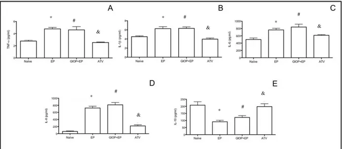
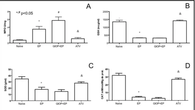
Results: ATV decreased bone loss, reduced MPO, TNF-α, IL-1β, -6, and -8, and increased IL-10, GSH, SOD and CAT levels. ATV reduced RANKL and DKK-1, increased OPG, WNT10b and β-catenin expressions and BALP activity.
Conclusions: ATV reduced inflammation, oxidative stress and bone loss in rats with EP and GIOP, with participation of WNT signaling pathway.
Clinical relevance
Scientific rationale for the study: There are conflicting clinical findings regarding the benefits of Atorvastatin in the treatment of osteoporosis and there is no study evaluating the effects of Atorvastatin on periodontitis associated with osteoporosis.
Principal finding: Atorvastatin demonstrated antiresorptive, anti-inflammatory and anti-oxidative effects in rats with GIOP and EP.
Introduction
Periodontitis is an inflammatory chronic infectious disease that affects the supporting structures of teeth and can lead to loss of the periodontal ligament (PDL) and alveolar bone and eventually to tooth loss (Pihlstrom et al. 2005). Its pathogenesis is multifactorial involving the microbial challenge that leads to an exacerbated inflammatory response, the main cause of tissue destruction (Giannobile et al. 2009).
Recent studies have shown that several systemic diseases, which affect the host immune response, can modify the progression rate of periodontitis (Di Benedetto et al. 2013, Rosa et al., 2014). Among them, osteoporosis stands out. It is well established the correlation between postmenopausal osteoporosis and periodontitis, with emphasis on the explanation of possible relations between premature tooth loss and decrease of length and density of jaw bones (Straka et al. 2015). However, the effects of glucocorticoid (GCs)-induced bone loss, the most common form of secondary osteoporosis (Saag et al. 1994), on periodontal inflammation it is still unclear.
Statins are cholesterol-lowering drugs capable of inhibiting cholesterol synthesis in humans and animals. In addition, studies have suggested that statins may have other effects beyond lipid reducing function, due to the inhibition of the isoprenoid intermediates of the mevalonate biosynthetic pathway, so-called pleiotropic effects (Maeda et al. 2004). These effects are able to modulate inflammation by lowering cytokine concentrations and inhibiting recruitment, migration and cell adhesion to endothelium. Statins have also shown anabolic effects on bone tissue (Mundy et al. 1999).
However, there are conflicting clinical findings regarding the benefits of statins for bone diseases treatment, such as osteoporosis. Although some clinical studies show the positive effect of statins on bone mineral density (Chen et al. 2014), a recent clinical trial, which included data of 17,802 patients, does not support the role of statin in reducing the risk of fractures and bone resorption (Peña et al. 2015).
no study evaluating the effects of ATV on periodontitis experimental associated with osteoporosis.
In this study, the effect of the ATV on rats associated with glucocorticoid-induced osteoporosis (GIOP) and experimental periodontitis (EP) is addressed, analyzing its action on various parameters of bone metabolism, inflammation, and oxidative stress.
Material and Methods Animals
All experimental protocols were approved by the Federal University of Ceará (UFC) Ethical Committee for Animal Research (number 78/2014). Surgical procedures and animal treatments were performed in agreement with the Ethical Principles for Animal Research.
Forty-eight male Wistar rats (180 to 220 g), from our animal facilities, were housed in temperature-controlled rooms and received water and food ad libitum.
A power calculation was performed to determine the sample size. The animal was considered the study unit. The sample size was determined to provide 80% power to recognize a significant difference of 20% among groups and the standard deviation of 15% with a 95% confidence interval (a = 0.05), considering the change in alveolar bone loss (ABL) as the primary outcome variable. Therefore, a sample size of 6 animals per group was required.
Treatment and experimental design