UNIVERSIDADE FEDERAL DO CEARÁ
FACULDADE DE FARMÁCIA ODONTOLOGIA E ENFERMAGEM DEPARTAMENTO DE CLÍNICA ODONTOLÓGICA
CURSO DE ODONTOLOGIA
PROGRAMA DE PÓS GRADUAÇÃO EM ODONTOLOGIA
PAULA GOES PINHEIRO
EFEITO ANTIINFLAMATÓRIO DA ATORVASTATINA NA PERIODONTITE
INDUZIDA POR LIGADURA EM RATOS
PAULA GOES PINHEIRO
EFEITO ANTIINFLAMATÓRIO DA ATORVASTATINA NA PERIODONTITE
INDUZIDA POR LIGADURA EM RATOS
Dissertação submetida à Coordenação do Programa de Pós-Graduação em Odontologia, da Universidade Federal do Ceará, como requisito parcial para a obtenção do grau de Mestre em Odontologia. Área de Concentração: Clínica Odontológica. Orientadora: Profª Drª Vilma de Lima
Dedicatória
À Valeria Goes (minha mãe)
“ ...Vê-la me faz crescer Vê-la me faz ter fé Vê-la me faz viajar Vê-la me faz pensar em tanta coisa que eu nunca vim pensar Vê-la me faz viver, vê-la me faz querer mudar Ela é quem me dá asas pra voar...”
AGRADECIMENTOS ESPECIAIS
Agradeço especialmente aos meus pais, Valéria Goes e Geraldo Uchôa, pelo amor incondicional e por todo o incentivo, confiança, e dedicação compartilhados, que certamente foram essenciais para a concretização dos meus objetivos.
Ao meu marido, Caio Dutra, exemplo de atitude e perseverança, companheiro de todas as horas com quem escolhi dividir minha vida até a eternidade.
Aos meus irmãos, João Vitor, Pedro Henrique e Thaís Andréa, parceiros fiéis e indissolúveis.
AGRADECIMENTOS
À minha orientadora de mestrado Profª Drª Vilma de Lima, por todo empenho, sabedoria, compreensão, exigência e acima de tudo por sempre me incentivar tanto na vida acadêmica quanto na vida pessoal.
À professora Norma Maria Barros Benevides, do Laboratório de Bioquímica do Departamento de Bioquímica e Biologia Molecular, por sua inestimável contribuição na realização de diversas fases desse estudo.
Aos professores Nylane Maria Nunes de Alencar, Gerly Anne de Castro Brito e Ronaldo de Albuquerque Ribeiro, pela pronta cessão de seus espaços laboratoriais no Departamento de Fisiologia e Farmacologia.
Aos professores dos Programas de Pós-Graduação em Odontologia (PPGO) e Farmacologia (PPGF), que muito contribuíram em minha formação acadêmica.
Aos meus colegas do Laboratório de Farmacologia Oral, a mestranda Ana Patrícia Souza de Lima, e os estudantes de Iniciação Científica Iracema Matos de Melo, Neiberg Alcântara Lima e Kharla Rabelo Patoilo, pela colaboração em vários experimentos.
Aos colegas dos Laboratórios de Bioquímica (PPGB) Luana Maria Castelo Silva, e de Bioquímica da Inflamação (PPGF) Flávio da Silveira Bitencourt, pela colaboração inicial nos ensaios bioquímicos.
Aos funcionários da secretaria do PPGO Germano Mahlmann Muniz Filho e Lúcia Ribeiro, pela atenção prestada.
Aos bioteristas do Departamento de Fisiologia e Farmacologia Francisco Haroldo Pinheiro e Carlos Pereira de Oliveira pela dispensação e cuidado dos animais laboratoriais.
Ao técnico de laboratório José Ivan Rodrigues (Departamento de Morfologia) por sua assistência técnica.
À Fundação Cearense de Apoio ao Desenvolvimento Científico e Tecnológico (FUNCAP), pela concessão de bolsa de mestrado.
Ao Conselho Nacional de Desenvolvimento Científico e Tecnológico (CNPq - Projetos Renorbio e Universal) e à Coordenação de Aperfeiçoamento de Pessoal de Nível Superior (Capes - Projeto Pró-equipamentos), pelo suporte financeiro a este estudo.
À Clínica Perboyre Castelo - Radiologia Odontológica, pela cessão gentil das radiografias digitais.
RESUMO
Efeito Antiinflamatório da Atorvastatina na Periodontite induzida por ligadura em ratos. PAULA GOES PINHEIRO. Dissertação apresentada ao Curso de Pós-graduação em Odontologia do Departamento de Clínica Odontológica da Faculdade de Farmácia Odontologia e Enfermagem da Universidade Federal do Ceará, como pré-requisito para a obtenção do título de Mestre em Odontologia. Aprovação em 30 de Janeiro de 2009. Orientadora: Profª Drª Vilma de Lima.
A periodontite é uma doença caracterizada por infiltração de leucócitos, perda de tecido conjuntivo e reabsorção óssea. Estatinas são fármacos amplamente usados para o tratamento da hiperlipidemia, com destaque à Atorvastatina (ATV), dada seus efeitos pleiotrópicos importantes, como atividade antiinflamatória e capacidade anabólica óssea, com potencial para modificação do curso de doenças inflamatórias crônicas. O objetivo desse trabalho foi avaliar o efeito antiinflamatório da ATV, utilizando modelo de periodontite induzido por ligadura em ratos Wistar machos, distribuídos em grupos experimentais: controle (Salina a 0,9%), e 5 subgrupos (ATV 0,3; 1; 3; 9 ou 27 mg/kg), administrados por via oral, diariamente, 30 min antes da colocação do fio de náilon 3.0 em torno dos segundos molares superiores esquerdos dos animais, durante 11 d, quando, então, foram sacrificados, e os seguintes parâmetros, analisados: 1) Perda Óssea Alveolar (POA), avaliada através de estudos morfométrico, histológico e radiográfico; 2) Avaliação Sistêmica através de: a) Leucogramas realizados antes e após a ligadura (6 h; 2, 7 e 11 d); b) Variação de massa corpórea; c) Análises hepáticas e renais, por dosagens séricas bioquímicas e estudo histológico; e d) Avaliação sérica de Fosfatase Alcalina Óssea (FAO). Os animais submetidos a 11 d de periodontite apresentaram intensa reabsorção óssea. Baixa dose de ATV (0,3 mg/kg) não foi capaz de prevenir a POA (p>0,05), contudo, todas as demais (ATV 1, 3, 9 ou 27 mg/kg) foram, de forma significante, capazes de reduzir a POA em 35%, 39%, 53%, 56%, respectivamente. Tal inibição foi corroborada pela análise histopatológica, onde se observou que a ATV (27 mg/kg) causou maior preservação do tecido periodontal [Mediana: 1,5 (0-2)], quando comparada à Salina [Mediana: 3 (2-3)]. Adicionalmente, animais submetidos a 11 d de periodontite apresentaram redução significante de densidade radiográfica periodontal (58%). ATV (1, 3 ou 9 mg/kg) preservou tal densidade em 5%, 9% e 20%, respectivamente. O leucograma dos animais com periodontite apresentou pico de leucocitose na 6ª h, mediado por neutrófilos, e nova leucocitose a partir do 7º d, à custa de mononucleares. ATV (27 mg/kg) foi capaz de reduzir a leucocitose, reduzindo o número de neutrófilos ou mononucleares, respectivamente (p<0,05), bem como foi capaz de reduzir a perda inicial de massa corpórea vista na periodontite. As análises bioquímicas séricas e histológicas de fígado e rins dos animais com 11 d de periodontite tratada (ATV 27 mg/kg) ou não (Salina) não apresentaram alterações (p>0,05). Observou-se aumento nas variações de dosagens séricas de FAO dos animais com 11 d de periodontite (Salina: 63,4±10,8 U/l), enquanto que ATV (27 mg/kg) previniu tal aumento (13,6±3,5 U/l) (p<0,05). Dessa forma, os resultados demonstram que o modelo de periodontite em ratos reproduziu os principais aspectos da doença em humanos, e ATV reduziu a destruição periodontal, sem causar alterações significantes hepáticas ou renais, além de manter os níveis de FAO, o que sugere que a ATV pode ser uma abordagem farmacológica importante como adjuvante à terapia periodontal a ser ensaiada clinicamente, devido a sua eficácia e segurança.
ABSTRACT
Antiinflammatory Effect of Atorvastatin on Ligature-Induced Periodontitis in rats. PAULA GOES PINHEIRO. Dissertation presented to Dentistry Post-graduation course from Clinical Dentistry Department of Pharmacy, Dentistry and Nursing Faculty of Federal University of Ceara, as pre-requisite for Master Degree on Dentistry. Approved in January 30th 2009. Supervisor: Prof. Dr. Vilma de Lima.
Periodontitis is a disease characterized by leukocyte influx, loss of connective tissue and bone resorption. Statins are drugs widely used to hyperlidemia treatment, in which stand out Atorvastatin (ATV) due to its important pleiotropic effects, such as antiinflammatory activity and anabolic bone capacity, with great potential to modify chronic inflammatory disease course. In this way the aim of this work was to evaluate the aniinflammatory effect of ATV, through ligature-induced periodontitis model in rats. Wistar male, located in experimental groups: control (0.9% Saline), and 5 subgroups (ATV 0.3, 1, 3, 9 or 27 mg/kg), given orally daily, 30 min before nylon thread 3.0 aroud cervix of second left upper molars during 11 d, when then, rats were sacrified, and the following parameters were analyzed: 1) alveolar bone loss (ABL), evaluated through morphometric, histologic and radiographic studies; 2) Sistemic evaluation through a) leucograms performed before and after ligature (6h and 2, 7, and 11 d); b) corporal mass variation; c) of liver and kidney analysis, by serum biochemical dosage and histological study; and d) serum evaluation of Bone-Specific Alkaline Phosphatase (BALP). Animals submitted to 11 d periodontitis presented intense bone resoption. Low dose of ATV (0.3 mg/kg) was not able to prevent ABL (p>0.05), meanwhile the other dose ATV (1, 3, 9 or 27 mg/kg) were , in a significant way able to reduce ABL by 35%, 39%, 53%, 56%, respectively. Such inhibition was corroborated by histological analysis where was observed that ATV (27 mg/kg) caused greater periodontal tissue preservation [Mean 1.5 (0-2)], when compared to Saline [Mean 3 (2-3)] . In addition, animals submitted to periodontitis presented a significant reduction on periodontal radiographic density (58%). ATV (1, 3 ou 9 mg/kg) preserved such density in 5%, 9% e 20%, respectively. The leucogram of animals submitted to periodontitis presented leukocytosis peak on the 6th h mediated by neutrophils and new leukocytosis after 7th d due mononuclear cells. ATV (27 mg/kg) was able to reduce leukocytosis, decreasing neutrophils or mononuclear cells respectivelly (p<0.05), as well as, it was able to reduce initial corporal mass loss seen in periodontitis. Serum biochemical and histological analysis of liver and kidneys of animals with 11 d periodontitis treated with (ATV 27 mg/kg) or not (Saline), did not show alterations (p>0.05). It was observed a raise on serum BALP dosage variation of animals with 11 d periodontitis (Saline: 63.4±10.8 U/l), while ATV (27 mg/kg) prevented that increase (13.6±3.5 U/l) (p<0.05).In this way, the results demonstrated that this periodontitis model in rats reproduced the main aspects of periodontal disease in humans, and ATV reduced periodontal destruction, without cause significant alterations on liver and kidneys, besides of keeping BALP activitys, what suggests that ATV may be an important pharmacological approach as an adjuvant to periodontal therapy, to be evaluated clinically, due to its efficacy and safety.
Keywords: Periodontitis, Atorvastatin, Inflammation, Radiography, Rats
RESUMO... 7
ABSTRACT... 8
I - INTRODUÇÃO GERAL... 10
II - PROPOSIÇÃO... 14
III – ARTIGOS CIENTÍFICOS... 15
ARTIGO 1... 16
Anti-resorptive Effect of Atorvastatin on Ligature-induced Periodontitis in Rats... 16
ARTIGO 2... 42
Effect of Atorvastatin in Radiographic Density on Alveolar Bone Loss in Rats... 42
V - DISCUSSÃO GERAL... 57
VI - CONCLUSÕES GERAIS... 64
VII - REFERÊNCIAS... 65
1. INTRODUÇÃO GERAL
A doença periodontal encontra-se entre as duas maiores doenças orais que afetam a população humana e, em todo o mundo, apresenta-se com altas taxas de prevalência (PETERSEN & OGAWA, 2005). Esse termo usualmente se refere a uma variedade de patologias que acometem tecidos de proteção, como a gengiva, ocasionando as chamadas gengivites, e os tecidos de sustentação, que incluem o osso alveolar, cemento radicular e o ligamento periodontal, determinando as variadas formas de periodontites (PIHLSTROM et
al., 2005).
À medida que a periodontite evolui, pode-se observar destruição progressiva de tecidos e conseqüente perda dentária. Diversos estudos têm sido realizados, e atualmente está bem descrito que para o desencadeamento destes eventos é fundamental a presença de periodontopatógenos nos sítios periodontais (TELES et al., 2006). Embora a colonização periodontal por bactérias destruidoras de tecido seja importante para o estabelecimento da periodontite, sabe-se, contudo, que a susceptibilidade do hospedeiro é extremamente necessária para o aparecimento dos sinais clínicos da doença, inerentes ao desequilíbrio nos processos de homeostase óssea (PIHLSTROM
et al., 2005).
O processo de remodelagem óssea compreende um equilíbrio dinâmico entre osteoblastos, envolvidos na formação óssea, e osteoclastos, os quais são responsáveis pela reabsorção óssea. Tais fenômenos são mediados pelo sistema constituído pelo Receptor Ativador do Fator Nuclear-κB (RANK), pelo Ligante do Receptor Ativador do Fator Nuclear-κB (RANKL) e pela Osteoprotegerina (OPG) (RANK/RANKL/OPG) (XING et al., 2005; REID & HOLEN, 2009).
balanço entre formação e reabsorção ósseas, a interação RANKL-RANK é inibida pela OPG, visto que a OPG atrai receptor e se liga como homodímero à estrutura homotrimérica de RANKL, prevenindo, assim, a ativação osteoclástica. Em outras palavras, a homeostase óssea é mantida através da OPG que inibe a osteoclastogênese por meio de ligação competitiva com RANKL (REID & HOLEN, 2009; SOEDARSONO et al., 2006).
Entretanto, durante um processo inflamatório crônico, onde se observam, dentre vários mediadores químicos, altos níveis de citocinas como TNF, ocorre a expressão abundante de RANKL. A superexpressão de RANKL determina uma inibição da ação da OPG, provocando, portanto, um maior desequilíbrio a favor de reabsorção óssea (REID & HOLEN, 2009). A periodontite é uma doença inflamatória crônica, onde se observa uma intensa desordem, conduzindo à perda óssea em torno dos dentes. Estudos têm demonstrado, inclusive, que um dos principais agentes causais da doença, a
Porphyromonas gingivalis, é capaz de liberar proteases que tem sido
relacionadas à degradação da forma recombinante de OPG (KOBAYASHI-SAKAMOTO et al., 2004).
O processo inflamatório periodontal tem como objetivo inicial a eliminação de bactérias e toxinas presentes nos tecidos subjacentes, diminuindo os danos decorrentes da inflamação mantida ou não controlada. Paradoxalmente, nas formas crônicas de periodontite, observa-se a superexpressão de mediadores químicos e a exacerbação das respostas imunoinflamatórias, o que conduz à destruição de tecido de suporte dentário e a alterações potencialmente irreversíveis (SHAPIRA et al., 2005).
VCAM) na parede endotelial (HUANG et al., 2008), as quais medeiam a migração de leucócitos para o sítio infectado (KANTERS et al., 2008). Ainda no periodonto, os neutrófilos contribuem para a destruição tecidual, induzindo novamente a produção de espécies reativas de oxigênio (ROS), como NO, e outras citocinas, que amplificam a resposta inflamatória (SALVEMINI et al., 2003).
Um aspecto importante relacionado às periodontites consiste em seu diagnóstico, tratamento, controle e manutenção. Assim, exames clínicos, especialmente associados a exames complementares auxiliares, são imprescindíveis para o diagnóstico precoce e preciso sobre o estágio da gravidade da doença. Parâmetros diversos como presença de sangramento à sondagem periodontal, profundidade de bolsas, cálculo dentário, biofilme bacteriano, bem como aspecto clínico do periodonto e o nível clínico de inserção, dentre outros, podem ser observados através do exame de sondagem e análise visual (PIHLSTROM et al., 2005).
Entretanto, como forma auxiliar de se avaliar o grau de perda óssea periodontal, imagens radiográficas podem ser obtidas, visto que apresentam e propiciam maior riqueza de detalhes quanto à qualidade e quantidade de suporte ósseo (KHOCHT et al., 2003). Entre os tipos de exames, as imagens radiográficas digitais vêm assumindo posição de destaque na odontologia e, principalmente, na periodontologia (VAN DER STELT, 2005), pois apresentam maior capacidade de detecção de sítios com perdas ósseas ainda sutis, quando comparadas às imagens radiográficas convencionais (KHOCHT et al., 2003), o que favorece, conseqüentemente, diagnóstico e terapia precoces.
Durante muito tempo, a base do tratamento periodontal objetivou o controle da placa bacteriana (BOEHM & SCANNAPIECO. 2007). No entanto, em alguns casos de periodontite tratados de forma convencional, através de controle de placa bacteriana juntamente com raspagem e alisamento radiculares, não se mostram com prognóstico favorável ao controle da progressão da doença, requerendo, portanto, terapias adjuvantes (BUDUNELI
et al., 2007). Considerando o papel proeminente do hospedeiro, como principal
hospedeiro, têm se sobressaído como uma nova abordagem de tratamento (BUDUNELI et al., 2007; PRESHAW et al., 2004).
Inibidores da enzima 3-hidroxi-3-metilglutaril coenzima A (HMG-CoA) redutase ou estatinas são fármacos amplamente utilizados no tratamento da hiperlipidemia e aterosclerose, por causar redução nos níveis sanguíneos de colesterol (KRONMANN et al., 2007). Alguns estudos vêm demonstrando que as estatinas, por sua vez, também apresentam efeitos pleiotrópicos não relacionados a sua capacidade hipolipemiante (KRONMANN et al., 2007), dentre eles, destacam-se a atividade antiinflamatória (NICHOLLS et al., 2006) e a capacidade anabólica em tecido ósseo (MUNDY et al., 1999). Tais propriedades oferecem grande potencial para estatinas modificarem o curso de doenças inflamatórias crônicas (BARSANTE et al., 2005), dentre as quais podem ser incluídas as periodontites crônicas.
Os efeitos secundários das estatinas estão intimamente relacionados ao seu grau de solubilidade. Estatinas lipofílicas apresentam maior potencial osteogênico (IZUMO et al., 2001), bem como, exercem maior influência na via regulatória de monócitos que regulam a produção de citocinas, induzindo uma reposta inflamatória mais controlada tanto in vivo como in vitro (KIENER et al., 2001). Dentre as estatinas, a Atorvastatina (ATV) é o agente que mais tem se destacado (SCHACHTER, 2005), não apenas por sua lipofilicidade, mas, também pelos poucos efeitos adversos apresentados e melhor relação custo-benefício (COSTA-SCHARPLATZ et al., 2008), justificando, portanto, o amplo uso da ATV na prática clínica (PLOSKER & LYSENG-WILLIAMSON, 2007), inclusive em doenças como, por exemplo, artrite reumatóide (McCAREY et al., 2004).
2. PROPOSIÇÃO
Os objetivos do presente trabalho, segundo cada um dos artigos relacionados adiante, foram:
1. Avaliar o efeito antirreabsortivo da Atorvastatina na periodontite experimental induzida por ligadura em ratos, através de:
a. Análises macroscópica e histológica da perda óssea alveolar b. Avaliação de parâmetros sistêmicos como leucograma, variação
de massa corpórea, alterações hepáticas e renais, e dosagens séricas de Fosfatase Alcalina Óssea.
2. Avaliar o efeito da Atorvastatina na densidade radiográfica na perda óssea alveolar induzida em ratos, através de:
III – ARTIGOS CIENTÍFICOS
Esta dissertação está baseada no Artigo 46 do Regimento Interno do Programa de Pós-graduação em Odontologia da Universidade Federal do Ceará que regulamenta o formato alternativo para dissertações de Mestrado e teses de Doutorado e permite a inserção de artigos científicos de autoria ou co-autoria do candidato.
Por se tratar de pesquisa envolvendo animais, os protocolos utilizados neste trabalho foram submetidos à apreciação e devidamente aprovados pelo Comitê de Ética em Pesquisa com Animais da Universidade Federal do Ceará (Anexo 1).
Dessa forma, a presente dissertação é composta por dois artigos científicos redigidos de acordo com as revistas científicas escolhidas para as devidas publicações, como apresentados adiante:
Artigo 1:
"Anti-resorptive Effect of Atorvastatin on Ligature-induced Periodontitis in Rats”. Goes P, Lima APS, Lima NA, Melo IM, Benevides NMB, Brito GAC, Alencar NMN, Rego ROCC, Lima V.
Este artigo seguiu normas de publicação do periódico European Journal
Oral Science(ISSN 1600-0722).
Artigo 2:
“Effect of Atorvastatin in Radiographic Density on Alveolar Bone Loss in Rats”. Goes, P, Lima APS, Melo IM, Rego ROCC, Lima V.
Este artigo seguiu normas de publicação do periódico Brazilian Oral
ARTIGO 1
Anti-resorptive Effect of Atorvastatin on Ligature-induced Periodontitis in
Rats.
Paula Goes1, Ana Patrícia Souza Lima1, Neiberg Alcântara Lima2, Iracema Matos Melo2, Norma Maria Barros Benevides3, Gerly Anne de Castro Brito4, Nylane Maria Nunes Alencar2, Rodrigo Otávio César Citó Rêgo5, Vilma Lima2,*.
1
Department of Dentistry Clinical, Federal University of Ceara (UFC), Fortaleza, Ceará, Brazil
2
Department of Physiology and Pharmacology, Federal University of Ceará (UFC), Fortaleza, Ceará, Brazil
3
Department of Biochemistry and Molecular Biology, Federal University of Ceará (UFC), Fortaleza, Ceará, Brazil
4
Department of Morphology, Federal University of Ceará (UFC), Fortaleza, Ceará, Brazil
5
Faculty of Dentistry, Federal University of Ceara (UFC), Sobral, Ceará, Brazil
Running title: Atorvastatin effect on rat periodontitis
*Corresponding Author: Prof Dr Vilma Lima
Universidade Federal do Ceará - Departamento de Fisiologia e Farmacologia Faculdade de Medicina
Rua Coronel Nunes de Melo, 1127 - Rodolfo Teófilo -
Goes P, Lima APS, Lima NA, Melo IM, Benevides NMB, Brito GAC, Alencar NMN, Rego ROCC, Lima V. Effects of Atorvastatin on Ligature-induced Periodontitis in Rats. Eur J Oral Sci.
ABSTRACT
Periodontitis is an inflammatory disease, characterized by alveolar bone loss (ABL). Atorvastatin (ATV), or HMG-CoA reductase inhibitor, is widely used on hyperlipidemia treatment, and has shown pleiotropic effects, as antiinflammatory and anabolic bone activity. This study aimed to evaluate the anti-resorptive effect of ATV on periodontitis. Periodontitis was induced by ligature in molar of rats for 11d. Animals received orally 0.9% Saline (0.5 ml) or ATV (0.3, 1, 3, 9 and 27 mg kg-1). ABL was evaluated through morphometric and microscopical analysis. To verify possible systemic repercussions, leukogram, corporal mass variation, liver and kidneys conditions, as well as, serum bone-specific alkaline phosphatase (BALP) activity were analyzed. Animals submitted to periodontitis presented intense ABL on 11th d. The low dose of ATV (0.3 mg kg-1) did not show bone protection (p>0.05), however ATV (1, 3, 9 or 27 mg/kg) significantly reduced ABL, by 35%, 39%, 53%, 56%, respectively. This inhibition was corroborated by histological analysis. ATV (27 mg/kg) also reversed leukocytosis, maintained the serum BALP activity, did not affect either liver and kidney, or body mass weight. In conclusion, ATV efficiently and safety, reduced ABL, suggesting that ATV may be an important tool as an adjuvant on periodontal therapy.
INTRODUCTION
Periodontitis is an inflammation that extends deep into the tissues and causes loss of supporting connective tissue and alveolar bone (1). This disease is one of the two major dental problems that affect human population at high prevalence rates (2). Nowadays, periodontal disease is understood as a result of a complex interplay between bacterial infection and host response, modified by behavioral factors (3). Periodontal inflammatory process initially has a protective role against bacterial invasion, but then, becomes destructive due to prolonged overexpression of harmful mediators (4), such as interleukin (IL-1) and tumoral necrosis factors (TNF) (5,6), and reactive oxygen species (ROS) (7), among others. These mediators, therefore, stimulate expression of leukocyte adhesion molecules. Selectins, intercellular and vascular adhesion molecules (ICAM and VCAM) act promoting neutrophil transmigration (8) that contributes to tissue destruction inducing de novo production of ROS and cytokines, which further amplify inflammatory response (9).
Statin or 3-hidroxy-3-methylglurayl coenzyme A (HMG-CoA) reductase inhibitor is a well-established pharmaceutical agent that effectively lower serum cholesterol levels, being therefore, widely prescribed for hypercholesterolemia treatment and atherosclerosis (10,11). In fact, recent studies have focused on the ability of statins to modulate chronic inflammatory diseases, such as multiple sclerosis (12). In animal models of the latter condition, atorvastatin, a statin of long duration, prevented or reverted chronic and relapsing paralysis and inhibited the secretion of cytokines IL-2, IL-12,
TNF-α, and IFN-γ (13).
(18). In the midst of various statins, Atorvastatin (ATV) stand out not only due to its lipofilicity, which is closely linked to pleiotropic effects (19), but also due to few adverse effects and better cost-effectiveness relationship (20) when compared to other statins (21) being, therefore, widely used on clinical practice (22).
Considering mainly pleiotropic effects of statins, this study was designed to evaluate the anti-resorptive effect of Atorvastatin on the inflammatory response and bone loss in an experimental model of periodontitis in rats.
MATERIAL AND METHODS
Animals
Forty-eight male Wistar rats (±200 g) (Rattus novergicus) from the Federal University of Ceará were used in this study. Animals were maintained on specific cages in temperature-controlled rooms, with free access to food and water during the whole experiment. All procedures and animal treatment conducted in order to reduce the number of animals and their suffering, were approved by Institutional Ethics Committee of Federal University of Ceará (Protocol number 74/07).
Experimental Protocol
Periodontitis Model
A model for experimental periodontal disease in rats was used as described previously (23). Briefly, rats were anesthetized with chloral hydrate (300 mg kg-1, i.p.), and a nylon (000) (Point Suture,Point Suture do Brasil Fortaleza-CE, Brazil) thread ligature was placed around the cervix of the second left upper molar. The ligature was then knotted on the vestibular side of the tooth. The contralateral right side was used as the unligated control. Rats were weighed daily until to 11th day, which demonstrated the apex of alveolar bone loss, and then, animals were killed (23).
Experimental Groups
periodontits. One of them (control) received 0.9% Saline solution (0.5 ml; v.o.) 30 min before the ligature. The other one (test) was subdivided into 5 more groups, which received Atorvastatin, on doses of 0.3, 1, 3, 9 and 27 mg kg-1, respectively, given orally 30 min before ligature. After this procedure, both groups received daily Saline or Atorvastatin, respectively, until the 11th d. Atorvastatin (Lipitor®, Pfizer; São Paulo-SP, Brazil), presented as 10 mg tablet, was macerated and dissolved in distilated water.
1. Alveolar bone structure loss
1.1 Morphometric analysis of alveolar bone
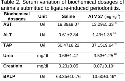
On the 11th day, animals were sacrificed under anesthesia (10% Chloral Hydrate), and had their maxillae removed and fixed in 10% neutral formallin for 24 h. Following, maxillae were splited in half, dissected, and stained with 1% methylene blue in order to differentiate bone from teeth (6, 23) In order to quantify alveolar bone loss (ABL), hemimaxillae were adjusted in microscope slides to be photographed with digital camera (Sony Cyber-Shot® model DSC-W80; Hong Kong, China). The acquired image was sent to the computer programm Image J® (ImageJ 1.32j, National Institute of Health; EUA) for horizontal alveolar bone loss analysis, which was measured using a modification of the area method of KUHR et al., 2004 (24). For this, measurements were made along the region between the molar cusp tip and the alveolar bone crest (Fig 1B), and subtracted from the respective area of contralateral normal hemimaxilla (unligated control) (Fig. 1A). All obtained images were compared to well-known area (0.5x0.5 mm2).
1.2. Histological analysis of alveolar bone
by light microscopy (x40). Parameters such as inflammatory cell infiltration, osteoclast number, and alveolar bone and cementum integrity, were determined in a single-blind manner and graded, on a score of 0–3 based on the intensity of findings, as follows: Score 0: absence of or only discrete cellular infiltration, few osteoclasts, preserved alveolar process and cementum; Score 1: moderate cellular infiltration, presence of some osteoclasts, some but minor alveolar process resorption and intact cementum; Score 2: accentuated cellular infiltration, large number of osteoclasts, accentuated degradation of the alveolar process, and partial destruction of cementum; Score 3: accentuated cellular infiltrate, total destruction of alveolar process and cementum (23).
2. Sistemic Parameters Analysis
2.1. Hematologic Study and Corporal Mass Variation
The method used for the analysis of white blood cell counts was as follows: 20 µl of blood, taken from the rat tail, was added to 380 µl Turk solution. Total and differential white blood cell counts were performed using a Neubauer chamber and stained smears by rapid Instant Prov Stain Set (Newprov Produtos para Laboratório; Pinhais-PR, Brazil), respectively. Leukogram of the groups of animals (Saline and ATV 27 mg kg-1) was performed before periodontitis induction, 6 h and 2, 7 and 11 d after the ligature. Also, animals from group Saline and ATV 27 mg kg-1 had their body mass measured before periodontitis induction, and after that daily until the 11th d. Values were expressed as body mass variation (g) compared to initial body mass.
2.2. Evaluation of Liver and Kidney Function
A. Serum Biochemical Parameters
expressed as serum dosage variation obtained on 11th d and compared to baseline of each animal.
B. Histological analysis of liver and kidney
On the 11th day, the animals (Saline and ATV 27 mg kg-1) killed for maxillae removal, also had their liver and kidney collected and fixed in 10% neutral formallin for 24-48 h period. Specimens were included in paraffin and serial sections of 4 µm thickness were obtained for hematoxillin and eosin (H&E) staining. Analyses were made through optical microscopy.
Liver parameters based on presence and amout of collagen fibers were determined in a single-blind manner and graded, on a score of 0-4 based on the findings intensity, as follows: Score 0: normal; Score 1: fibrosis present, collagen fibers present that extend from the portal triad or central vein to the peripheral region; Score 2: mild fibrosis, some extended collagen fibers present without compartmental formation; Score 3: moderate fibrosis, moderate amounts of collagen fibers present with some pseudolobe formation; Score 4: severe fibrosis, abundant collagen fibers present with a thickening of the partial compartments and frequent pseudolobe formation (25).
Kidneys parameters, such as protein/cellular casts in proximal tubule; cortical proximal convoluted tubule necrosis; pallor of outer stripe of proximal tubule; intracellular mineralization; nuclear pyknosis; interstitial nephritis were also determined in a single-blind manner and graded, on a score of 0-3 based on the findings intensity, as follows: Score 0: normal; Score 1: Mild; Score 2: Moderate; Score 3: Severe (26).
2.3. Serum dosage variation of Bone-Specific Alkaline Phosphatase
(BALP) activity
Bone Alkaline Phosphatase (BALP). Methodology to evaluate the enzymes followed the manufactor orientations (Labtest: Lagoa Santa-MG, Brasil). Values were expressed as serum variation activity compared to baseline.
3. Statistical Analysis
The data are presented as means±standard error of the mean (SEM) or medians (and range), where appropriate. Univariate analysis of variance (Anova), followed by Bonferroni’s test, was used to compare means, and Kruskal-Wallis and Mann Whitney tests were used to compare medians. A P-value of <0.05 was considered as indicating significant differences.
RESULTS
1. Alveolar bone structure
1.1 Morfometric analysis of bone tissue
In preliminary experiments, we confirmed that the bone changes observed peaked at 11 days of periodontitis (data not shown), as showed by other authors (23). Therefore, for the analysis of drug treatment, alveolar bone loss in the buccal side was measured at this time. Analysing of bone tissue through morphometric measurement (Fig. 2) we showed that ligature-induced periodontits in rats receiving only Saline during 11 d caused intense alveolar resorption (4.19±0.3 mm²) (Fig. 2), when compared to the normal hemimaxillae (Fig. 3A). The hemimaxilla submitted to ligature (Saline group) presented classical clinical signs of periodontitis such as root exposure, furcation lesion, intense alveolar resorption, and lack of proximal contact (Fig. 3C). In the other hand, Atorvastatin (ATV 1, 3, 9, or 27 mg kg-1) treatment tended to elicited a significant (P<0.05) alveolar bone protection in a dose-dependent manner (Figs. 2 and 3E), reducing alveolar bone loss by 35%, 39%, 53%, 56%, respectively. The minor dose of ATV (0.3 mg kg-1) .ATV 0.3 mg kg-1 of ATV was not able to protect alveolar bone (4.16±0.3 mm²) (Fig. 2).
1.2. Histopathological analysis of alveolar bone
were processed for histopathological analysis (Table 1). Some alterations (P<0.05) were observed on Saline group, characterized by alveolar bone and cementum resorption, associated to important inflammatory influx of leukocytes, [Median score of 3 (2-3)] (Table 1; Fig. 3D), when compared to normal periodontium [Median score of 0 (0-0)] (Table 1; Fig. 3B). ATV (27 mg kg-1) treatment significantly (P<0.05) showed an absence cellular inflammatory infiltration, and a preservation of the periodontal ligament, alveolar process and cementum [Median score of 1.5 (0-2)] (Table 1; Fig. 3F), when compared to the Saline group.
2. Sistemic Parameters Analysis
2.1. Hematologic Study and Corporal Mass Variation
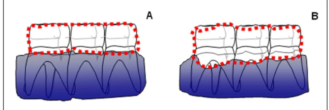
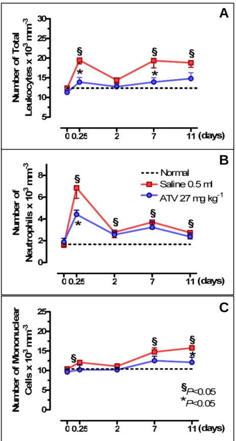
In order to verify a possible systemic repercussion of ligature-induced periodontitis, leukocyte counts were performed, and body weight was measured. All experimental groups presented the similar leukocyte levels or corporal mass on day 0 (P>0.05) (Figs. 4 and 5). It was observed that ligature-induced periodontitis caused a leukocytosis (P<0.05), at 6 h (19.5±0.9 leukocytes x 103 mm-3) (Fig. 4A), marked by neutrophils (6.8±0.9 x 103 mm-3) (Fig. 4B). On the 2nd d leukocytes tended to normal levels (Fig. 4A). Following, the new leucocytosis (P<0.05), at 11th d (18.8±1.5 leukocytes x 103 mm-3) was represented by mononuclear cells (15.8±0.6 x 103 mm-3) (Fig. 4C). ATV (27 mg kg-1) reduced (P<0.05) the leukocytosis at 6 h occurring in rats submitted to periodontitis (14.8±1.5 leukocytes x 103 mm-3), as well as neutrophil cells (4.4±0.4 x 103 mm-3) (Figs. 4A and B)., and also reduced the mononuclear cells at 11th d (12.1±1.3 x 103 mm-3), when compared to Saline (Figs. 4A and C). Figure 5 shows that periodontitis caused a significant loss (P<0.05) in body weight starting on day 2, which persisted during the 11 d of observation, in comparison to normal animals. ATV did not alter the loss in body weight observed in animals submitted to periodontitis.
2.2. Serum Biochemical Parameters and Histological analysis of liver and
kidney
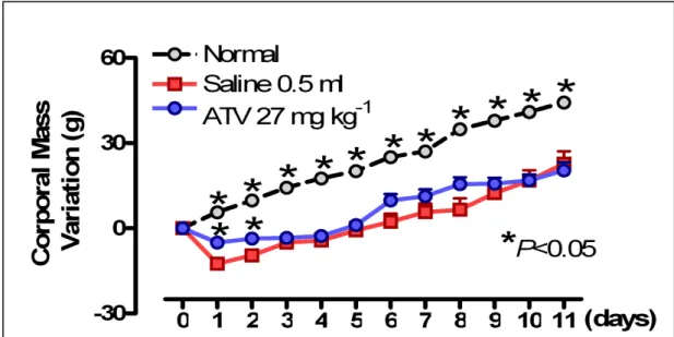
serum biochemical dosage variation analyzed either in liver (AST and ALT) or kidney (Urea and Creatinin) activity. ATV 27 mg kg-1 did not alter these serum dosages in animals submitted to periodontitis, when compared to non-treated rats (Saline), except for serum Creatinin activity (Table 2). So, to confirm the non-toxicity of ATV, histological analysis of liver and kidneys were performed. It was observed that any alterations were found, since that evaluating serial slices of both organs after 11 d of ATV (27 mg kg-1) therapy, it as possible to notice total absence of liver fibrosis, as well as the normal aspect of kidney, when compared to Saline or normal animal organs (data not shown).
Besides, serum TAP activity variation after 11 d periodontitis showed, although not significant, lower variation, when compared to baseline.
2.3. Serum dosage of Bone-Spefic Alkaline Phosphatase (BALP)
TABLES
Table 1. Microscopic analysis of rat hemimaxillae submitted to periodontitis.
Normal Saline ATV 27 (mg kg-1)
Scores 0 (0-0) 3 (2-3)* 1.5 (0-2)#
Ligature-periodontitis was induced in rats. Animals were examined at day 11. Data represent the median values (and range) of microscopic scores in 6 animals per group. *P<0.05 compared to normal contralateral; #P<0.05 compared to Saline
(Data were analysed by using Kruskal-Wallis and Mann Whitney tests).
Table 2. Serum variation of biochemical dosages of animals submitted to ligature-induced periodontitis.
Biochemical
dosages Unit Saline ATV 27 (mg kg
-1
)
AST U/l 19.89±9.07 13.29±5.33ns
ALT U/l 0.61±2.84 1.43±1.35 ns
TAP U/l 50.47±6.22 37.15±9.64ns
Urea mg/dl 0.66±1.47 3.53±1.25 ns
Creatinin mg/dl 0.23±0.05 0.07±0.10*
BALP U/l 63.35±10.76 13.60±3.46*
FIGURES
Fig. 1. Representation of the demarked area for alveolar bone resorption measurement. Area of
contralateral hemimaxilla (A) and its hemimaxilla with periodontitis (B), whose difference was considered as value of alveolar bone resorption (mm2).
Fig. 2. Effect of Atorvastatin (ATV) on Alveolar Bone Resorption of rats submitted to
periodontitis. Periodontitis was induced by ligature around second right upper molars. Animals received orally (v.o.) ATV (27 mg kg-1) or 0.5 ml Saline 30 min before periodontitis induction, and daily for 11 d. The hemimaxillae were dissecated and photographed after the animal was killed, and measured for alveolar bone resorption (mm2). Barrs represents the mean value ± standard error of the mean (SEM). *P<0.05 represents statistical differences compared to the group with ligature-induced periodontitis receiving Saline; #P<0.05 indicates statistical diference
Fig. 3. Macroscopic and microscopic aspects of normal hemimaxillae (A and B) or hemimaxillae
of rats submitted to periodontitis, receiving Saline (C and D) or 27 mg kg-1 Atorvastatin (ATV) (E and F), respectivelly. Periodontitis was induced by ligature around second right upper molars. Animals received orally (v.o.) ATV (27 mg kg-1) or 0.5 ml Saline 30 min before periodontitis induction, and daily for 11 d. D=dentin; C=cementum; AB=alveolar bone; G=gingival and PL=periodontal ligament. The hemimaxillae were dissecated and photographed, or processed for hematoxylin & eosin (H&E) staining (x 40) after the animal was killed.
Fig. 4. Effect of Atorvastatin (ATV) on leukocyte counts of rats. Saline (0.5 ml) or ATV (27 mg
kg-1) was injected orally daily for 11 d after ligature placement. Blood was taken from the rat tail immediately before experimental periodontitis and afterwards at 6 h and 2, 7 and 11 d. Each point represents the mean value ± standard error of the mean (SEM) of total leukocytes (A), neutrophils (B) and mononuclear cells (C) x 103 mm-3 of group. *P<0.05 represents statistical differences compared to the group with ligature-induced periodontitis receiving Saline. The number of animals in each group was at least six [data were analysed by using analysis of variance (ANOVA) and Bonferroni tests].
A
B
Fig. 5. Atorvastatin (ATV) does not reduce weight loss in rat with periodontitis. Periodontitis was
DISCUSSION
Periodontitis is considered the second major oral pathology that most affect human population world wide (2). For this reason, a better understanding about its origin, development, diagnosis and treatment is necessary in order to lower this disease prevalence rates. Several aetiological factors have been associated to periodontitis, nevertheless recent research about its pathogenesis have shown an important paradigm shift on disease progression and severity (28). Although bacterial biofilms have been shown to be primary aetiological factor to periodontal destruction, its presence alone accounts for a relatively small proportion being to sufficent to explain periodontitis progression and severity (29). Therefore, the major component of soft- and hard-tissue destruction associated with periodontitis is the result of activation of the host immuno-inflammatory response to bacterial challenge (29).
In fact, the undelying biological mechanisms of this response are characterized by expression of endothelial cells and intercellular adhesion molecules (29), as well as excessive production and persistence of inflammatory mediators, like tumor necrosis factor (TNF) and interleukin-1 (IL-1) (30), and -6 (31), matrix metalloproteinases (MMPs) (32); nitric oxide (NO) (33), prostaglandins (PGs) (34), along with inflammatory cells like, neutrophils monocytes, lymphocytes and fibroblasts (29), that lead to destruction of periodontal tissue and results in irrevesible pathological changes such as clinical condition of periodontitis (4).
human periodontitis, and is already well-stablished in literature (1).
Nowadays, we have used a periodontitis model described by LIMA et
al.(6), with some modification with respect to the taking of macrocospic
measures of the bone loss. Similarly to other studies (6, 23, 36, 37), our results have shown that the placement of nylon thread during 11 d caused intense alveolar bone destruction, root exposition and lack of proximal contact.
Although the exact role of the bacteria or the host response in periodontal destruction observed in rodents is not clear yet, it has been proposed that accumulus of bacterial plaque on nylon thread induces host response that leads to inflammatory cell infiltration, osteoclast formation, bone loss and loss of attachment (4, 38). Corroborating these findings, our histopathological analysis of periodontitis showed alveolar bone resorption, intense inflammatory cellular infiltration, cementum injury and soft tissue destruction. In fact, these data are in accordance with previous reports (39, 40), and probably can be explained by release of several inflammatory mediators (4, 38).
accompained by chemical mediator release caused by ligature placement was responsible for leukocytes peak.
In the present and unpublished study, we aimed to demonstrate that Atorvastatin (ATV), an agent known to have pleiotropic effects, including antiinflammatory action and anabolic effect on bone tissue, could alter the evolution of a periodontitis in rats. Our resuts showed that Atorvastatin (ATV) was able to significantly reduce alveolar bone loss (ABL) in this periodontitis model. This effect was associated with a reduction of inflammatory parameters seen by histological analysis, besides ATV reversed peripheral leukocytosis in 11 days of ligature-induced periodontitis, without affect systemic parameters.
It has been described that alveolar bone protection exerted by ATV is linked to the ability of statins on increasing up to 50% new bone formation and in promoting osteoblastic differentiation and mineralization, by enhancing production of Vascular Endothelial Growth Factor (VEGF) in osteoblasts stimulating bone growth and repair (18, 44). Such bone anabolic property seems be related to the drug lipophilicity and this ATV chemical characteristic may elicit greater mineralization process (18, 19) In addition, ATV presents antiinflammatory activity, once it was shown that ATV was to be able to inhibit important mediators involved on recruitment and transmigration of leukocytes and alveolar bone resorption as IL-6 (15, 45), TNF (46), nuclear transcriptional factor-κB (NF-κB) (47), ICAM-1 and VCAM (15), monocyte chemoattractant protein 1 (MCP-1) (48) and P-selectin expression (49). Additionally, analysing corporal mass variation, we have seen that periodontitis induced important loss of weight in the first two days of experiment, probably due to ligature placement trauma, as seen in previous reports (23). Meantime, it was found that ATV prevented initial loss of weight, but was not able to recover corporal mass lost thoughout the experiment, indicanting that statin therapy does not intefere significantly on body mass index (50).
(51), nevertheless ATV is a lipophilic statin, what suggests an explanation for transaminases findings in this study (52). Moreover, a recent clinical trial demonstrated that only 1% of patient presented upper levels of transaminases after 12 month-ATV therapy (53). Additionally, in spite of urea and creatinine are considered both sensitive biochemical markers employed in the diagnosis of renal damage because urea and creatinin are excreted through the kidney (54), it has been related that creatinin activity is not good enough to stabilish a definitive diagnostic of renal function, been necessary to associate more analytical exams (55, 56, 57). Although some assays had demonstrated that statin therapy does not induce tubular disfunction (58) or alter glomerular filtration even in higher doses (59), our findings suggested a significant alteration on serum Creatinin activity. It is worthy to notice that despite of creatinin reflect renal filtration (WU, 2008) (50), it is not linearly related to glomerular filtration rate, being often linked to bias (55), and being considered as low specific biomarker (56). In fact, when analyses of kidneys were performed, we observed that any macroscopic or histological alterations were seen in the rat kidneys that received ATV, there was normal arrangement of the medulla and cortex with the glomeruli and blood vessels neatly arranged, when compared to control rats. So, in this context, we considered that ATV in fact did not alter liver or kidneys.
Considering that the bone-specific alkaline phosphatase (BALP), an isoenzyme of total alkaline phosphatase (TAP), is a bone formation marker due to its linkage with osteoblastic differention (60) and mineralization of newly formed bone (61), we also aimed is enzyme as bone marker in rat periodontitis. Our data suggested that periodontitis induced great variation on serum BALP activity, from day 0 to day 11, indicated by low BALP activity, probably related to bone resorption (62). In the other hand, ATV maintained, with less variation, the serum BALP activity, corroborating with some clinical trials on hipercholesterolemic patients using ATV, which demonstrated low variation on serum BALP activity, when compared to baseline (63, 64, 65), contributing in this way to bone homeostasis.
leukocytosis in ligature-induced periodontitis in rats. Moreover, ATV therapy kept serum BALP activity, and did not promoted significant alteration on liver or kidney, nor affected, significantly, corporal mass when compared to control animals. Therefore, ATV reduced bone loss in an efficient and safety manner, suggesting that ATV may be an important tool as an adjuvant on periodontal therapy, besides it merits further investigation.
ACKNOWLEDGMENTS
The authors gratefully acknowledge to J. Ivan R. Sousa, by technical assistance. This work was supported by “Ceará State Foundation for Scientific and Technological Development-FUNCAP” and “National Counsel of Technological and Scientific Development-CNPq”, Brazil.
REFERENCES
1. PIHLSTROM BL, MICHALOWICZ BS, JOHNSON NW. Periodontal disease. The lancet 2005; 366: 1809-20.
2. PETERSEN PE & OGAWA H. Strengthening the prevention of Periodontal Disease: The WHO Approach. J Periodontol 2005; 76: 2187-93.
3. VAN DYKE TE & DAVE S. Risk Factor for Periodontitis. J Int Acad
Periodontol 2005; 7(1): 3-7.
4. SHAPIRA L, WILENSKY A, KINANE DF. Effect of genetic variability on the inflammatory resposnse to periodontal infection. J Clin Periodontol 2005; 32(6): 72-86.
5. DELIMA AJ, OATES T, ASSUMA R, SCHWARTZ Z, COCHRAN D, AMAR S, GRAVES DT. Soluble antagonists to interleukin-1 (IL-1) and tumor necrosis factor (TNF) inhibits loss of tissue attachment in experimental periodontitis. J Clin Periodontol 2003; 28: 233-240.
6. LIMA V, VIDAL FD, ROCHA FA, BRITO GA, RIBEIRO RA. Effects of tumor necrosis factor-alpha inhibitors pentoxifylline and thalidomide on alveolar bone loss in short-term experimental periodontal disease in rats.
J Periodontol 2004; 75: 162-8.
BRITTI D, DE MAJO M, GENOVESE T, CUZZOCREA S. Effect of Aminoguanidine in Ligature-Induced Periodontitis in Rats. J Dent Res 2004; 83 (4): 343-8.
8. WEEPLER A & ISSEKUTZ AC. Alveolar epithelium down-modulates endotoxin-but not tumor necrosis factor alpha-induced activation of endothelium and selectively inhibits neutrophil transendothelial migration.
Exp Lung Res 2008; 34: 425-53.
9. SALVEMINI D, ISCHIROPOULOS H, CUZZOCREA S. Roles of nitric oxide and superoxide in inflammation. Methods Mol Biol 2003; 225: 291-303.
10. KRONMMAN L, HATFIELD C, KRONMANN K. Statins therapy. Not Just used to lower cholesterol? Crit Care Nurs Q 2007; 30(2): 154-160.
11. MAHER M, DHONNCHU TN, BURKE JP, SOO A, WOOD AE, WATSON RWG. Statins alter neutrophil migration by modulating cellular Rho activity – a potential mechanism for statins-mediated pleiotropic effects?
J Leukocyte Bio 2008; 85:1-8
12. YOUSSEF S, STÜVE O, PATARROYO JC, RUIZ PJ, RADOSEVICH JL, HUR EM, BRAVO M, MITHCELL DJ, SOBEL RA, STEINMAN L, ZAMVIL SS. The HMG-CoA reductase inhibitor, atorvastatin promotes a Th2 bias and reverses paralysis in central nervous system autoimmune disease.
Nature 2007, 420: 78-84.
13. JASIÑSKA M, OWCZAREK J, ORSZULAK-MICHALAK D. Statins: a new insight into their mechanisms of action and consequent pleiotropic effects. Pharmacological reports 2007; 59: 483-499.
14. HORIUCHI N & MAEDA T. Statins and bone metabolism. Oral Disease 2006; 12: 85-101
15. NAWAWI H, OSMAN NS, ANNAUAR R, KHALID BAK, YUSOFF K. Soluble intercellular adhesion molecule-1 and interleukin-6 levels reflect endothelial dysfunction in patients with primary hypercholesterolemia treated with atorvastatin. Atherosclerosis 2003; 16: 283-91.
downregulatory effects on HMG-CoA Reductase Inhibitors. Circulation 2004; 109: 1966-72.
17. HEEBA G, HASSAN MKA, KHALIFA M, MALINSKI T. Adverse balance of nitric oxide/peroxynitrite in the dysfunctional endothelium can be reversed by statins. J Cardiovasc Pharmacol 2007; 50: 391-8.
18. MAEDA T, KAWANE T, HORIUCHI N. Statins augment vascular endothelial growth factor expression in osteoblastic cells via inhibition of protein prenylation. Endocrinol 2003; 144: 681-92.
19. IZUMO N FUJITA T, NAKAMUTA H, KOIDA M. Lipophilic statins can be osteogenic by promothing osteoblastic cacification in Cbfa1-and BMP-2-independent manner. Methods Find Exo Clin Pharmacol 2001; 23: 389-94.
20. COSTA-SCHARPLATZ M, RAMANATHAN K, FRIAL T, BEAMER B, GANDHI S. Cost-effectiveness analysis of rosuvastatin versus atorvastatin, simvastatin, and pravastatin froma Canadian health system prespective. Clin Ther 2008; 30: 1345-57.
21. NEWMAN CB, SZAREK M, COLHOUN HM, BETTERIDGE DJ, DURRINGTON PN, HITMAN GA, NEIL HAW, DMICCO DA, AUSTER S, FULLER JH. The safety and tolerability of atorvastatin 10 mg in the collaborative atorvastatin diabetes study (CARDS) Diabetes Vasc Dis
Res 2008; 5: 177-83.
22. PLOSKER GL, LYSENG-WILLIAMSIN KA. Atorvastatin: a pharmacoeconomic review of its use in the primary and secondary prevention of cardiovascular events. Pharmacoeconomics 2007; 25: 1031-53
23. LIMA V, BEZERRA MM, ALENCAR VBM, VIDAL FDP, BRITO GAC, RIBEIRO RA. Effects of chlorpromazine on alveolar bone loss in experimental periodontal disease in rats. Eur J Oral Sci 2000; 108: 123-9.
24. KUHR A, POPA-WAGNER A, SCHMOLL H, SCHWAHN C, KOCHER T. Observation on experimental marginal periodontitis in rats. J Periodontal
Res 2004; 39: 101-6.
ZF. Protective effects of emodin and astragalus polysaccharides on chronic hepatic injury in rats. Chin Med J (Engl). 2008 Jun 5;121(11):1010-4.
26. WALLACE SJ, LI J, NATION RL, RAYNER CR, TAYLOR D, MIDDLETON D, MILNE RW, COULTHARD K, TURNIDGE JD. Subacute toxicity of colistin methanesulfonate in rats: comparison of various intravenous dosage regimens Antimicrob Agents Chemother. 2008 Mar;52(3):1159-61.
27. MOSS DW & WHITBY LG. A simplified heat-inactivation method for investigating alkaline phosphatase isoenzymes in serum. Clin Chim Acta
1975; 61: 63-71.
28. PRESHAW PM, HEFTI AF, JEPSEN S, ETIENNE D, WALKER C, BRADSHAW MH. Subantimicrobial dose doxycycline as adjunctive treatment for periodontitis. A review. J Clin Periodontol. 2004 Sep;31(9):697-707.
29. SALVI GE, LANG NP. Host response modulation in the management of periodontal diseases. J. Clin. Periodontol. 2005; 32(suppl. 6):108-29. 30. ASSUMA R, OATES T, COCHRAN D, AMAR S, GRAVES DT. IL-1 and
TNF antagonists inhibit the inflammatory response and bone loss in experimental periodontitis. J. Immunol 1998 Jan; 160(1):403-409.
31. RADVAR M, TAVAKKOL-AFSHARI J,.BAJESTAN MN, NASEH MR, ARAB HR. The effect of periodontal treatment on IL-6 production of peripheral blood monocytes in aggressive periodontitis and chronic periodontitis patients. Iran. J. Immunol, 2008 Jun; 5(2):100-106.
32. ACHONG R, NISHIMURA I, RAMACHANDRAN H, HOWELL TH, FIORELLINI JP, KARIMBUX NY. Membrane type (MT) 1-matrix metalloproteinase (MMP) and MMP-2 expression in ligature-induced periodontitis in the rat. J. Periodontol. 2003 Apr; 74(4):494-500.
33. DI PAOLA R, MARZOCCO S, MAZZON E, DATTOLA F, ROTONDO F, BRITTI D, DE MAJO M, GENOVESE T, CUZZOCREA S. Effect of aminoguanidine in ligature-induced periodontitis in rats. J. Dent. Res. 2004 Apr; 83(4):343-348.
LUNDERGAN W, DUZGUNE N. Longitudinal evaluation of
prostaglandin E2 (PGE2) and periodontal status in HIV+ patients. Arch.
Oral. Biol. 2007 Nov; 52(11):1102-1108.
35. WEINBERG MA, BRAL M. Laboratory animal models in periodontology.
J Clin Periodontol. 1999 Jun;26(6):335-40.
36. BEZERRA JP, DA SILVA LR, DE ALVARENGA LEMOS VA, DUARTE PM, BASTOS MF. Administration of high doses of caffeine increases alveolar bone loss in ligature-induced periodontitis in rats. J Periodontol. 2008 Dec;79(12):2356-60.
37. NAPIMOGA MH, BENATTI BB, LIMA FO, ALVES PM, CAMPOS AC, PENA-DOS-SANTOS DR, SEVERINO FP, CUNHA FQ, GUIMARÃES FS. Cannabidiol decreases bone resorption by inhibiting RANK/RANKL expression and pro-inflammatory cytokines during experimental periodontitis in rats. Int Immunopharmacol. 2008 Dec 11 [in press]. 38. MADIANOS PN, BOBETSIS YA, KINANE DF. Generation of
inflammatory stimuli: how bacteria set up inflammatory responses in the gingiva. J. Clin. Periodontol. 2005; 32 (suppl. 6): 57-71.
39. CAI X, LI C, DU G, CAO Z. Protective effect of baicalin on ligature-induced periodontitis in rats. J. Periodontal Res. 2008 Feb;43(1):14-21. 40. JIN Q, CIRELLI JA, PARK CH, SUGAI JV, TABA M JR, KOSTENUIK PJ,
GIANNOBILE WV. RANKL inhibition through osteoprotegerin blocks bone loss in experimental periodontitis. J Periodontol. 2007 Jul;78(7):1300-8.
41. HYDAR ALI, BODDULURI HARIBABU, RICARDO M. RICHARDSON, RALPH SNYDERMAN. MECHANISMS OF INFLAMMATION AND LEUKOCYTE ACTIVATION . Medical Clinics of North America, 1997 Jan; 81(1):1-28.
42. ZARBOCK A, LEY K. Mechanisms and consequences of neutrophil interaction with the endothelium. Am J Pathol., 2008 Jan;172(1):1-7. 43. WAGNER JG, ROTH RA. Neutrophil migration mechanisms, with an
emphasis on the pulmonary vasculature. Pharmacol Rev., 2000 Sep;52(3):349-74.
1999; 24 (Suppl): 35-8.
45. HALOUI M, MEILHAC O, JANDROT-PERRUS M, MICHEL JB. Atorvastatin limits the pro-inflammatory response of rat aortic smooth muscle cells to thrombin. Eur J Pharmacol 2003;474: 175-84
46. MOZAFFARIAN D, MINAMI E, LETTERER RA, LAWLER RL, MCDONALD GB, LEVY WC. The effects of atorvastatin (10mg) on systemic inflammation in heart failure. J Am Cardiol 2005; 96: 1699-704. 47. ORTEGO M, BUSTOS C, HERNÁNDES-PRESA MA, TUÑÓN J, DÍAZ
C, HERNÁNDEZ G, EGIDO J. Atorvastatin reduces NF-κB activation and chemokine expression in vascular smooth muscle cells and mononuclear cells. Atherosclerosis 1999; 147: 253-61
48. BLANCO-COLIO LM, MARTÍN-VENTURA JL, TERESA E, FARSANG C, GAW A, GENSISI G, LEITER LA, LANGER A, MARTINEAU P, EGIDO J. Elevated ICAM-1 and MCP-1 plasma levels in subjects at high cardiovascular risk are diminished by atorvastatin treatment. Atorvastatin on inflammatory markers study: A substudy of achieve cholesterol targets fast with atorvastatin stratified titration. Am Heart J 2007; 153: 881-8.
49. SOUZA-COSTA DC, SANDRIM VC, LOPES LLF, GERLACH RF, REGO EM, TANUS-SANTUS JE. Antiinflammatory effects of atorvastatin: Modulation by the t-786C polymorphism in the endothelial nitric oxide synthase gene. Atherosclerosis 2007; 193: 483-44.
50. GEORGESCU EF & GEORGESCU M. Therapeutic options in non-alcoholic steatohepatitis (NASH). Are all agents alike? Results of a preliminary study. J Gastrointestin Liver Dis 2007; 16: 39-64.
51. DALE KM, WHITE CM, HENYAN NN, KLUGER J, COLEMAN CI. Impact of statin dosing intensity on transaminase and creatine kinase. Am. J. Med. 2007 Aug; 120(8):706-712.
52. STOLLEY SN, ITO MK. Lipophilic nature of atorvastatin. Am. J. Health
Syst. Pharm. 1999 Out; 56(19):1988-1989.
disease. Prilozi 2007; 28: 137-48.
54. GARBA SH, ADELAIYE AB, MSHELIA LY. Histopatological and biochemical changes in the rats kidney following exposure to a pyrethroid based mosquito coil. J Appl Sci Res., 2007; 3(12):1788-1793.
55. SOLOMON R, SEGAL A. Defining acute kidney injury: what is the most appropriate metric? Nat Clin Pract Nephrol. 2008 Apr;4(4):208-15.
56. VAIDYA VS, FERGUSON MA, BONVENTRE JV. Biomarkers of acute kidney injury. Annu Rev Pharmacol Toxicol. 2008;48:463-93.
57. WU I, PARIKH CR. Screening for kidney diseases: older measures versus novel biomarkers. Clin J Am Soc Nephrol. 2008 Nov;3(6):1895-901.
58. PAULSEN L, HOLM C, BECH JN, STARKLINT J, PEDERSEN EB. Effects of statins on renal sodium and water handling. Nephrol dial
Transplant 2007; 0:1-6.
59. EPSTEIN M, LEARY F, LASKEY R, ZUCKERMAN A, REED D, LOWE W. Atorvastatin does not induce glomerular or tubular dysfunction even at high doses. J Cardiometab Syndr 2007; 2: 163-7.
60. COUTTENYE MM, D´HAESE PC, VAN HOOF VO, LEMONIATOU E, GOODMAN, VERPOOTEN GA, et al. Low serum levels of alkaline phosphatase of bone origin: a good marker of adynamic bone disease in haemodialysis patients. Nephrol Dial Transplant 1996; 11: 1065-72. 61. WATTS NB. Clinical utility of biochemical markers of bone remodeling.
Clinical Chemistry 1999; 45: 1359-68.
62. KELES GC, ACIKGOZ G, AYAS B, SAKALLIOGLU E, FIRATLI E. Determination of systemically & locally induced periodontal defects in rats. Indian J Med Res 2005; 121: 176-184.
63. STEIN EA, FARNIER M, WALDSTREICHER J, MERCURI M. Simvastatin/Atorvastatin study group. Nutr meatb Cardiovasc Dis 2001; 11: 84-7.
ARTIGO 2
Effect of Atorvastatin in Radiographic Density on Alveolar Bone Loss
in Rats (Periodontics Section)
1
Paula Goes 1
Ana Patrícia Souza Lima 2
Iracema Matos Melo 3
Rodrigo César Otávio Citó Rego 4
Vilma Lima
*Corresponding author:
Prof Dr Vilma Lima
Federal University of Ceará
Department of Physiology and Pharmacology Laboratory of Oral Pharmacology
1127 Coronel Nunes de Melo St., Rodolfo Teófilo CEP: 60.420-270, Fortaleza - Ceará - Brasil
Tele/fax: 55 (85) 3366.83.33 and 55 (85) 9989.21.99 E-mail: vilma@ufc.br and vilma-lima@hotmail.com
1
Master’s degree in Dentistry, Department of Clinical Dentistry of Federal University of Ceará (UFC), Fortaleza, Ceará, Brazil;
2Academic in Dentistry, Department of Clinical Dentistry of Federal University of Ceará (UFC),
Fortaleza, Ceara, Brazil;
3
Doctor's degree in Periodontics, Professor of Periodontics, Department of Clinical Dentistry of Federal University of Ceara (UFC), Sobral, Ceará, Brazil;
4
Abstract
The use of digital radiographic images has contributed in alveolar bone loss analysis. Atorvastatin (ATV), a hypolipemiant drug has demonstrated pleitropic effects, standing out an anti-inflammatory action and an anabolic bone potential. We aimed to study ATV activity on alveolar bone loss radiographic density induced in rats. Periodontitis was induced throughout ligature around the upper molar of male Wistar rats (±200 g). Groups of six animals received via oral gavage 0.9% Saline solution or ATV (1, 3 and 9 mg/kg), over 11 days, when were sacrificed and their maxillae were removed, dissected, stained, radiographed through Digora System®, andphotographed using digital camera. It was verified that ATV (9 mg/kg) caused a significant increase over 48% on gray tone variation (ATV 9= 118.25±11.97), when compared to Saline animals (Saline= 79.79±6.22 gray tones), indicating greater radiographic density. Those data were corroborated by macroscopic findings, where ATV (9 mg/kg) lowered ABL over 47% (p<0.05), when compared to untreated animals (Saline). ATV was able to protect alveolar bone loss seen on periodontitis, and that bone increments, even the most delicate ones, can be well visualized by digital radiographic analysis, in a very secure manner.
Introduction
The bone tissue is an organ that it is continuously in remodeling process through a coordinated activity of osteoblasts and osteoclasts. Meanwhile, in front of pathological process, this homeostasis suffers an unbalance allowing resorptive events overlap the formative ones, resulting in loss of bone structure.1 Periodontitis is characterized by inflammatory response and alveolar bone loss, which chemical mediators, such as cytokines, prostaglandins, metalloproteinases, among others, have been identified as immunoinflammatory process regulators.2 For this reason, this disease is among oral problems that extensively affect the human population, being one of the greatest responsible for teeth loss in adults.3
Different approaches have been used to treat periodontal diseases. Mechanical therapy, as scaling and root planning, and surgical procedures, reduce microbial burden being effective on the control of periodontitis progression.4 Nevertheless, this regulation is not always satisfactory, possibly due to the prominent role of immune response on periodontal destruction and, then, adjunctive therapies may be required in some cases.5
Statins gather a class of agents, which inhibit Hydroxymethylglutaryl Coenzyma A (HMG-CoA) reductase enzyme, leading to reduction on cholesterol production.6 Among theses drugs, Atorvastatin has been widely used on clinical practice with the aim of preventing cardiovascular accidents.6 Beyond hypolipemiant function, statins have stood out by its additional secondary effects, including anti-inflammatory, immunomodulatory, antioxidant, antithrombotic and endothelium stabilization action,7 besides of angiogenesis promotion and increase on osteoblastic differentiation, inducing formation of bone tissue.8 Still, statins slow down atherosclerosis progression by inhibition of monocyte activation, synthesis of vascular metalloproteinases and cytokine production, such as tumor necrosis factor (TNF) and interleukins (IL)-6 and IL-1β.9