ABSTRACT: The aim of this study was to evaluate, in rats, the role of the systemic use of dexamethasone in the pathogenesis of induced alveolar bone loss. In 26 female Wistar rats, ligatures were placed around the second up-per molars, and the contralateral ones served as intra-group controls. Two groups were formed. The test group re-ceived 0.5 mg/kg of dexamethasone subcutaneously every third day during thirty days. The control group rere-ceived the same amount of saline solution. After thirty days, the animals were sacriiced and their maxillae were removed. Sodium hypochlorite was used to prepare the specimens, and the cementum-enamel junction was stained with 1% methylene blue. Morphometric analysis of the alveolar bone loss was performed with standardized digital photo-graphs, and the distance between the cementum-enamel junction and the alveolar bone crest was measured with the software ImageTool 3.0. Intra-examiner calibration revealed a Pearson correlation coeficient of 0.99. Statisti -cal analysis was performed by paired or independent sample t tests, as appropriate (α = 0.05). Dexamethasone increased the mean alveolar bone loss in ligature-induced periodontitis in relation to the control group (0.77 and 0.61 buccally, and 0.65 and 0.56 palatally, respectively). No signiicant differences were observed intergroups in the teeth without ligatures. In the animal model used here, the use of dexamethasone increased the progression of ligature-induced alveolar bone loss.
DESCRIPTORS: Alveolar bone loss; Dexamethasone; Periodontal diseases; Rats.
RESUMO: O objetivo deste estudo foi avaliar o papel do uso sistêmico da dexametasona na patogênese da perda óssea alveolar induzida em ratos. Em 26 ratos Wistar fêmeas, foram colocadas ligaduras em torno do seu segundo molar superior, servindo o lado contralateral como controle intragrupo. Foram formados dois grupos. O grupo teste recebeu 0,5 mg/kg de dexametasona subcutaneamente a cada três dias durante um período de 30 dias. O grupo controle recebeu a mesma quantidade de solução salina. Após 30 dias, os animais foram sacriicados e suas maxi -las removidas. Para o preparo das peças foi usado hipoclorito de sódio e, para corar a junção cemento-esmalte, foi usado azul de metileno. A análise morfométrica da perda óssea alveolar foi realizada por fotograias digitais padro -nizadas, e a distância entre a junção cemento-esmalte e a crista óssea alveolar foi medida pelo programa ImageTool 3.0. A calibração intra-examinador revelou um coeiciente de Pearson de 0,99. A análise estatística foi realizada pelo teste t pareado ou teste t para amostras independentes, conforme apropriado (α = 0,05). A dexametasona au-mentou a média de perda óssea alveolar na periodontite induzida por ligadura em relação ao grupo controle (0,77 e 0,61 por vestibular e 0,65 e 0,56 por palatino, respectivamente). Não foram observadas diferenças signiicativas intergrupos nos dentes sem ligadura. No modelo animal usado neste estudo, a dexametasona aumentou a progres-são de perda óssea alveolar induzida por ligadura.
DESCRITORES: Perda óssea alveolar; Dexametasona; Doenças periodontais; Ratos.
INTRODUCTION
The pathogenesis of destructive periodontal disease is currently understood as the response given by an individual to the bacterial challenge of subgingival dental biofilm. This response is modu-lated by different mechanisms including genetic,
environmental and acquired factors. Thus, the way the host responds to the bacterial challenge is going to be reflected or not in periodontal breakdown14.
The role of anti-inflammatory drugs in the pathogenesis of destructive periodontal disease
* DDS; **MS – School of Dentistry, Lutheran University of Brazil.
*** DDS; ****PhD – School of Dentistry, Federal University of Rio Grande do Sul.
The effect of dexamethasone in the pathogenesis of
ligature-induced periodontal disease in Wistar rats
Efeito da dexametasona na patogênese da doença
periodontal induzida por ligadura em ratos Wistar
Juliano Cavagni* Ana Cristina Soletti** Eduardo José Gaio***
has been studied, especially the use of non-steroi-dal anti-inflammatories. These drugs are linked to the inhibition of the metalloproteinase matrix and the reduction in prostaglandin expression, by act-ing in the cyclooxygenase pathway. The results of these studies have demonstrated either less peri-odontal breakdown4,10,15 or absence of significant
differences2,16.
Additionally, studies with patients that used non-steroidal anti-inflammatories5,20,23 revealed, at
least in some study periods, differences in peri-odontal tissue destruction. However, studies that have analysed the role of steroidal anti-inflamma-tory drugs are scarse and were performed under research paradigms different from those of today. Addittionally, studies involving dexamethasone and periodontites are lacking1,8,12. Dexamethasone
inhibits the production of some inflammatory cy-tokines activated by LPS, diminishing macrophage activation. This is a clear demonstration that it is capable of modulating inflammation13.
The aim of the present study was to evaluate the role of the systemic use of dexamethasone in the pathogenesis of induced alveolar bone loss in Wistar rats.
MATERIALS AND METHODS
Animals
Twenty-six female Wistar rats, 2 months old, were used in the present study. A 12 hour light and dark cycle was applied. Four to five rats were housed in each cage at a temperature of around 20°C. Standard rat chew pellets (Nuvilab, Curi-tiba, Brazil) and water ad libitum were available. Sample size calculation was performed with the standard deviations of the data of this study, con-sidering a mean difference of 0.3 mm as signifi-cant, admitting an alpha error of .05 and a beta of .1, and a sample of 11 animals was considered necessary per group.
Experimental groups
The animals were randomly assigned by the flip of a coin into two groups as follows:
• Test Group - was submitted to ligature-induced periodontal disease and received dexametha-sone (Merck Sharp & Dohme, São Paulo, Bra-zil), 0.5 mg/kg of body weight subcutaneously every third day during 30 days (n = 13). • Control Group – was submitted to
ligature-in-duced periodontal disease and received the same
amount of saline solution (Texon, Viamão, Bra-zil) subcutaneously every third day for the same time period as the Test Group (n = 13).
Experimental procedures
Cotton ligatures (Ethicon, Johnson & John-son, São Paulo, Brazil) were placed around the second maxillary molars on the right side at the first day. Sacrifice took place 30 days after the placement of the ligatures.
Body weight was measured weekly in order to verify systemic conditions of the animals. The Ethical Committee of the Lutheran University of Brazil approved the present study.
Laboratory procedures
Following sacrifice, the left and right segments of the maxillae were dissected out and fixed in 10% neutral buffered formalin (Sigma-Aldrich, Saint Louis, USA) for 24 hours. The specimens were then immersed in sodium hipochlorite (Q’Boa, Osasco, Brazil) with 9% of active chlorine during 5 hours. Then the specimens were defleshed. 1% Methilene blue (Sigma-Aldrich, Saint Louis, USA) was used during 1 minute to stain the cementum-enamel junction, followed by rinsing with water.
Measurements
Standardized pictures were taken of each specimen with a Nikon D100 Camera (Nikon, Ayuthaya, Thailand) and Medical Lenses (Nikon, Ayuthaya, Thailand). A minimal focal distance was used and the specimen was placed with the occlu-sal surfaces parallel to the floor. A tripod was used to minimize errors. Pictures were taken from the buccal and palatal aspects of the specimens. Pic-tures were measured with the aid of the ImageTool 3.0 computer software (UTHSCSA ImageTool 3.0, San Antonio, USA). The bone level at the mesial aspect of the mesial root of the second maxillary molar, buccally and palatally, on both sides (with or without ligatures), was measured. Bone level was measured between the enamel-cementum junction and the alveolar bone crest in the pic -ture. This measure was considered in the present study as the alveolar bone loss. This method was performed according to Gaio et al.6 (2004).
Reproducibility
20 specimens, with a one-week interval. Paired
t test statistics was run and no differences were observed in the mean values for comparison (p value = 0.51). Additionally, Pearson’s correlation coefficient was obtained between the 2 measure-ments and revealed a very high correlation (0.99, p = 0.000).
Statistical analysis
A normal distribution of the data was verified. Mean values of animal weight were obtained at baseline, 7, 14, 21 and 30 days of the experiment. Independent sample t test was used to compare weight between tests and controls. Mean values of bone level were obtained for buccal and palatal aspects and compared by independent sample t
test between both groups and by paired sample t
test between the sides with and without ligatures within the same animals. The level of significance was set at 5%.
RESULTS
The results of the present study concern 2 ap-proaches: first, the systemic effect of dexametha-sone, demonstrated by the analysis of body weight of the animals; and second, the main outcome – al-veolar bone loss – in teeth with or without ligature for groups test and control.
Graph 1 shows the mean body weight values for the experimental groups. The absence of sta-tistically significant differences between tests and controls observed at baseline was not maintained throughout the study. At day 7, animals from the control group gained weight, whereas the ones from the test group (dexamethasone) lost weight, revealing statistically significant differences. At days 14, 21 and 30, test animals showed a ten-dency to gaining weight, although the body mass of the controls revealed statistically significant higher values (p < .05).
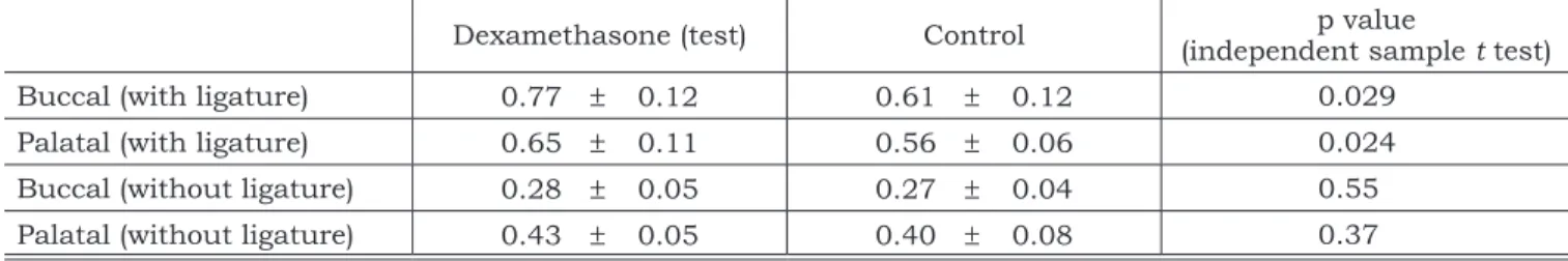
Table 1 demonstrates the main outcome of the present study (alveolar bone level 30 days after baseline) for both test and control groups during the study period. It can be observed that for the analysis performed buccally in teeth with ligature, groups test and control showed a mean value of 0.77 (± 0.12) and 0.61 (± 0.12) millimeters. This difference was statistically significant (p = 0.029). The same result was seen palatally with 0.65 (± 0.11) and 0.56 (± 0.06) for tests and controls, respectively, with a p value of 0.024.
Statistically significant differences were not detected between test and control groups neither buccally nor palatally in teeth without ligatures.
An intra-group comparison was performed between teeth with and without ligature and it is demonstrated in Graph 2. Independently from the experimental group, teeth with ligatures always showed statistically significant higher amounts of bone loss both buccally and palatally.
TABLE 1 - Mean (± standard deviation) of alveolar bone loss, in millimeters, for test and control groups, in teeth with or without ligature, 30 days after ligature placement.
Dexamethasone (test) Control p value
(independent sample t test)
Buccal (with ligature) 0.77 ± 0.12 0.61 ± 0.12 0.029
Palatal (with ligature) 0.65 ± 0.11 0.56 ± 0.06 0.024
Buccal (without ligature) 0.28 ± 0.05 0.27 ± 0.04 0.55
Palatal (without ligature) 0.43 ± 0.05 0.40 ± 0.08 0.37
GRAPH 1 - Mean body weight (g) for the experimental groups and time periods.
2
1
4
.4
6
2
1
6
.6
9
2
0
6
.0
7
2
2
6
.6
9
2
1
0
.2
3
2
3
9
.1
5
2
1
2
.7
6
2
4
2
2
1
5
.4
6
2
4
5
.7
6
180 190 200 210 220 230 240 250
Baseline
Me
a
n
b
o
d
y
w
e
ig
h
t
(g
)
Day 7 Day 14 Day 21 Day 30
Dexamethasone Control
DISCUSSION
The present study evaluated the effect of a steroidal anti-inflammatory (dexamethasone) on the pathogenesis of ligature-induced periodontal breakdown in Wistar rats. The results demonstrat-ed a higher bone loss in sides with ligature and under the intake of dexamethasone.
In the study of the pathogenesis of periodontal diseases, animal models are useful because hu-man studies are difficult to be performed for ethical reasons. Ligature-induced periodontal disease is considered to be one of the possible ways of study-ing this subject7,18,19.
Most of the studies have used histometric measurements for this purpose. However, the limi-tation of the number of slides evaluated as well as the possibility of not representing the complete pattern of alveolar bone loss should be kept in mind3,11,21. For this reason, in the present study,
dry bone was used to demonstrate bone level. Gaio
et al.6 (2004) have validated this dry bone
quanti-fication of alveolar bone loss in relation to histo-metric measurements.
Additionally, blinding of the examiner, ran-domization, utilization of a sufficient number of animals and use of comparative groups were prin-ciples followed by this study in order to better gen-erate evidence17. Our sample size calculation, with
a power of .90 indicated a minimum number of 11
rats in each group. Thus, we consider the sample of our study adequate in terms of quantity.
The first observed outcome in this study was body weight of the animals, which is a known way of evaluating systemic health7. At baseline, no
sta-tistically significant differences were observed be-tween test and control groups, which means they were comparable in this aspect at the beginning of the study. However, the use of dexamethasone caused an initial loss in body weight, which was recovered subsequently, but led test animals to always display statistically lower weight. A pos-sible explanation for this fact is that some loss of weight (of approximately 5% of body mass, which is considered low) has been reported in the first week of dexamethasone intake. The elevated dose of dexamethasone used in this study was chosen in order to better verify the potential of interfer-ing in pathogenesis. However, the concentration used was equivalent to that used in human ste-roid treatments of short duration13. It has to be
reminded that the use of dexamethasone is wide-spread. Adaptation of the animals to the medicine was associated with the ceasing of the diarrhea noted in the first days, including gain in weight from day 7 on.
Dexamethasone is suggested to impair wound healing and to diminish bone mineralization22. The
first effect could be reflected in more attachment loss, and the second effect could be connected to the observed fact that the maxillae from the test group could be easily fractured, as compared to those of controls.
The main outcome of this study, alveolar bone loss, was different between the studied groups both in the analyses performed buccally and palatally. Hence, no differences were observed in teeth with-out ligatures (Table 1). Moreover, Graph 2 demon-strates that teeth with ligature, regardless of the group, displayed more alveolar bone loss, which guarantees that the ligature-induced model pro-moted periodontal breakdown.
Thus, the potential role of steroidal antiin-flammatories in enhancing tissue loss can be dis-cussed. The results of our study indicate a differ-ent outcome as compared to that observed with the use of non-steroidal antiinflammatories5,9,10.
The greater alveolar bone loss observed in the present study is an important finding, which deserves some interest. The anti-inflammatory ef-fect can minimize clinical signs of inflammation at first by reducing host response. This impaired host response could be responsible for more
tis-0
.7
7
0
.2
8
0
.6
1
0
.2
7
0
.6
5
0
.4
3
0
.5
6
0
.4
0
0 0.1 0.2 0.3 0.4 0.5 0.6 0.7 0.8
Buccal Dexamethasone
Al
ve
o
la
r
b
o
n
e
l
o
ss
(mm)
Buccal Control
Palatal Dexamethasone
Palatal control
With Ligature Without Ligature
GRAPH 2 - Alveolar bone loss for test and control groups, according to location and the presence of liga-ture, 30 days after baseline.
sue breakdown. One of the suggested side effects of dexamethasone is enhancing potential of infec-tions22.
Studies with other anti-inflammatory drugs presented diverse results from these, howev-er, the host response modulation is always ob-served2,4,5,15,16.
Taking this into consideration, the results of the present study may add to the knowledge of the biological plausibility of pathogenesis of peri-odontal disease, suggesting association between the used medication and increase in alveolar bone loss. This information does not have the
inten-tion of being of direct clinical impact, but to show the potential of dexamethasone, as well as other steroidal antiinflamatory drugs to be a modifier of periodontal breakdown. However, patients and practitioners should be aware of this fact in clini-cal approaches.
CONCLUSION
It may be concluded that the use of systemic dexamethasone is associated with higher bone loss in ligature-induced periodontal disease in rats.
REFERENCES
1. Applebaum E, Seelig A. Histologic changes in jaws and teeth of rats following nephritis, adrenalectomy, and cortisone treat-ment. Oral Surg Oral Med Oral Pathol 1955;8(8):881-91. 2. Bissada NF, Ng VW. Clinical evaluation of systemic doxycycline
and ibuprofen administration as an adjunctive treatment for adult periodontitis. J Periodontol 1998;69(7):772-6.
3. Bjornsson MJ, Velschow S, Stoltze K, Havemose-Poulsen A, Schou S, Holmstrup P. The influence of diet consis-tence, drinking water and bedding on periodontal disease in Sprague-Dawley rats. J Periodontal Res 2003;38(6):543-50. 4. Ciancio SG. Systemic medications: clinical significance in
periodontics. J Clin Periodontol 2002;29 Suppl 2:17-21. 5. Feldman RS, Szeto B, Chauncey HH, Goldhaber P.
Non-ste-roidal anti-inflammatory drugs in the reduction of human alveolar bone loss. J Clin Periodontol 1983;10(2):131-6. 6. Gaio EJ, Fernandes MI, Oppermann RV, Rados PV, Rösing
CK. Analysis morphometric and hystometric bone high in induced periodontitis. J Dent Res [periódico on line] 2004. Available from: URL: http://iadr.confex.com/iadr/brazil04/ preliminaryprogram/abstract_55977.htm.
7. Galvão MPA, Chapper A, Rösing CK, Ferreira MBC, Souza MAL. Methodological considerations on descriptive studies of induced periodontal disease in rats. Pesqui Odontol Bras 2003;7:56-62.
8. Glickman I, Shklar G. The effect of systemic disturbances on the pulp of experimental animals. Oral Surg Oral Med Oral Pathol 1954;7(5):550-8.
9. Heasman PA, Benn DK, Kelly PJ, Seymour RA, Aitken D. The use of topical flurbiprofen as an adjunct to non-surgi -cal management of periodontal disease. J Clin Periodontol 1993;20(6):457-64.
10. Holzhausen M, Rossa Junior C, Marcantonio Junior E, Nassar PO, Spolidorio DM, Spolidorio LC. Effect of se-lective cyclooxygenase-2 inhibition on the development of ligature-induced periodontitis in rats. J Periodontol 2002;73(9):1030-6.
11. Klausen B, Evans RT, Sfintescu C. Two complemen-tary methods of assessing periodontal bone level in rats. Scand J Dent Res 1989;97(6):494-9.
12. Labelle RE, Schaffer EM. The effects of cortisone and induced local factors on the periodontium of the albino rat. J Periodontol 1966;37(6):483-90.
13. Metzger Z, Klein H, Klein A, Tagger M. Periapical lesion development in rats inhibited by dexamethasone. J Endod 2002;28(9):643-5.
14. Page RC, Kornman KS. The pathogenesis of human periodon-titis: an introduction. Periodontol 2000 1997;14:9-11. 15. Paquette DW, Willians RC. Modulation of host
inflam-matory mediators as a treatment strategy for periodontal disease. Periodontol 2000 2000;24:239-52.
16. Pauletto N, Silver JG, Larjava H. Nonsteroidal anti-in -flammatory agents: potential modifiers of periodontal disease progression. J Can Dent Assoc 1997;63(11):824-9,832. 17. Sacket D. Evidence-based medicine: how to practice
and teach EBM. New York: Churchill Livingtone; 2000. 18. Sallay K, Sanavi F, Ring I, Pham P, Behling UH,
No-wotny A. Alveolar bone destruction in the immunosup-pressed rat. J Periodontal Res 1982;17(3):263-74. 19. Susin C, Rösing CK. O rato como modelo para o estudo
das repercussões do estresse nas doenças periodontais. Revista Periodontia 2002;13(6):7-17.
20. Taiyeb Ali TB, Waite IM. The effect of systemic ibupro-fen on gingival inflammation in humans. J Clin Periodontol 1993;20(10):723-8.
21. Tatakis DN, Guglielmoni P. HLA-B27 transgenic rats are susceptible to accelerated alveolar bone loss. J Perio-dontol 2000;71(9):1395-400.
22. Wannmacher L, Ferreira MBC. Farmacologia clínica para dentistas. 2ªed. Rio de Janeiro: Guanabara Koogan; 1999. 23. Williams RC, Jeffcoat MK, Howell TH, Rolla A, Stubbs
D, Teoh KW, et al. Altering the progression of human al-veolar bone loss with the non-steroidal anti-inflammatory drug flurbiprofen. J Periodontol 1989;60(9):485-90.