Cyclin D1 expression and
cervical metastases in
squamous cell carcinoma of the
mouth
Summary
Gerson Schulz Maahs 1, Denise Cantarelli Machado 2, Emilio Antonio Jeckel-Neto 3, Vinicius Schenk Michaelsen 4
[1][1] Otorhinolaryngologist/Head and Neck Surgeon. M.S. in Medicine - PUCRS. Member of the Otorhinolaryngology/Head and Neck Surgery Department - PUCRS - Porto Alegre.
[2][2] PhD. Coordinator of the Cellular Biology and Respiratory Diseases Laboratory - Biomedical Research Institute. Professor at the School of Medicine - PUCRS. [3][3] Biologist and Master in Education (PUCRS), PhD in Medicine (Aichi Medical University, Japan), Full Professor at the School of Biosciences.
[4][4] PhD Student in Clinical Medicine and Health Sciences. Pharmaceutic. Biochemist. Otorhinolaryngologist/Head and Neck Surgeon. Porto Alegre RS PUCRS - Hospi-tal São Lucas e HospiHospi-tal Mãe de Deus - Porto Alegre RS.
Mailing Address: Gerson Schulz Maahs - Av. Palmeira 95/402 Bairro Petrópolis 90470-300 Porto Alegre RS.
Paper submitted to the ABORL-CCF SGP (Management Publications System) on January 24th, 2006 and accepted for publication on November 2nd, 2006. cod. 1693.
C
ervical metastasis represent the most relevant prognostic factor in carcinomas of the mouth. Clinical and histological factors are associated with the development of cervical metastasis; however, research on molecular factors has been broadly carried out in recent years. Aim: The aim of this study is to analyze the association of the Cyclin D1 as a risk factor for the presence of cervical metastasis. Materials and methods: Cyclin D1 expression was measured and the association between such substance and metastasis was found in 45 patients with mouth cancer treated by the author of this paper. Cyclin D1 presence was checked through the stereological method. Clinical and histological characteristics have been analyzed and associated with metastasis. Results:Cyclin D1 expression was found in 15 patients but it was not associated with clinical and histological factors related to the presence of metastasis. Such conclusion indicates that Cyclin D1 is an independent protein. The most important predictive aspects related to metastasis development have been clinical staging and vascular embolization. Conslusions: Cyclin D1, although independent, is not associated with cervical metastasis, while staging and vascular embolization are.
Keywords: cyclin d1, squamous cell carcinoma, mouth cancer metastases immunohistochemistry.
original article
INTRODUCTION
Epidermoid carcinoma corresponds to more than 90% of malignant mouth tumors, and its survival is mo-dified according to disease staging. When the disease is local, survival reaches 79%, loco-regional diseases yields 42%; and 19% is the survival rate when there are distant metastasis1. Clinical, histological, and most recently biolo-gical or molecular characteristics are considered prognostic factors, being predictive of cure and survival. The most important prognostic factor in mouth cancer is the pre-sence of neck metastasis, and this makes the diagnosis of distant metastasis fundamental for the oncology treatment planning2,3.
Cervical metastasis identification is carried out through physical exam and complementary tests, which fail in the diagnosis of 20 to 35% of the clinically negative patients2,3. Such false negative rate is deemed high and
justifies preventive neck treatment. When the primary tumor is treated exclusively, and there is hidden neck metastasis, there is a relevant reduction in patient survival. Patient selection for neck clearance is fundamentally ba-sed on the tumor clinical characteristics, as to its size and location3. Histopathological characteristics, such as cellular
differentiation level and tumor thickness also help in this selection; however, still keeps high failure rates. Tumor biological aspects are being researched in an attempt to identify which patients have a higher chance of developing neck metastasis3,4.
D1 cyclin is a protein coded by gene CCND1 located in chromosome 11q13. This protein acts on the cellular cycle, accelerating phase G1 and was described as an oncogene in 1991 by Motokura5, who observed that an increase in cyclin deregulates the cell cycle and contri-butes to tumor genesis. D1 cyclin presence is shown by immunohistochemistry in head and neck cancer, and the higher its expression, the lower patient survival is, thus being considered a prognostic factor by many researchers in carcinomas of the esophagus, breast, uterine cervix, colon, rectum and melanomas5,6.
In the present investigation, we assessed D1 cyclin expression in a series of 45 patients with mouth epider-moid carcinoma, and its association with neck metastasis. The goal of our study was to try and relate D1 cyclin expression as a risk factor for neck metastasis.
PATIENTS AND METHODS
1. Patients
We studied 45 patients with mouth epidermoid carcinomas who were operated by the author during the period of January 1991 and December of 2001. The patients suffered tumor surgical resection in its place of origin and neck lymphnode clearance. The neck lymphnodes surgery
included patients who did not present clinical metastasis (N-), and those from patients who did present clinically diagnosed metastasis (N+).
D1 cyclin expression was carried out in histologic slides obtained from the primary tumor. We reviewed patients’ charts and collected pre-surgical clinical data, pathologic exam data from the surgical specimen and post-operative development.
Patients who presented one or more of the follo-wing situations were taken off the study:
+ Surgery without curative intention.
+ Other concomitant neoplasias or history of prior tumors.
+ Neo-adjuvant therapy (chemo and radiothera-py).
+ Patients whose chart data were incomplete. + Impossibility of defining clinical staging
2. Immunohistochemistry
We then obtained the paraffin blocks that had the primary tumors from the patients we were studying. From the main block of each tumor we obtained 4mm thick cross sections.
The sections were placed on 10% poly-L-lysine (Sigma, USA) pre-treated slides and taken to the oven at 60oC for 24 hours.
Paraffin was taken off by means of 10 minute xy-lene incubation for three times, followed by rehydration in ethanol sequence in decreasing concentrations, starting by absolute ethanol, 90%, 80% and 70% for 3 minutes in each dilution. Following that, the sections were washed three times in distilled water.
Antigenic sites recovery with trypsin digestion was carried out after heating at 37oC in a 0.1% trypsin solution with 0.1% calcium chloride in 7.8 pH. The slides were then incubated in this solution for 30 minutes at 37oC, and were then washed in distilled water.
High temperature antigen exposure was carried out by incubation in glass cradle, with a buffer containing 1mM of EDTA arranged in a 45o angle, in mean power for five minutes and fifty seconds, followed by eight minutes in low power (Panasonic, 1.500W microwave oven).
with peroxidase. After 10 minutes of incubation, the slides were washed in PBS and a solution with diaminobenzidine (Dakoâ Liquid DAB Substrate Chromogen System, USA). After washing in distilled water, the slides were counter-dyed with Harris hematoxylin for 15 seconds, washed in three water baths and dipped in an ammonia 37 mM so-lution. After counter-dye, the slides were washed in three baths of warm water, followed by dehydration in growing ethanol concentrations (80%, 90% and pure, respectively) for three minutes in each dilution. After two treatments for five minutes in xylene, the sections were covered by thin slides assembled in Entellanâ (Merck, Germany).
The slides were analyzed under light microscopy aiming at assessing D1 cyclin expression.
D1 cyclin expression measure was carried out by means of a method called stereologic, which aim is to determine quantitative parameters from bi-dimensional sections of three-dimensional microscopic anatomical structures, by means of geometry and statistics7,8.
D1 cyclin expression was measured by the count of dyed nuclei divided by the area in mm2 and the value indicated by Qa 9.
Slide analysis was carried out under Olympus AX-70 (Japan) light microscopy, coupled to a video camera. The images obtained were transferred to a computer and counts made with the help of the Image Pro Plus version 4.1 software (Media Cybernetics, USA). On an image taken from a 20x lens we applied a grit that divided it in nine equal fields, according to Figure 1.
From each slide we obtained five values and cal-culated the average of five Qa obtained.
3. Statistical analysis
The quantitative variables were described by avera-ge and standard deviation, and percentaavera-ges were used for the qualitative variables. The group’s quantitative variables were compared by means of the Student t test, and for the qualitative variables we used the chi-squared test. Associa-tion measures such as the odds ratio and the relative risk with its respective confidence intervals were completely performed. In order to adjust the association between cyclin and metastasis in front of so diverse potential risk factors, we used the multivariate logistical regression.
Survival curves were calculated using the Kaplan-Meyer’s method. Death that did not occur because of the primary tumor or from its metastasis were not considered treatment failures, but rather censored observations. The differences between the survival curves were compared by the log rank test. In order to simultaneously adjust the effects of many factors considered and their relation with death, we used Cox regression. The P values below 0.05 were considered statistically significant, and the multiva-riable models considered P values below 0.1.
For the statistical analysis, data was processed and
analyzed with the help of the SPSS (Statistical Package for Social Science) software for Windows and Excel 97, version 11.1.
4. Ethics
The study is in compliance with items III.3.i and III.3.t from the Research Regulatory Guidelines and Stan-dards involving Human Beings (Resolution CNS 196/96), as well as Guideline 12 from the International Ethics Gui-delines for Biomedical Research Involving Human Beings (CIOMS 1993).
The principles present in the Helsinki Declaration were abided (WORLD MEDICAL ASSOCIATION, 1996), keep confidential the patients’ identities, whose data were identified only by the record number in the project.
The project was submitted and approved by the Research Ethics Committee of the Pontifícia Universidade Católica do Rio Grande do Sul (Protocol number 298/01 - CEP).
RESULTS
D1 cyclin clinical and histologic characteristics of the 45 patients studied may be seen on Table 1.
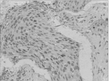
D1 cyclin was positive in 15 (33.3%) cases and ne-gative in the remainder 30 (66.7%). The average of labeled nuclei (Qa) varied between 0.15 and 1.24 in the positive cases. The protein presence in the cell nucleus may be seen in Figure 2.
The protein expression in the cell cytoplasm was also observed in three patients.
The associations and correlations between patients with metastasis (pN+) and those without it (pN-) were
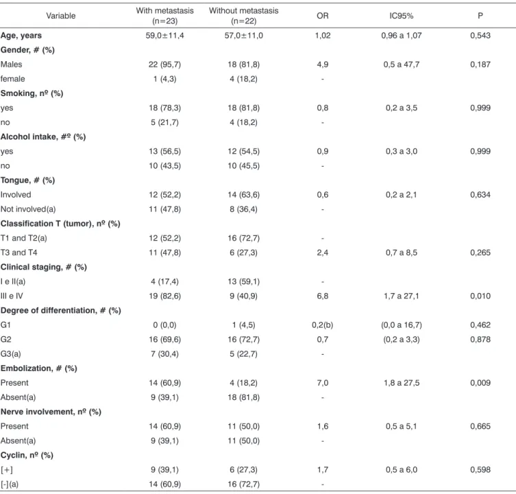
Table 1. Comparison between metastatic and non-metastatic groups as to the following variables: demographics, habits and clinical traits, histology and tumor molecular characteristics.
Variable With metastasis (n=23)
Without metastasis
(n=22) OR IC95% P
Age, years 59,0±11,4 57,0±11,0 1,02 0,96 a 1,07 0,543
Gender, # (%)
Males 22 (95,7) 18 (81,8) 4,9 0,5 a 47,7 0,187
female 1 (4,3) 4 (18,2)
-Smoking, nº (%)
yes 18 (78,3) 18 (81,8) 0,8 0,2 a 3,5 0,999
no 5 (21,7) 4 (18,2)
-Alcohol intake, #º (%)
yes 13 (56,5) 12 (54,5) 0,9 0,3 a 3,0 0,999
no 10 (43,5) 10 (45,5)
-Tongue, # (%)
Involved 12 (52,2) 14 (63,6) 0,6 0,2 a 2,1 0,634
Not involved(a) 11 (47,8) 8 (36,4)
-Classification T (tumor), nº (%)
T1 and T2(a) 12 (52,2) 16 (72,7)
-T3 and T4 11 (47,8) 6 (27,3) 2,4 0,7 a 8,5 0,265
Clinical staging, # (%)
I e II(a) 4 (17,4) 13 (59,1)
-III e IV 19 (82,6) 9 (40,9) 6,8 1,7 a 27,1 0,010
Degree of differentiation, # (%)
G1 0 (0,0) 1 (4,5) 0,2(b) (0,0 a 16,7) 0,462
G2 16 (69,6) 16 (72,7) 0,7 (0,2 a 3,3) 0,878
G3(a) 7 (30,4) 5 (22,7)
-Embolization, # (%)
Present 14 (60,9) 4 (18,2) 7,0 1,8 a 27,5 0,009
Absent(a) 9 (39,1) 18 (81,8)
-Nerve involvement, nº (%)
Present 14 (60,9) 11 (50,0) 1,6 0,5 a 5,1 0,665
Absent(a) 9 (39,1) 11 (50,0)
-Cyclin, nº (%)
[+] 9 (39,1) 6 (27,3) 1,7 0,5 a 6,0 0,598
[-](a) 14 (60,9) 16 (72,7)
-Data presented as average ± standard deviation and frequency (percentage). OR: odds ratio; CI95%: Confidence Interval of 95%. (a) Referen-ce category in the calculation of odds ratio; (b) Odds ratio adjusted by Agresti’s method.
carried out base don clinical and histological traits, habits, demographics and D1 cyclin, are depicted on Table 1. The multivariate analysis used to asses the risk of D1 cyclin association with other potential risk factors was carried out and is depicted on Table 2.
Survival was assessed based on D1 cyclin expression and the presence of metastasis in months, by means of a
survival curve of which outcome considered was death due to mouth cancer, which occurred in seven situations. Results may be seen in Figures 3 and 4.
Figure 2. D1 cyclin expression - D1 cyclin expression dyed in Brown in the nucleus of neoplastic cells.
Table 2. Multivariable logistic regression model which adjusts the cyclin effect [+] when facing many potential risk factors for its asso-ciation with metastasis.
Variable OR CI90% P
Cyclin [+] 2.7 0.6 a 11.6 0.267
Clinical staging III and IV 16.4 2.8 a 95.5 0.009
Tongue involvement 0.2 0.1 a 0.9 0.068
Vascular embolization 4.3 1.1 a 17.5 0.084
Cellular differentiation G3 8.1 1.2 a 53.4 0.069
Nerve involvement 0.5 0.1 a 2.1 0.460
OR: odds ratio; CI90%: Confidence Interval of 90%.
Figure 3. Metastasis and survival - Kaplan-Meier’s chart showing death occurrence stratified by metastasis presence or absence.
Figure 4. D1 cyclin and survival - Kaplan-Meier’s chart showing death occurrence stratified by positive ([+]) and negative ([-]) cyclin groups.
Table 3. Multivariable Cox regression model which adjusts the cyclin effect [+] when facing many potential risk factors for its association with death.
Variable RR CI90% P
Cyclin [+] 0.8 0.2 to 3.5 0.846
Clinical staging III and IV 4.9 0.6 to 37.9 0.198
Tongue involvement 1.0 0.2 to 5.7 0.964
Vascular embolization 1.6 0.3 to 7.2 0.625
G3 Cell differentiation G3 3.6 0.6 to 20.9 0.226
Nerve involvement 0.4 0.1 to 2.4 0.409 OR: odds ratio; CI90%: Confidence Interval of 90%.
involvement, cell differentiation, vascular embolization and nerve involvement were studied and are depicted on Figure 5.
DISCUSSION
D1 cyclin was expressed in 15 (33.3%) patients from the sample, which is in agreement with published results that vary between 30 and 40%10-12, although by a
distinct methodology of immunohistochemistry reading. The literature on D1 cyclin and cancer assesses the pre-sence of cyclin by counting the nuclei labeled by a group of neoplastic cells, which usually vary between 500 and 1000 cells, in percentages. By means of such method, the investigator needs to count a total of neoplastic cells, regardless whether they are marked or not, and after that, recounting the cells that express the protein, and calculate the percentage. Besides being a labor-intensive method, it bears a higher risk of distortions in counting. The choice of cutting point that separates the positive from the negative D1 cyclin expression is arbitrary. Han et al.13, consider it
positive when there is more than 5% of labeled nuclei. Ravi et al.14, Itami et al.11 and
Capaccio et al.12, consider the D1 cyclin expression
only significant when present in over 10% of the tumoral cells. El-Naggar et al.15 consider values above 50% as an overexpression of this protein. Mineta et al.16 consider values between 0-50% being of low expression, and those above 50% as being of high expression. Schoelch et al.10 use the negative score when there is no expression, + for up to 33% expression, ++ between 33 and 66% and +++ above 66% of protein expression in tumor cells.
The stereological method used in this investigation assess the protein expression in the nuclei by tumor area, is more reliable8,9. The protein expression in the cell
cyto-plasm was also observed as described in other studies17.
D1 cyclin may be expressed in different intensity levels. The term “high expression” is defined by the la-beling of more than 50% of the neoplastic cells10. In the
present study, Qa results obtained for the 15 positive pa-tients varied from 0.15 to 1.27, and the differences of the values found were not significant. It was not possible to establish the cyclin expression in low and high expression because of patient sample size; however, the quantitative analysis of Qa values was carried out.
D1 cyclin expression in the 15 positive patients was not constant in the whole tumor surface area. In all the cases, protein expression analysis was carried out in the region of greatest labeling in the slide, showing tumor areas that did not express the protein. This lack of labeling constancy may be justified by the fact that about 20% of the neoplastic cells are under mitosis18.
Conside-ring this asymmetrical expression distribution, one could question the studies in which the researchers used biopsy specimens, as reported by Michalides et al.19, where D1
cyclin expression false negatives were observed in biopsy material when compared to the surgical specimen; none-theless, further studies are required in order to measure which is the true sensibility of D1 cyclin expression in biopsy material compared to a surgical specimen. This is relevant, since false negatives occurring in the biopsy material would compromise the clinical application of such marker when a treatment decision is to be made, for example, whether or not to indicate neck clearance in a patient clinically classified as N0.
As to gender, smoking and alcohol intake, there was no relation with the presence of neck metastasis and the invasion of lymphnode capsule, which confirms that they do not represent risk factor for neck metastasis16.
Age was also not related to the presence of neck metastasis and lymphnode capsule invasion, although very young and very old patients develop tumors with more aggressive behavior20.
As to location, there was also no association with the presence of metastasis. The 14 T1 and T2 patients with tongue epidermoid carcinomas presented metastasis in six cases, while the remaining 14 T1 and T2 patients classified as having cancer in other parts of the mouth presented metastasis in six cases. Location is not impor-tant when the tumor is large, as is the case with T3 and T4 tumors that already bear high metastasis development risk individually.
Tumor location in the mouth was associated to lym-phnode capsule invasion. The tumors that involved more than one location in the mouth had greater involvement of the lymphnode capsule; with a p value of 0.049.
Twelve of the 28 T1 and T2 patients presented histologic metastasis (42.8%), a value close to expected, which is of 20 to 40%. As to the 17 T3 and T4 patients, 11 had metastasis (64.7%) in agreement with the literature21. There was no statistical relation between tumor size and metastasis (P = 0.265). We also did not find significant association between tumor size and metastatic lymphnode capsule invasion (P = 0.089).
Regarding the classification N, histology showed that of the 25 negative cases, eight (32%) were false negatives and 17 (68%) were true negatives. As to the 20 N+ patients, histology revealed five (25%) false positive patients, and in 15 (75%) neck metastasis was confirmed. The P value found was of 0.001 for neck metastasis and 0.047 for cap-sule invasion. The number of false positives was of 25%, corresponding to what has been described in the literature, which is 20%, while the rate of false negatives was of 32%, while the value reported in the literature lays between 20 and 35%22,23. Metastasis patients’ stratification was not
stu-died in the present investigation, due to the small number of patients allocated in each subdivision N.
can-cer patient is defined, it is considered tumor size and the clinical presence of metastasis by neck palpation. Since patients classified as positive N are in stages III and IV, the value found is not surprising and is directly associated with the incidence of false positives and false negatives on neck palpation, which in the present study was of 25% and 32% respectively. Neck palpation and the T classification alone were not significant, and this by itself justifies the use of a staging process.
The degree of cell differentiation was not associated to the presence of neck metastasis or with lymphnode capsule invasion. The presence of only one patient with well differentiated epidermoid cancer made it impossible to observe the relationship between the degree of diffe-rentiation and the risk of metastasis development.
Nerve sheath invasion was not associated to the pre-sence of neck metastasis, nor to the invasion of lymphnode capsules. Nerve sheath invasion happened in 14 (60.9%) of the 23 metastatic patients; however without statistical meaning. There was also no association between nerve sheath invasion and lymphnode capsule invasion.
Vascular embolization was predictive as far as metastasis presence is concerned. Woolgar24 found 78%
of vascular invasion in N positive patients and 70% nerve sheath invasion in another sample also of 45 patients with mouth epidermoid carcinoma. In the present study we found 60.9% for both situations; however with different meaning when we observe the results of the proper sta-tistical significance tests, and this shows the relevance of data analysis tests, thus avoiding misinterpretations.
D1 cyclin expression is not associated to metastasis. D1 cyclin quantified by Qa value also did not change the results. The lack of studies published in the literature using the stereological method makes it impossible to compare quantitative results. Cyclin was also not associated to me-tastatic lymphnode capsule invasion.
In the literature, the association between D1 cyclin expression and the presence of neck metastasis is ambiguous. The results of the present investigation agree with those presented by Han et al.13, Michalides et
al.19,25 and Bellacosa et al.26. However, the papers from
Frachiolla27, Itami11, Masuda28, Tapia et al.29, Capaccio et
al.30 and Mineta et al.16 showed that D1 cyclin is indeed
associated with the presence of neck metastasis. Results are contradictory and the reasons may be the following: D1 cyclin expression differ in the different organs of the body; the very condition of the surgical specimen such block tumor and formaldehyde time until it is fixed; cutting point used to define what is protein overexpression and a standard reading methodology avoiding interpretation subjectiveness.
D1 cyclin multivaried analysis with the most relevant prognostic factors did not change the results, showing that D1 cyclin expression does not depend on the other
factors studied, agreeing with the findings from Capaccio et al.30 and Mineta et al.16.
The survival curve of the 45 patients in the sam-ple shows that D1 cyclin positive patients have a lower tendency to survive when compared to negative patients, although the statistical significance was weak.
Survival analysis concerning metastasis reveals that neck metastasis patients present lower survival rates, and confirms the meaning of neck metastasis as a prognostic factor associated with mouth cancer. 3,31-33.
Multivaried analysis shows that D cyclin and neck metastasis are independent factors as to their relation with survival, agreeing with the results from Han et al.13. These
authors did not find associations between D1 cyclin and the presence of metastasis in gastric cancer, and found an association with patient survival. Michalides et al.19,25
studied head and neck carcinomas and Bellacosa et al.26
studied patients with larynx cancer and also did not re-port correspondence between metastasis and D1 cyclin expression; nonetheless, they did find relations with pa-tient survival.
In the present investigation, neck metastasis was significant in survival reduction, while D1 cyclin did not show the same statistical significance, only showing a trend that patients with higher cyclin expression had worse survival rates.
CONCLUSIONS
The present investigation leads us to conclude that:
a. D1 cyclin expression seems to be independent from mouth epidermoid carcinoma.
b. It is very likely that D1 cyclin is not associated with the presence of neck metastasis.
c. Vascular embolization and clinical staging were the most significant factors as neck metastasis predictors in the cases hereby studied.
d. Neck metastasis was significant in the survival of our patients in this study, while the same can not be said about D1 cyclin.
REFERENCES
1. Gloecker Ries LA. SEER cancer statistics review, 1973-1991.Bethesda MD: US. Department of Health and Human Services, Public Health Service, National Cancer Institute 1994. Report no. NIH-94-2789. 2. Shaha AR, Spiro RH, Shah JP, Stron EW. Squamous carcinoma of the
floor of the mouth. Am J Surg 1984;148:455-9.
3. Russolo M, Giacomarra V, Papanikolla L, Tirelli G. Prognostic indica-tors of occult metastases in oral cancer. Laryngoscope 2002;112:449-52.
4. Mohit-Tabatabai M. Relation of Thickness of Floor of Mouth Stage I and II Cancers to Regional Metastasis. The Am J. of Surg1986;152:351-3.
5. Motokura T, Bloom T, Kim HG. A novel cyclin encoded by a bcl1-linked candidate oncogene. Nature 1991;350:512-5.
marker in head and neck squamous cell carcinomas. Head Neck 2001;23(2):147-59.
7. Mandarim-de-Lacerda CA. Métodos quantitativos em morfologia. Rio de Janeiro: UERJ; 1995. p 72-107.
8. Gundersen HJG et al. Some new, simple and efficient stereological methods and their use in pathological research and diagnosis. APMIS 1988;96:379-94.
9. Hamilton PW, Allen DC. Quantitative clinical pathology. Oxford: Blackwell Science Inc.; 1995, pp. 4-5.
10. Schoelch ML, Regezi JA, Dekker NP. Cell cycle proteins and the deve-lopment of oral squamous cell carcinoma. Oral Oncol 1999;35(3):333-42.
11. Itami A, Shimada Y, Watanabe G, Imamura M. Prognostic value of p27 and cyclin D1 expression in esophageal cancer. Oncology 1999;57:311-317.
12. Capaccio P, Pruneri G, Carboni N, Pagliari AV, Quatela M, Cesana BM, Pitnataro L. Cyclin D1 expression is predictive of occult metastases in head and neck cancer patients with clinically negative cervical lymph nodes. Head and Neck 2000;5:234-40.
13. Han S, Kim H, Park K. Expression of p27 and cyclin D1 proteins is inversely correlated and associated with poor clinical outcome in human gastric cancer. J Surg Oncol 1999;71:147-54.
14. Ravi D, Ramadas K, Mathew BS, Nalinakumari KR, Nair MK, Pillai MR. Angiogenesis during tumor progression in the oral cavity is related to reduced apoptosis and high tumor cell proliferation. Oral Oncology 1998;34:543-8.
15. El-Naggar AK, Lai S, Cayman GL, Zhou J, Tucker SA, Myers J, Luna MA, Benedict WF. Expression of p16, Rb and Cyclin D1 gene products in oral and laryngeal squamous carcinoma: biological and clinical implications. Human Pathol 1999;30:1013-8.
16. Mineta H, Miura K, Takebayashi S. Cyclin D1 overexpression cor-relates with poor prognosis in patients with tongue squamous cell carcinoma. Oral Oncology 2000;36:194-8.
17. Goodger NM, Gannon J, Hunt T, Morgan PR. Cell cycle regulatory proteins - an overview with relevance to oral cancer. Oral Oncology 1997;33:61-73.
18. Alberts B, Bray D, Lewis J, Raff M, Roberts K, Watson SD. Biologia molecular da célula. Porto Alegre: Artes Médicas;2002. p 1273-90. 19. Michalides RJAM, Van Veelen NMJ, Kristel PMP. Overexpression of
cyclin D1 indicates a poor prognosis in squamous cell carcinoma of the head and neck. Arch Otolaryngol Head Neck Surg 1997;123:497-502.
20. Carvalho FE, Papaleo NM. Geriatria, fundamentos, clínica e terapêu-tica. São Paulo: Atheneu; 1994, pp 9-30.
21. Jones AS. Prognosis in Mouth Cancer: Tumor Factors. Oral Oncol, Eur J Câncer 1994;30B: 8-15.
22. Carter LR. Patters and mechanisms of spread of squamous carcinomas of the oral cavity. Clinic Otolaryngology 1990;15:185-91.
23. Ali S, Tiwari RM, Snow G. False-positive and False-negative neck nodes. Head Neck Surg 1985;8:78-82.
24. Woolgar J, Path MRC, Scoot J, Path FRC. Prediction of cervical lymph node metastasis in squamous cell carcinoma of the tongue/floor of mouth. Head & Neck 1995;17:463-72.
25. Michalides R, Van Veelen N, Hart A, Lofturs B, Wientjens E, Balm A. Overexpression of cyclin D1 correlates with recurrence in a group of forty-seven operable squamous cell carcinomas of the head and neck. Cancer Res 1995;55:975-8.
26. Bellacosa A, Almadori G, Cavallo S. Cyclin D1 gene amplification in human laryngeal squamous cell carcinomas: prognostic significance and clinical implications. Clin Cancer Res 1996;2(1):175-80. 27. Fracchiolla NS, Pruneri G, Pignataro L, et al. Molecular and
immuno-histochemical analysis of the bcl-1/cyclin D1 gene in laryngeal squa-mous cell carcinomas: correlation of protein expression with lymph node metastases and advanced clinical stage. Cancer 1997;79:1114-21.
28. Masuda M, Hirakawa N, Nakashima T, Kuratomi Y, Komiyama S. Cyclin D1 overexpression in primary hypopharyngeal carcinomas. Cancer 1996;78:390-5.
29. Tapia JPR, Meana MVG, Gonzáles LAG. Inactivación de p53 y ampli-ficación de la ciclina D1 en los carcinomas epidermoides de cabeza e cuello. Acta Otorrinolaringol Esp 2002;53:225-32.
30. Capaccio P, Pruneri G, Carboni N. Cyclin D1 protein expression is related to clinical progression in laryngeal squamous cell carcinomas. J Laryngol Otol 1997;111(7):622-6.
31. Spiro RH, Alfonso AE, Farr HW, Stron EW. Cervical node metastases from epidermoid carcinoma of the oral cavity or oropharynx: a critical assessment of current staging. Am J Surg 1974;128,562-7.
32. Hibbert J, Marks NJ, Winter PJH, Shaheen OH. Prognostic factors in oral carcinoma and their relation to clinical staging. Clin Otolaryngol 1983;8:197-203.